Connected Productivity in the Materials Lab
ZEISS ZEN core
Your Software Suite for Connected Microscopy—from the Materials Lab to Production
› The Advantages › The Applications
The System

ZEISS ZEN core
Your Software Suite for Connected Microscopy—from the Materials Lab to Production
› The Advantages › The Applications
The System
ZEISS ZEN core is a powerful software suite for microscopy imaging, automated control of motorized ZEISS microscopes, and multi-modal workflows in material laboratory environments.
Use ZEN core to handle routine tasks on a wide range of ZEISS microscope and camera systems. While extracting the highest technical performance from your microscopes, ZEN core provides access to every parameter and function you might wish to optimize through an intuitive and configurable graphical interface.
Create task-specific workbenches that show only the required microscope controls on the screen. Combine these workbenches to create dedicated jobs that assist your operators through a defined flow of consecutive tasks, to ensure data repeatability in a multi-user environment.
ZEN core helps you to make your lab even more productive. With workflow and infrastructure solutions that connect data from different microscopes, ZEN core delivers more meaningful information in the form of correlated multiscale and / or multi-modal characterization data. And thanks to its database connectivity features, you keep your valuable data together across instruments, laboratories, and locations.
Easy to Configure. Easy to Use.
ZEN core gives you the benefit of an adaptive user interface tailored to the needs of industrial and research environments. The easy-to-follow GUI configuration accommodates tasks of all kinds and any complexity. ZEN core also offers you configurable user management, so you can specify users and user roles. Whatever their level of experience, operators will learn the software quickly. Using the ZEISS Word Add-In lets you easily create user configured report templates in MS Word ®. The reporting functionality exports reports to various file formats including PDF or DOCX.
ZEN core is the command center for automated imaging and analysis functions on compound light microscopes. By using built-in automated image acquisition routines, such as HDR or Best Image, you benefit from the consistency of an advanced and repeatable workflow. ZEN core provides automated image segmentation based on machine learning algorithms, as well as analysis functionality such as phase analysis or particle counting. Application-specific modules enhance your microscope to answer typical questions about the material structure in research and quality control.
ZEN core provides the infrastructure for connected laboratory environments, linking all your ZEISS imaging and microscope solutions to a single, familiar GUI. ZEN core is also the interface to the ZEISS Axiocam camera portfolio, safeguarding an open connected laboratory architecture for 3 rd party solutions. ZEN core bridges different forms of light and electron microscopy, improving productivity and multi-modal data integrity. Data management and database connectivity features help you to keep your valuable analysis data together across instruments, laboratories, and locations.
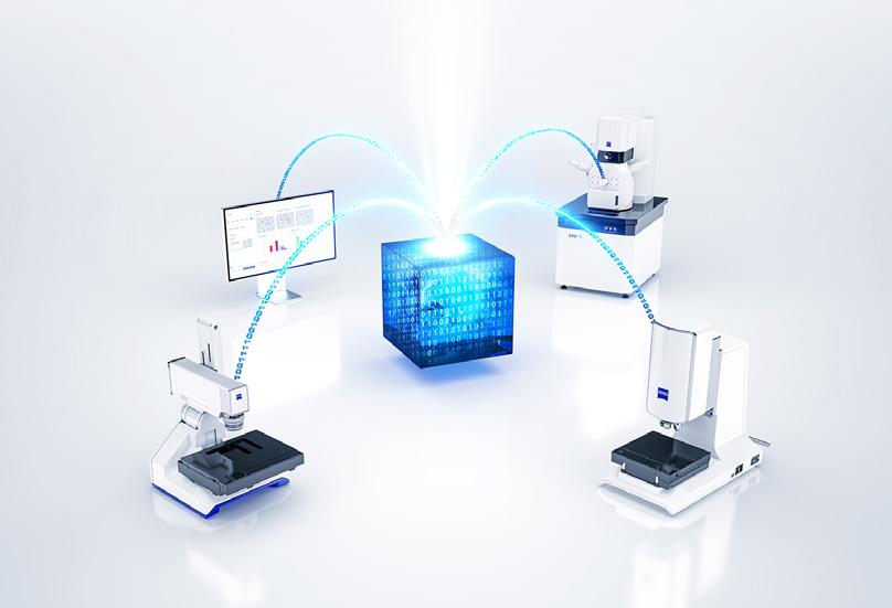
From entry-level stereo microscopes to fully automated imaging systems, ZEN core provides a unified user interface for ZEISS microscopes and cameras. ZEN core enables the correlation of light and electron microscopy in multi-modal workflows and provides connectivity between systems, laboratories and locations.
1 Microscope Control Manual and automated control of light microscopes and ZEISS Axiocam cameras
2 Data Acquisition and Analysis Light and electron microscopes
3 Correlative Microscopy Sample and data exchange between light, digital, and electron microscopes
4 Post Acquisition Analysis Image processing, analysis, and reporting on separate office workstations
5 Automated Segmentation Image segmentation based on machine learning algorithms
6 Contextual Analysis Data visualization and organization across scales and imaging modalities
7 Reporting Integrated reporting across connected images and datasets
8 Mobile Access Data exploration on mobile device
9 Central Data Management Central storage of images, analysis data, presets, workflows, and reports
10 Conntected Laboratory Data and workflow exchange between systems, labs, and locations
11 Interfaces
Exchange data for further analysis with other software solutions e.g. PiWeb, Zaphire, Confomap, GOM Inspect
In Brief
The Advantages
The Applications › The System
Image, Analyze and Connect your Data Using the Complete Software Suite
ZEN core
ZEN core, with its configurable user interface, gives you seamless control of your ZEISS microscopes and cameras as you perform data acquisition, analysis and reporting.
ZEN analyzer is the desktop version of ZEN core, designed for all those activities that can be done independently of the microscope. This is the ideal solution for analysis, reporting and creating job templates, providing remote access to ZEN Data storage. Your instrument is not blocked for post-processing tasks, but instead can be used to run other experiments—anywhere, anytime, and with genuine efficiency. Additionally, access to all workbenches available in ZEN core gives you full control of all data and templates, accessible from your desk.
Combine ZEN Data Explorer with ZEN Data Storage for mobile access to your data. This lets you use your tablet or smartphone to examine your results when you’re on the go.
ZEN Data Storage enables data and workflow exchange between systems, labs and locations. It allows server-based user management and secures central data handling for your lab and quality assurance. All your images, templates, forms and reports are accessible in one hub.
User Management Designed to Assure
Data Repeatability and Integrity
ZEN core comes with powerful built-in features for user management. Create accounts with defined privileges and roles such as administrator, supervisor and operator. If necessary, you can define precisely what each user is allowed to do, right down to the level of individual features. Create and manage users directly in ZEN core—or connect to ActiveDirectory and reuse those user accounts. Protect access with passwords and leverage extensive capabilities to put password rules and expiration times in place. Auto-logoff helps you prevent further unwanted access.

workflow multiple times and store a summarized report at the end. You can even set workbenches to “run silent” (that is, in the background and invisible to the operator) with preset options for implementing batch processing and, for example, analyzing multiple images in exactly the same way. › In Brief › The Advantages › The Applications › The System
Job Mode makes it easy to set up workflows for repetitive tasks. You can predetermine microscope parameters and specify them as individual workbenches to guarantee reproducible results. Then combine workbenches in a job template to set up individual workflows. Any parameter in the work -
benches of a job template (e.g. camera exposure time) can be locked or even preset and hidden from the operator to ensure consistency in sample examination. Combine Job Mode with user management functions to assign inspection tasks to certain users or user groups. Create loops for when you want an operator to execute the same
User form to enter sample information, project name or user data
Image acquisition combined with Tiles, EDF or 3D functionality
Workflow example for a job in Job Mode of ZEN core.
Interactive or automatic measurements
Repeat at several sample positions
Automated reporting
archiving of relevant data
Reporting
ZEN core´s powerful reporting functionality lets you create reports in MS Word or PDF format.
Combined with ZEN core’s Job Mode, the reporting process is fully automated without any customer interaction.
Report templates are created in MS Word using the plug-in ZEISS Word-In.

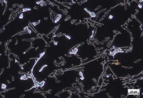
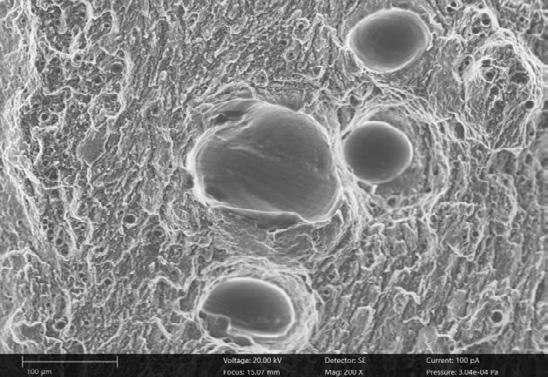
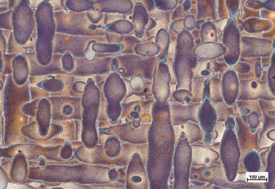
File Name AlSi10Mg_0017.czi Process 3D printed
Condition Fractured surface
Comment SEM_TP_22 File Name AlSi10Mg_04.tif
Process 3D printed
Condition As printed Comment LM_TP_00
› In Brief › The Advantages › The Applications › The System
So, you have just purchased a new microscopy system from ZEISS—or perhaps you now have access to several systems to deliver correlated data across length scales. The possibilities are endless and you are anxious to get to work. So where to begin?
If you are looking for a specific solution to your challenge, skim the website to see what is already available. The hub for the Solutions Lab offers apps for industrial R&D, Materials Science, investigation of metals and natural resources. Even if you do not find a suitable solution, something close to it may have been created for a similar application in another field and we can get you the rest of the way. Simply contact ZEISS via the contact form on the ZEISS Solutions Lab webpage or ask your ZEISS salesperson, explaining what you are looking for. You can always count on our support in developing your best solution.
• automation of your imaging process or workflow
• advanced image analysis and corrections
• automation of your ROI (region of interest) search
• tailoring of flexible and adaptable workflows for your configuration and of course, some handy small applications that turn 20 clicks into one.
The Advantages
The Applications
The System
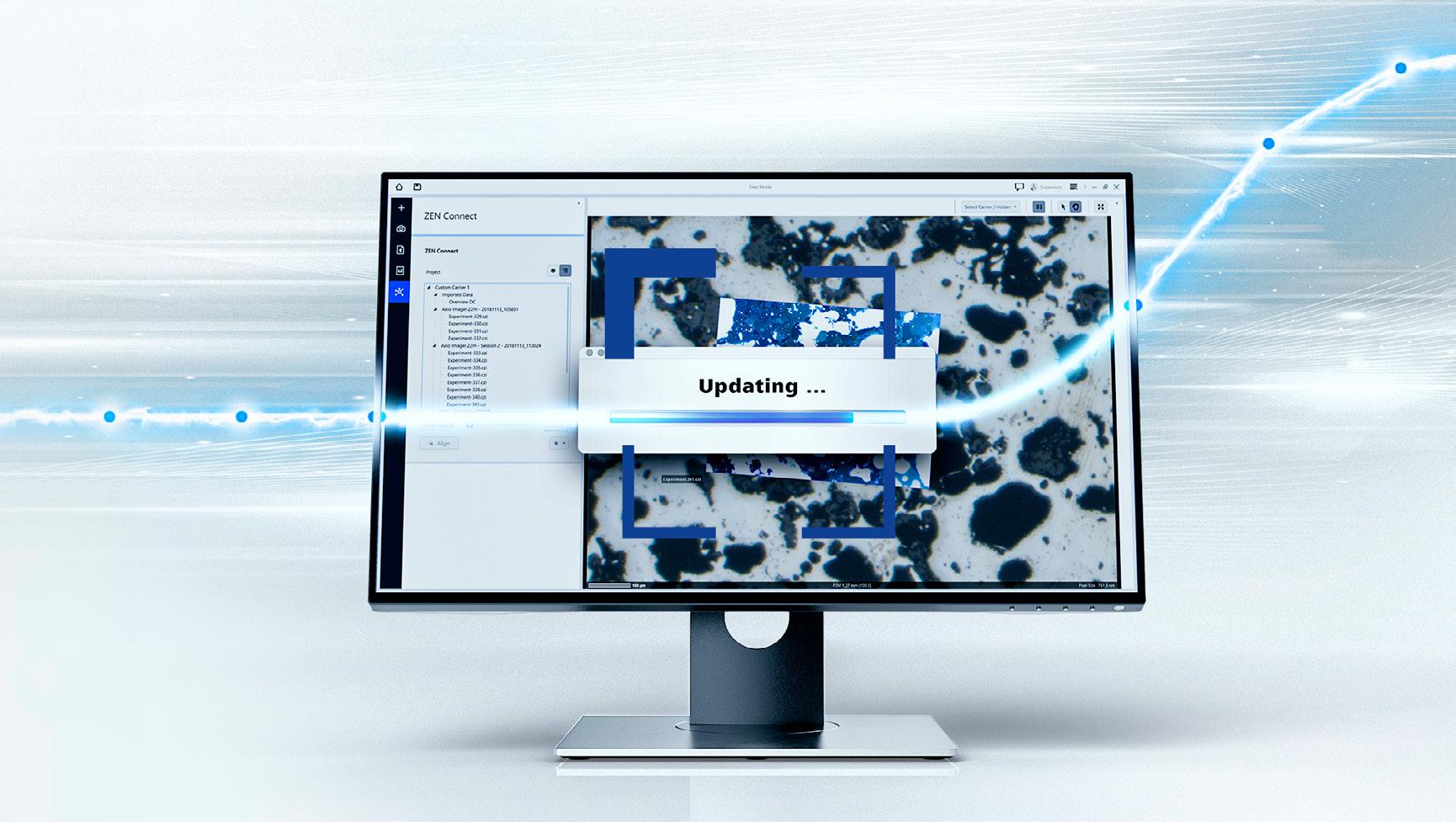
Sign up for an SMA (Software Maintenance Agreement) and get notified immediately via the ZEISS Portal when a new version for your system is ready to be downloaded. As a result, you will always keep all your systems up to date. Enjoy the latest features, newest possibilities and most recent security mechanisms.
The ZEISS Portal delivers everything you need in one platform:
• Keep an overview of your systems.
• Get the latest software every six months and thus take advantage of optimal instrument performance.
• Create tickets and benefit from fast and competent support.
• Learn and enhance your skills from deeply technical how-to videos.
• Never miss any ZEISS news. Visit us at: portal.zeiss.com
› In Brief
› The Advantages
› The Applications
› The System
Standard Features
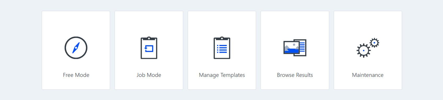
ZEN core home screen: Access to image acquisition and job functions
• Full operational control of ZEISS microscopes, cameras and components
• Workbenches for repetitive application tasks
• Single and automated panorama image acquisition
• High dynamic range (HER) image acquisition
• Creation and management of input forms
• Live image video recording
• Image and data information displayed in datazone below the image
• Sample-centric correlative display and organization of data and images
• Processing and analysis of any kind of image e.g. both light and electron microscopy
• User management functionality
• Best Image functionality
• Multi-image viewer
• Enhanced depth of field via manual focus
• Measurement functions
• Topography visualization including profile line measurement
• Microsoft Word ® and Adobe Acrobat ® PDF reports and report templates
• Data archive for images, documents and templates with searching and tagging
• Image exporting to all standard image formats such as JPG, BMP, TIFF
• Connection to ConfoMap
• Connection to Zaphire
• Connection to GOM Inspect
• Extended workflow capabilities, loop functionality
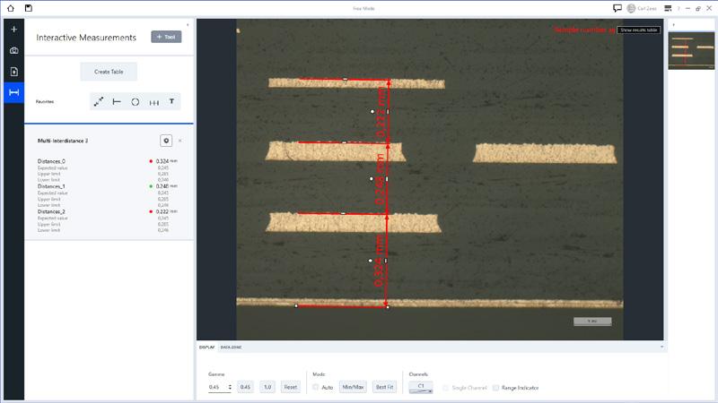
Measurement functions
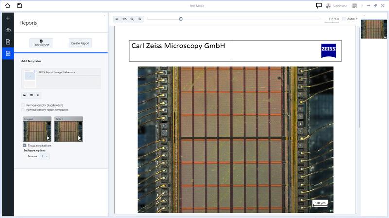
Reporting template
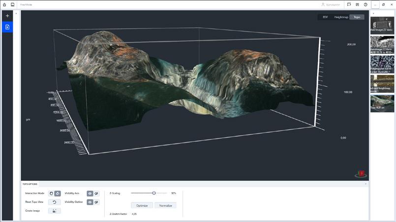
Topography visualization
› In Brief › The Advantages › The Applications › The System
Obtain results – rapidly and repeatably. ZEN core provides you with a wide range of choices for automated image acquisition:
• Best Image: assists you with the optimum microscope settings for image acquisition.
• HDR: ensures best image quality even with challenging light conditions.
• Panorama and Tiles: create stitched images in just a few clicks.
• Autofocus: automatically determine the perfect focus position for your sample.
• EDF: Automatically acquire multiple images at different focus positions and combine them to an image with enhanced depth of field.
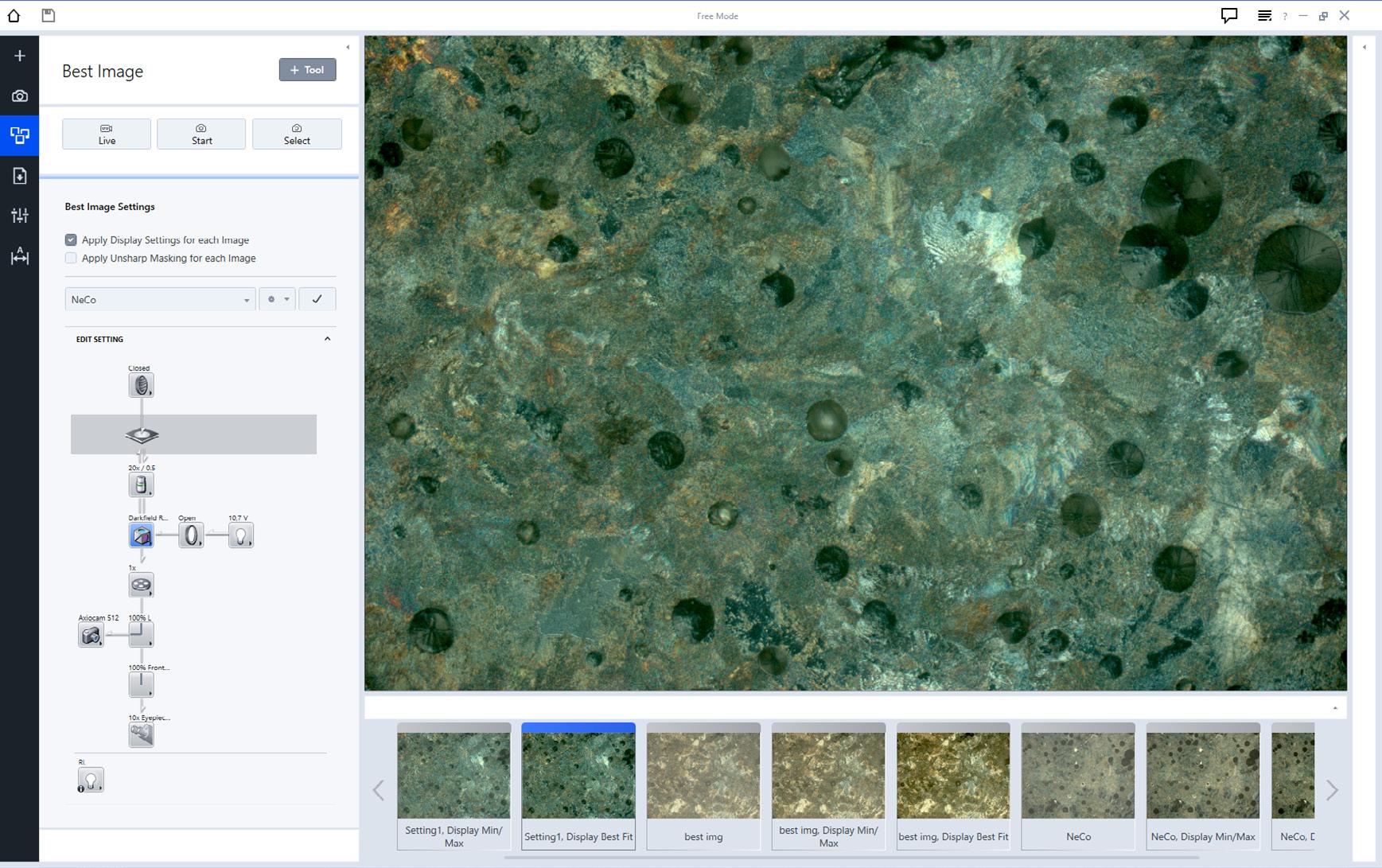
› In Brief
› The Advantages › The Applications › The System
ZEN Multi Channel Acquisition
Whether you work in materials or life sciences, ZEN core lets you acquire and overlay signals automatically in various individual channels. This is key for optimal visualization and data analysis. Prominent examples include polymer analysis in materials research—say, for investigating microand nanoplastics—and fluorescence imaging in biomedical disciplines. The latter supports the understanding of structure and dynamics of biological samples, the characterization of spatial distribution of proteins, cell-cell interactions and colocalization experiments.
ZEN Multi Channel Acquisition allows for:
• Easy and intuitive definition of your experiment using smart setup
• High reproducibility and ease-of-use for repetitive work using job mode
• Complete workflows including image acquisition, analysis and reports
• Combining with the GxP module to work in regulated environments such as pharmaceutical companies
• The combination of fluorescence, transmitted light and reflected light channels
• Channel-specific camera and light source settings
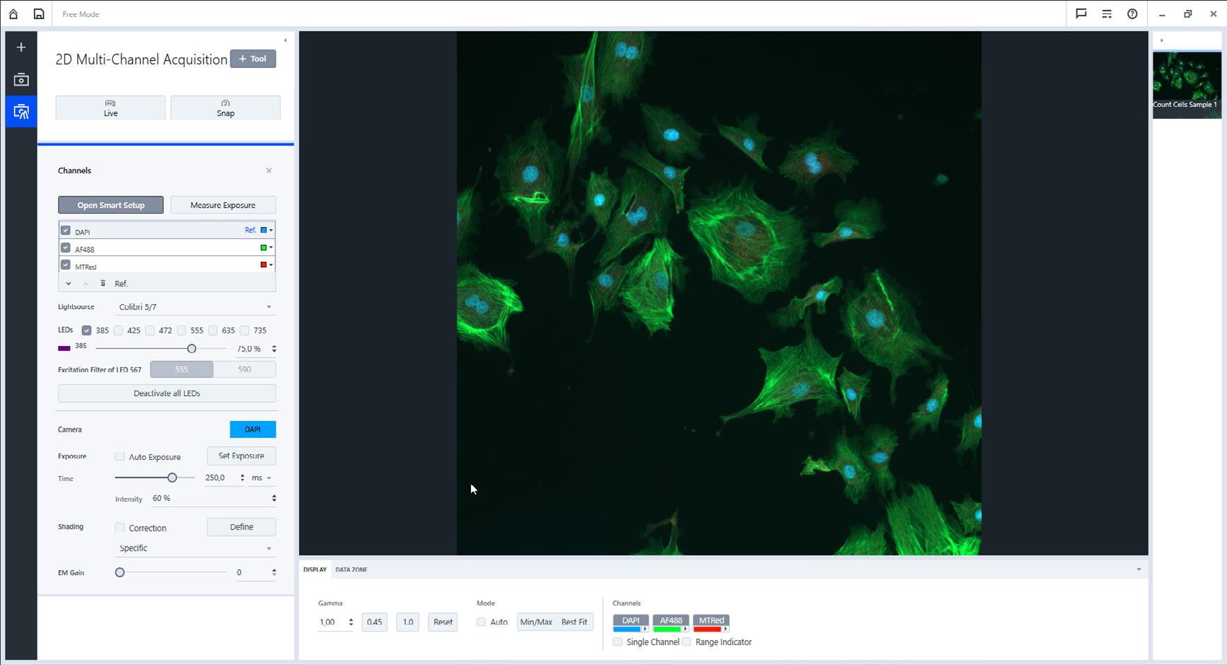
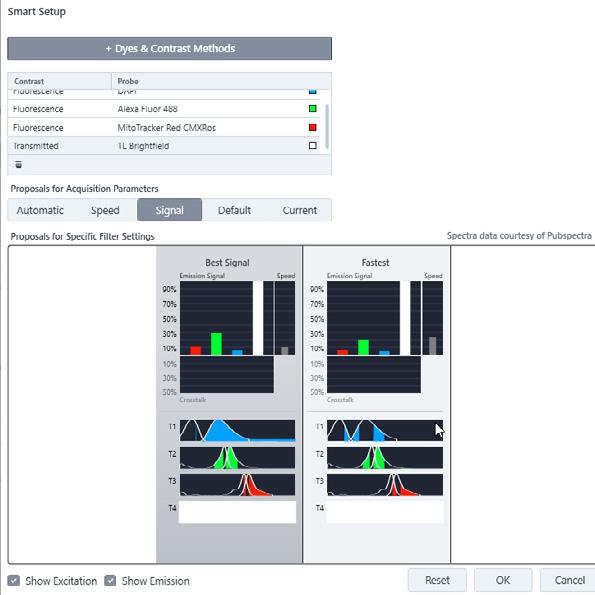
Smart Setup allows to easily define the channels of the acquisition.
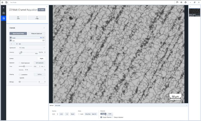
2D Multi-Channel Acquisition can also be used to setup a multichannel experiment without any fluorescence imaging, just using e.g. bright- and darkfield.
› In Brief
› The Advantages
› The Applications
› The System
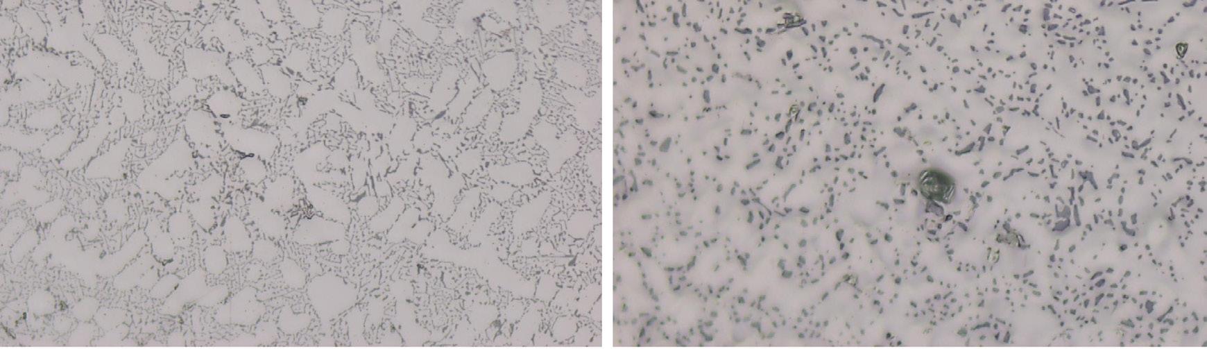
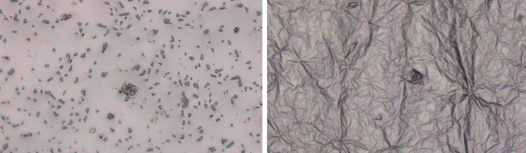
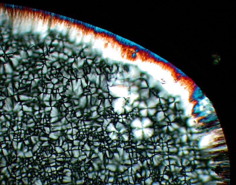
Observing materials in a static state can reveal useful insights into their microstructure, but there’s much more to be learned from observing them in situ under real service conditions. With Linkam heating/cooling stages you can perform experiments that reveal information about how a material behaves under temperature. Use ZEN core to set up and control the experiment directly—that guaranties a perfect interplay between image acquisition and heating stage control.
• Setup heating/cooling ramps
• Setup holding time
• Setup temperature
• Image acquisition triggered by time or temperature.
Typical Applications:
• Observation of melting and crystallization behavior
• Determination of the melting and solidification interval
• Investigation of the shape, structure, number and size of different phases
• Investigation of the influence of additives or fillers on crystallization behavior.
› In Brief
› The Advantages
› The Applications
› The System
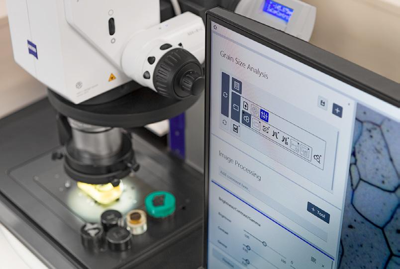
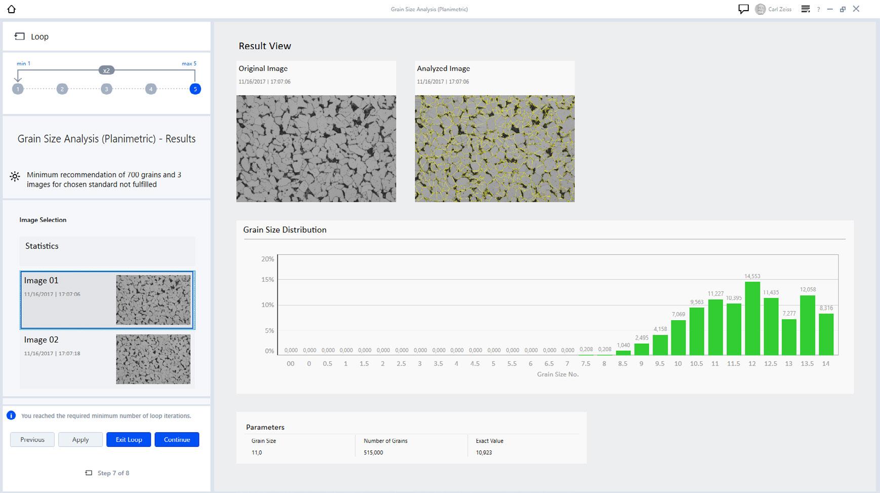
The size and distribution of grains are directly linked to the material properties. Quantify the crystallographic structure of your materialographic samples in accordance to international standards. Three evaluation methods allow you to characterize your material:
• Planimetric method for automatic grain boundary reconstruction
• Intercept method with a variety of different chord patterns to interactively recognize and count the intersections with grain boundaries
• Comparison method for manual image evaluation with comparative diagrams
Image segmentation can be performed by latest machine learning algorithms (see ZEN Intellesis).
Supported Standards:
• DIN EN ISO 643:2020
• ASTM E 112-13
• ASTM E 1382-97
• GB/T 6394 2017 Plate I-V
› In Brief
› The Advantages
› The Applications
› The System
Multiphase Analysis
Any part of the material with a distinct crystal structure can be taken as a “phase”. Different phases are separated from one another by distinct boundaries. Distribution and orientation of phases affect the material properties like hardness, strength or elongation at break.
Analyze the phase distribution in your samples. Determine size, shape or orientation precisely and fully automatically. Use this distribution analysis to gain information about porosity of additive manufactured material.
Image segmentation can be performed by latest machine learning algorithms (see ZEN Intellesis).
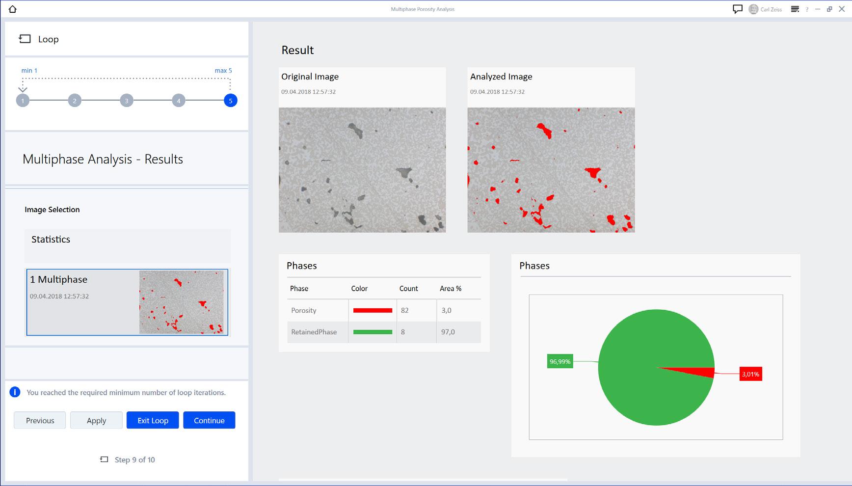
Multiphase Analysis – result view with distribution of different phases
› In Brief
› The Advantages
› The Applications
› The System
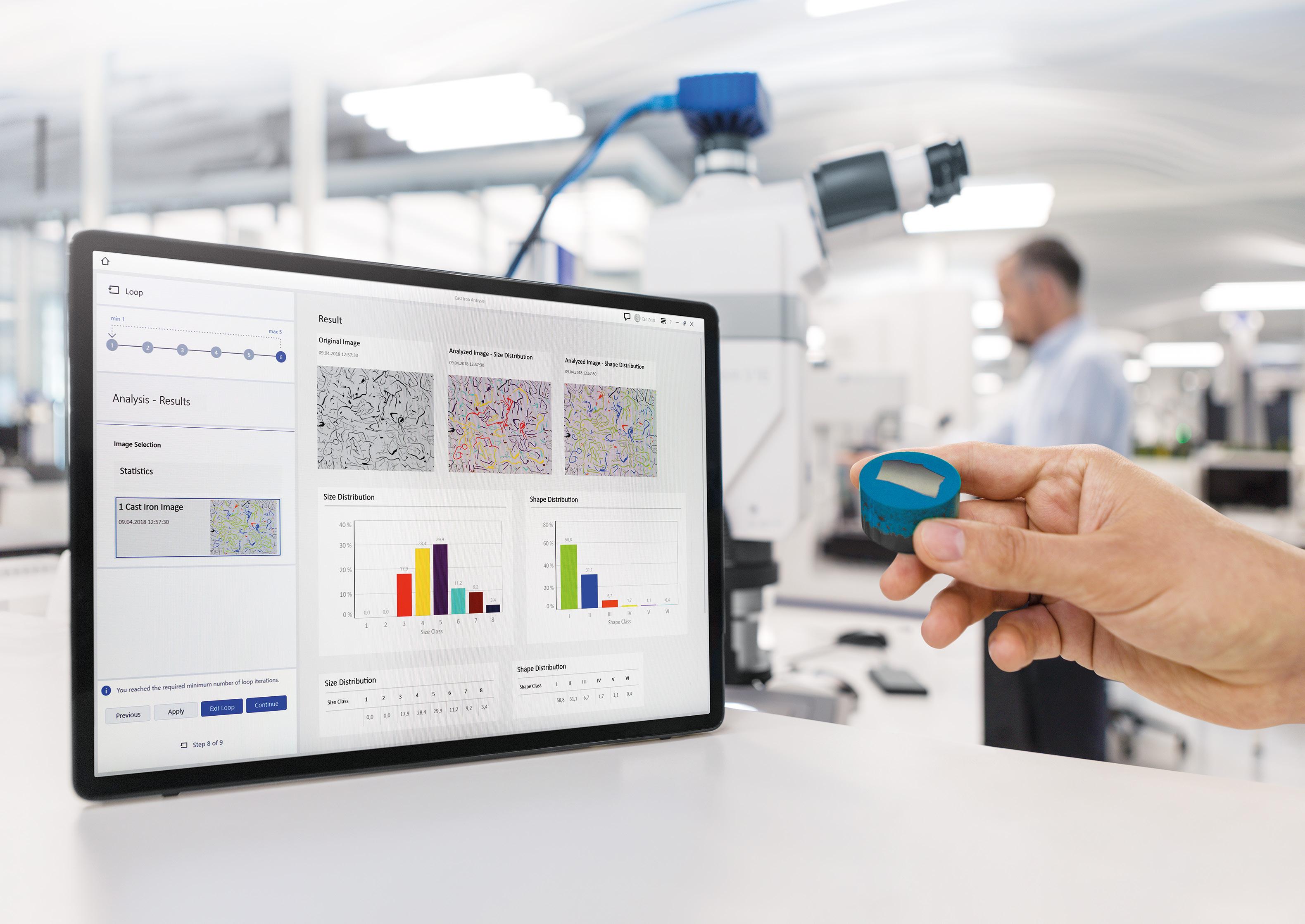
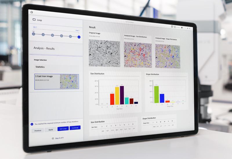
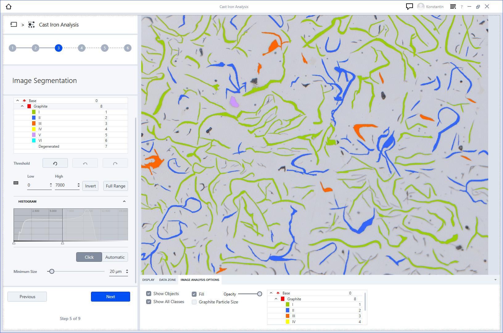
Cast Iron Analysis
Depending on process parameters and chemical composition of the material, graphite particles in cast iron can occur in different shape and distribution. This influences the mechanical properties of the material.
Analyze the shape and size of graphite particles fully automatically. Obtain the spheroid number according to DIN EN ISO 945 (2019). Determine the nodularity of vermicular graphite and examine the content of graphite particles in area percentage.
Supported Standards:
DIN EN ISO 945:2019
› In Brief
› The Advantages
› The Applications
› The System
Measure thickness of coatings and platings, or the depth of hardened surfaces in the cross section of a sample.
Evaluate complex layers systems either auto-matically or interactively. The module calculates the course of the measurement chords depending on the gradient present.
Get the results from your part in a clear report containing images, sample data and measurement values, such as the maximum and minimum chord lengths, mean, and standard deviation.
Image segmentation can be performed by latest machine learning algorithms (see ZEN Intellesis).
Supported Standards:
• DIN EN ISO 1463:2004
• ASTM B 487 - 2007
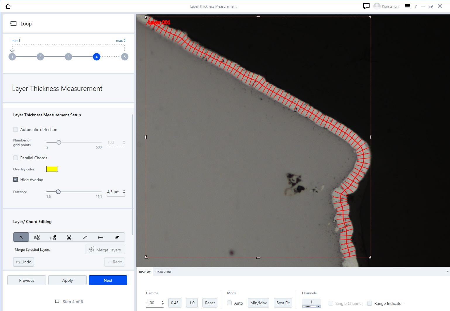
Layer Thickness Measurement – automatic detection of a layer
The Advantages
The Applications
The System
Comparative Diagrams
Make your Wall Charts digital. Compare your sample under the microscope with comparative diagrams directly on your screen. Choose between different schematic micrographs with specific characteristics. These change gradually from image to image and may relate to grain size, carbide precipitation in steel, or quality of sample preparation. The module also provides a chart series creator to design your own comparison diagrams, e.g. for pass-/fail criteria in quality control or best target preparation images for your individual material microstructures.
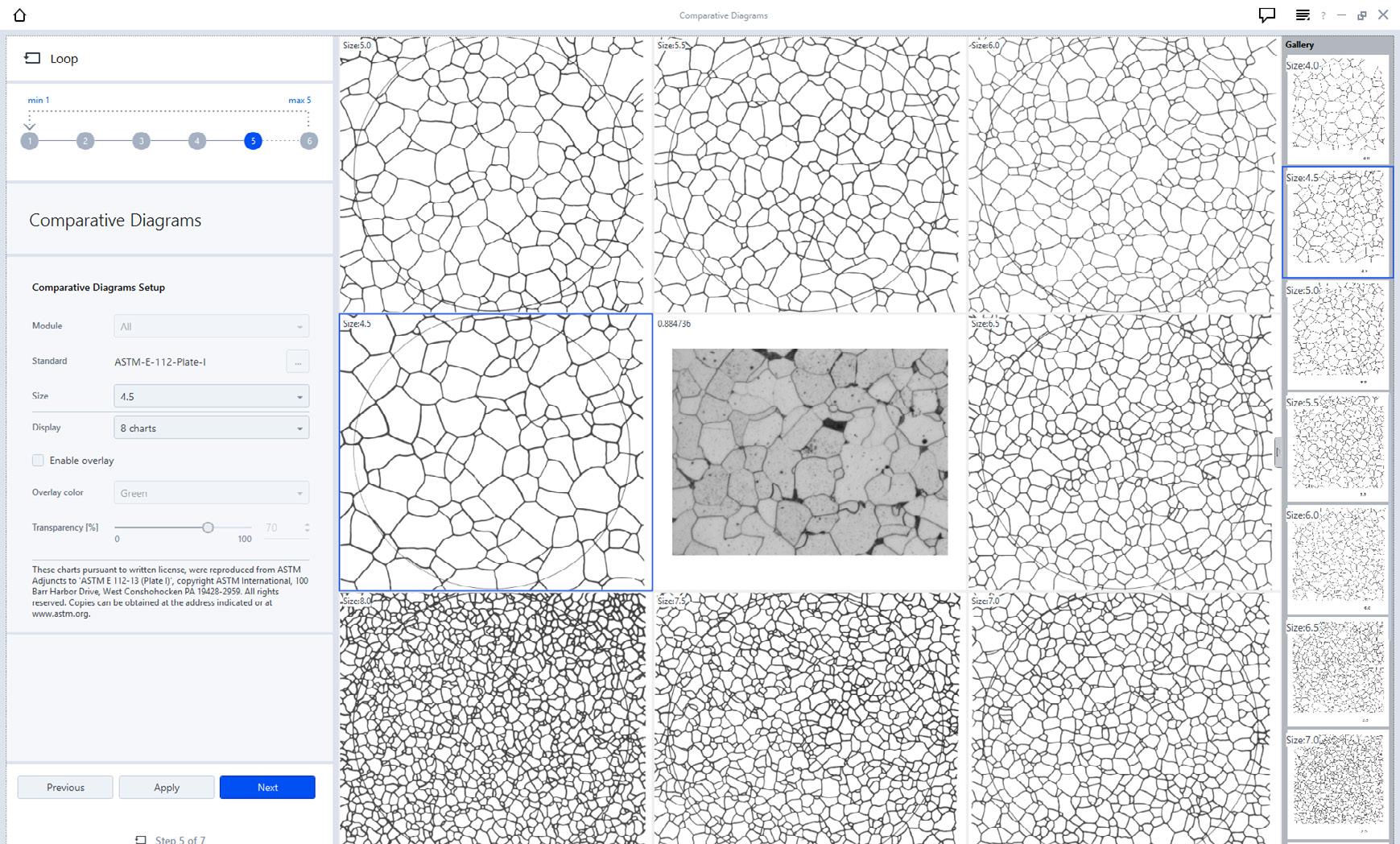
Comparative Diagrams: Compare the sample with standardized or customized wall charts.
Supported Standards
Grains:
DIN EN ISO 643:2020
ASTM E 112-13 Plate I-IV
GB/T 6394 2017 Plate I-IV
Graphite: EN ISO 945-1:2019
Non-metallic inclusion (NMI):
ASTM E45
ISO 4967
GB/T 10561
EN 10247
DIN 50602
› In Brief
› The Advantages
› The Applications › The System
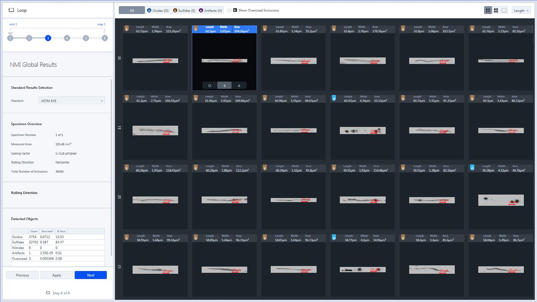
The type and amount of non-metallic inclusions (NMI) in steels strongly affect the mechanical and physical properties of these steels.
Metallographic analysis of NMI is governed by industry standards that are supported by the modular and customizable ZEN core software which guides the user quickly and easily through the workflow, generating a report and inclusion gallery compliant with the standards.
ZEISS ZEN module Non-Metallic Inclusion Analysis confirms that manufacturing processes, grade and quality of the product meet strict specifications for impurities or defects that can cause a component to fail or impact its tensile strength, toughness and fatigue.
Powerful inspection views and automated deformation axis detection features make analysis easy, intuitive and repeatable. With additional GxP functionality, ZEN core users are able to offer their customers full traceability and data integrity in NMI analyses, meaning that grade certification is auditable, particularly advantageous for customers in regulated industries.
ASTM E45
ISO 4967
JIS G0555
GB/T 10561
EN 10247
SEP 1571
DIN 50602
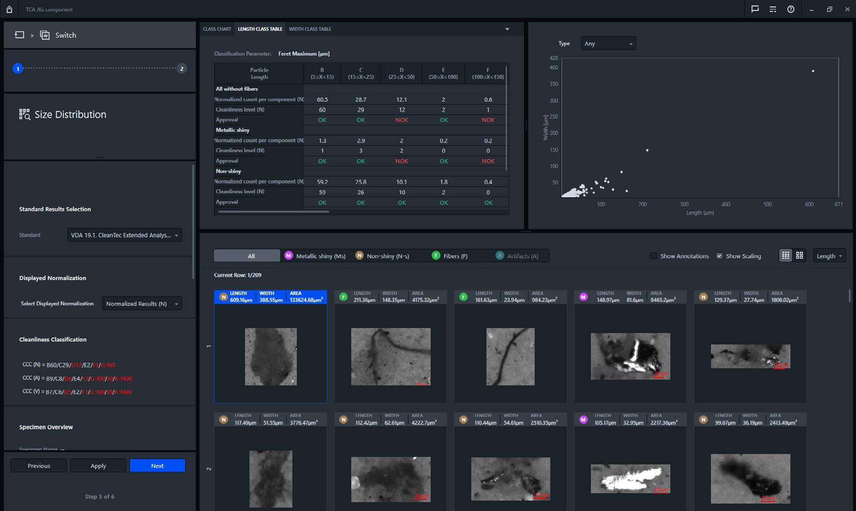
Technical cleanliness in production areas is vital for any company that aims to produce along globalized standards. Manufacturers and end users alike demand ever-higher quality standards to avoid contamination. Identifying critical contamination levels for components, parts, oil or hydraulic fluids—as well as for medical devices—is therefore essential to maintaining high quality production. Technical cleanliness analysis is your easy-to-use module enabling the identification, classification and height measurement of particles along standard-compliant analyses. Correlative microscopy lets you combine data from light and electron microscopes in a seamless workflow. At the click of a button, automated reporting puts major industry standards such as process data transfer of HYDAC extraction units at your disposal. And it can be customized to your own company standards equally well.
Acquiring brightfield and crossed polarization images in just one scan speeds up both your workflows and the identification of contamination sources. ZEN core gives you access to extended analysis workflows and central data management—for example, GxP options for CFR compliance such as electronic signatures and audit trails or global cleanliness structures.
› In Brief
› The Advantages
› The Applications
› The System
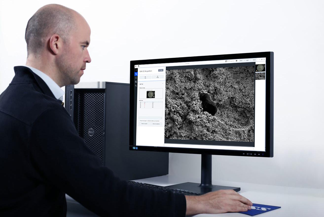
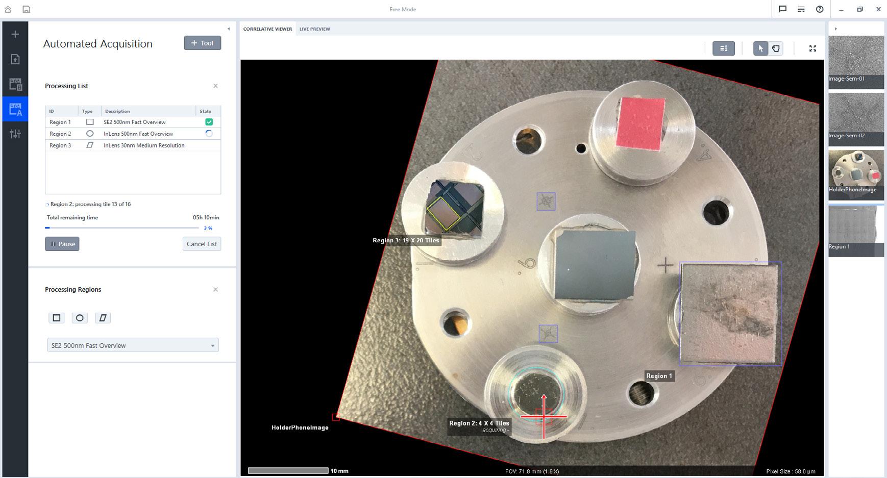
ZEN core helps you bridge light and electron microscopy in many ways. The SEM Automated Imaging module simplifies electron microscope operation for new users, enabling them to use predefined parameters to acquire single images as well as tiled images of larger regions.
• Manage protocols for all necessary acquisition parameters in a central location. Choose from predefined settings or adapt them to fit your imaging needs.
• Leverage existing image data in the ZEN Connect workspace to define regions for automated SEM mosaic acquisition.
• Add all regions to the acquisition queue and record images in one session.
• Take advantage of acquisition time estimates to plan when imaging will be finished.
• Use the live preview to verify image quality and acquisition settings per region.
• Acquire single image frames completely freely or based on settings from a saved protocol. Arrange multiple view containers to see different detector signals at the same time.
• Store and recall stage positions with ease.
• Rely on automated routines to stitch acquired SEM mosaics or use powerful interactive stitching to fine-tune individual seams.
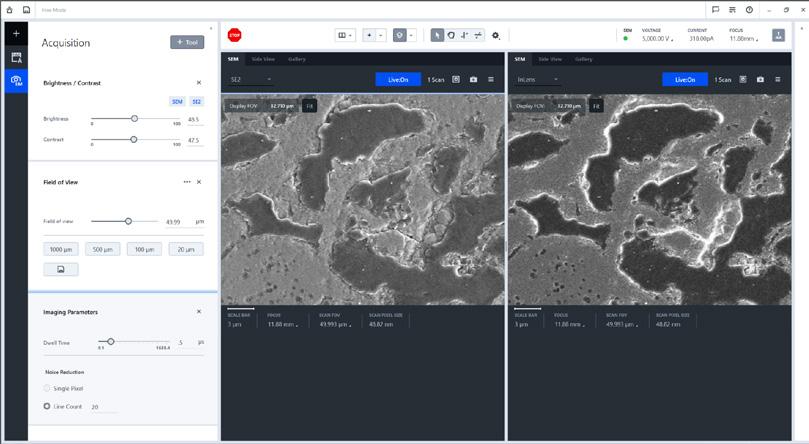
The Single Acquisition workbench allows multiple detector signals to be seen at the same time in configurable view containers including the chamber camera view. SEM imaging settings can be changed easily and single images get acquired with one click.
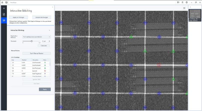
After an SEM mosaic acquisition the image is stitched by automated algorithms. In case these do not yield a perfect result, every individual seam can be reviewed and adjusted manually using the Interactive Stitching workbench
In Brief › The Advantages › The Applications › The System
TEM Sample Preparation for ZEISS Crossbeams
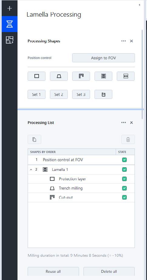
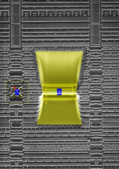
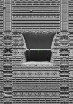
ZEN core takes full control of ZEISS Crossbeam for the fast, easy and reliable preparation of samples for transmission electron microscopy (TEM) studies.
• Speed up your workflow with a task-specific workbench that shows only the controls you need.
• Run automatic routines for the preparation of single or multiple regions of interest.
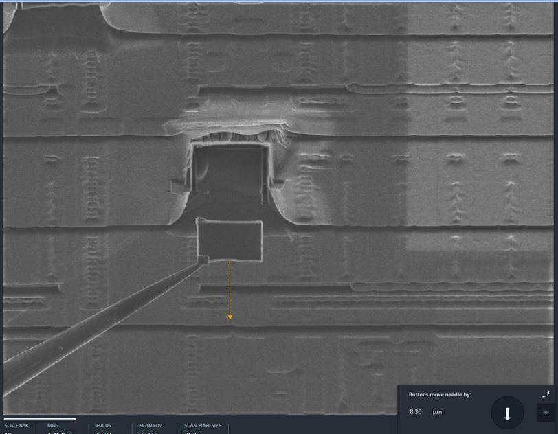
• Experience ease-of-use with fully integrated micromanipulator control. Intuitively drive the manipulator needle by just clicking on SEM and FIB images.
• As a novice, enjoy high success rates from start on using guided workflows.
• Increase the productivity of your advanced TEM sample workflows for planar view and backside preparation. These include flipping of the TEM sample support grid, which in combination with ZEISS Flip Holder, is now just one click away.
› The Advantages › The Applications
› The System
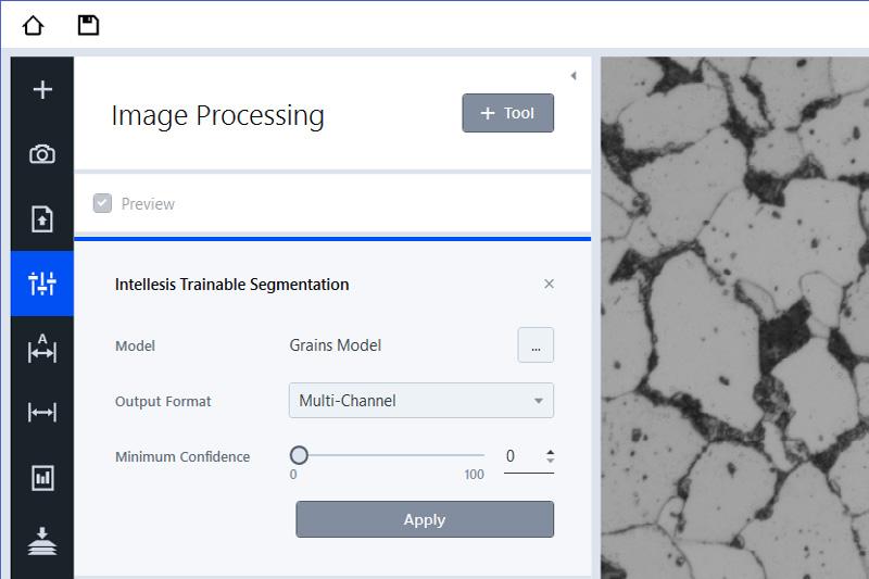
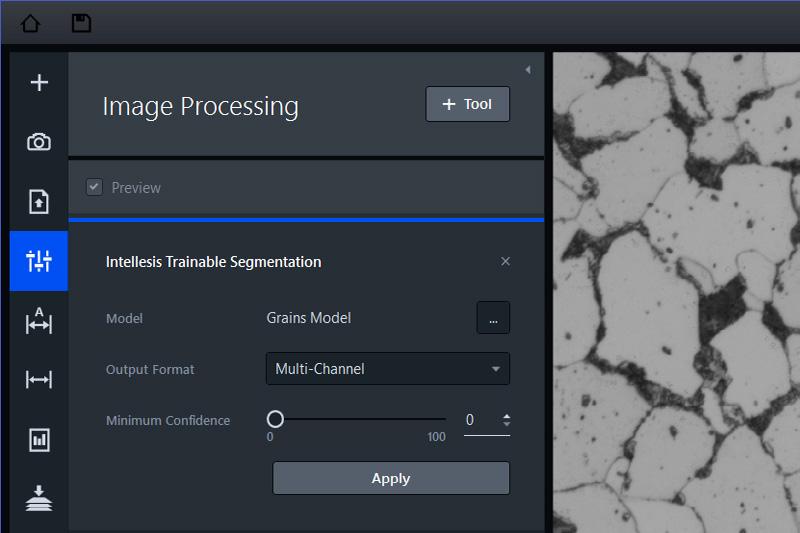
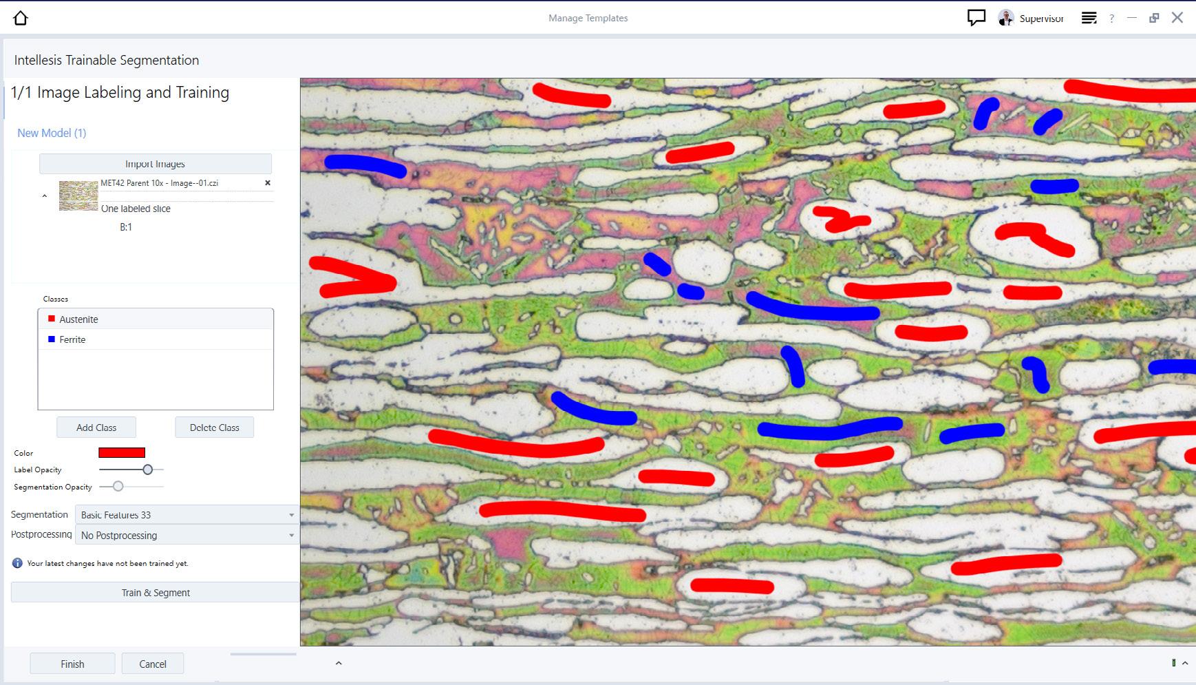
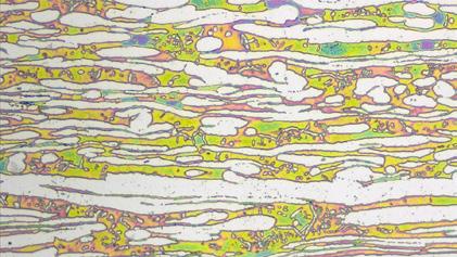
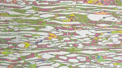
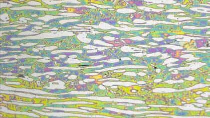
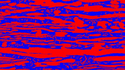
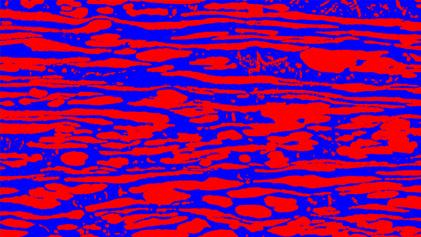
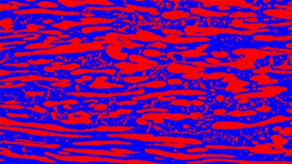
Segmentation is one of the biggest challenges faced by today’s microscopists, but you can avoid errors and user bias by using machine learning for image segmentation. The software module ZEN Intellesis Segmentation produces powerful machine learning segmentation of multidimensional images including 3D datasets. It’s designed for smooth integration of multiple imaging modalities and for achieving superior segmentation on any single image.
Images that once had to be processed manually can now be analyzed automatically by training a model to segment them for you, using the straightforward graphical interface. Use your own expertise to train the software and let it do the tedious segmentation. Or import dedicated networks trained elsewhere (for example, on www.APEER.com) or use your own software and apply them via ZEN Intellesis Segmentation. Both Tensorflow2 and ONNX model formats are supported.
You will also benefit from saving time in sample preparation as ZEN Intellesis Segmentation can adapt to your own preparation process. Reproducibility is guaranteed as the stored analysis program can be used again, sample by sample, or retrained to handle new samples.
› In Brief › The Advantages
› The Applications
› The System
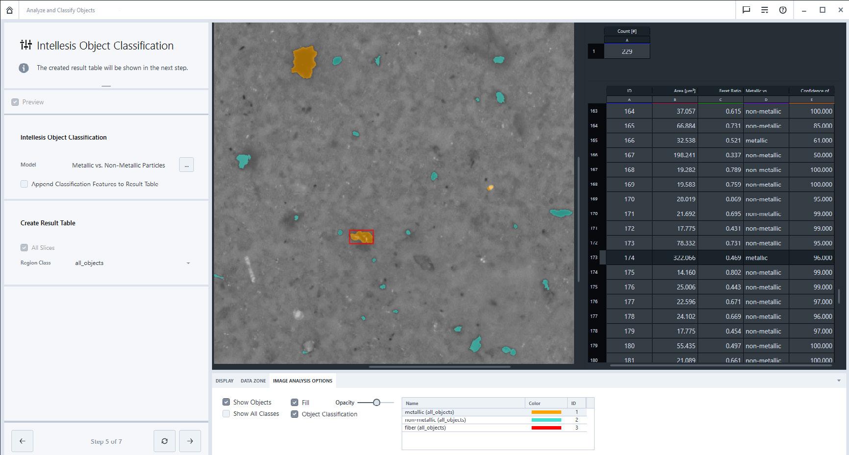
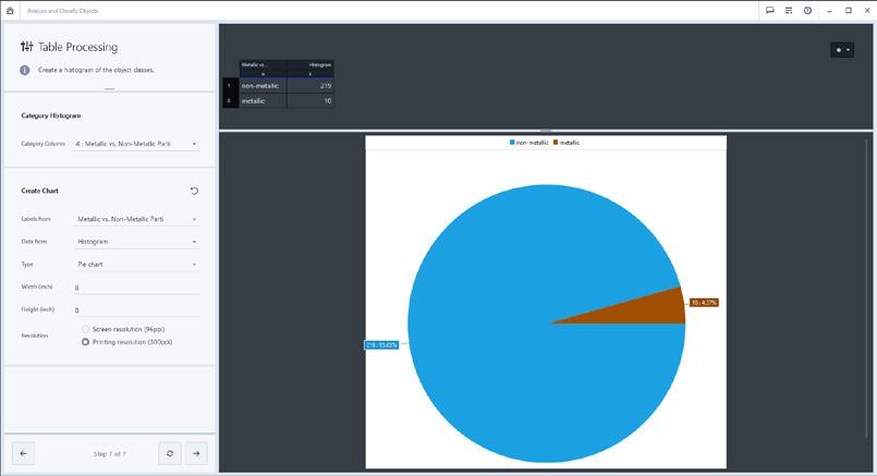
Sometimes segmenting objects like particles, inclusions or grains is straightforward, but it can still be hard to classify them further into different types. Even machine learning-based segmentation techniques may struggle in this case because they only take the appearance of pixels into account and are unable to consider derived properties of pixel clusters (objects) as well. ZEN Intellesis Object Classification now offers an easy way to classify already segmented objects into subclasses. Based on the same machine learning algorithms as ZEN Intellesis Segmentation, an object classification model can be trained to perform the classification automatically. Instead of looking at individual pixels, the model uses more than 50 properties measured per object to distinguish them. These derived measurements include all kinds of geometric as well as intensity-based features. Since ZEN Intellesis Object Classification works on tabulated instead of image data, the classification process is much faster than, for instance, segmentation by specifically trained deep neural networks. Additionally, it is independent of the prior segmentation, whether it was done by classic thresholding or using machine learning.
› In Brief
› The Advantages
› The Applications
› The System
Organize and visualize different microscopy images and data from the same sample in their context, all in one place. For sample-centric analysis, ZEN Connect workflows enable you to get from a quick overview image to advanced imaging with multiple modalities. The correlation between images at different scales can be explored in the workspace and easily used for navigation. The interdependencies of different datasets can be stored, exported and re-used in a Client Server Database. ZEN Connect also enables integrated reporting across the connected images, videos and datasets.
The correlative microscopy interface enables you to seamlessly transfer samples between different light and/or electron microscopes, then quickly and automatically relocate regions of interest to collect maximum relevant data with minimal effort.
• Organize and align data and images from multiple modalities in a sample-centric workspace.
• Import third-party images including even metadata when supporting the BioFormats standard.
• Import Horiba Raman maps and attach even non-image data at a chosen position in the workspace.
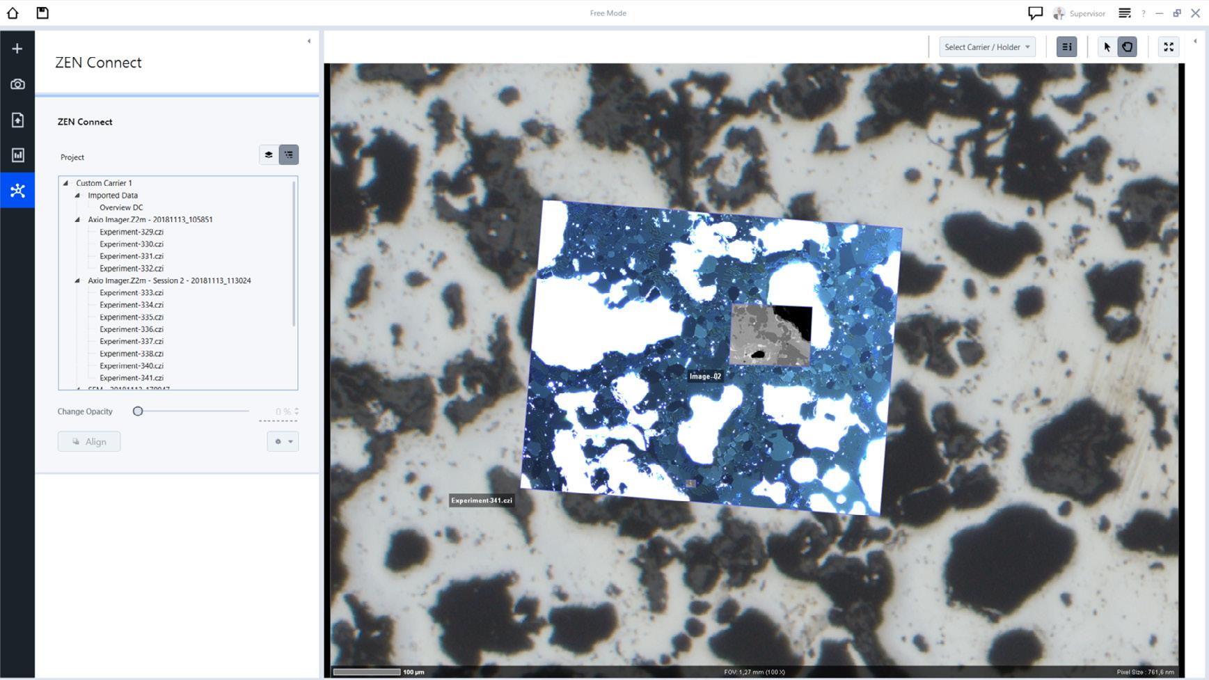
• Choose from different options to align your images precisely.
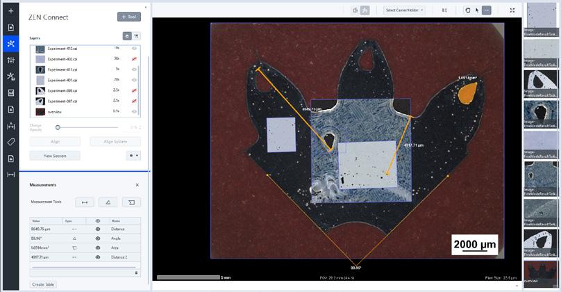
• Perform exportable line, angle and area measurements in the workspace within or across aligned images.
• Transfer samples and image data between ZEISS light and electron microscope systems.
• Relocate regions of interest automatically.
• Benefit from interpolated multichannel image export out of the workspace and easy-to-use video export.
› The Advantages › The Applications › The System
GxP Module
The GxP module enables traceable workflows through seamlessly integrated microscopy hardware and software to meet the requirements of industries such as pharmaceutical or medical. Every workflow available in ZEN core can be made GxP compliant.
The GxP module helps:
• Quality Managers who want to guarantee reproducible results and secured processes
• Validation Engineers who want to be able to validate their microscopy processes
• Lab Managers who need to document their microscopy processes
• IT Managers who need backup/disaster recovery functionality, and archive/database capability
• Pharmaceutical Companies who need to be compliant to e.g. FDA 21 CFR Part 11
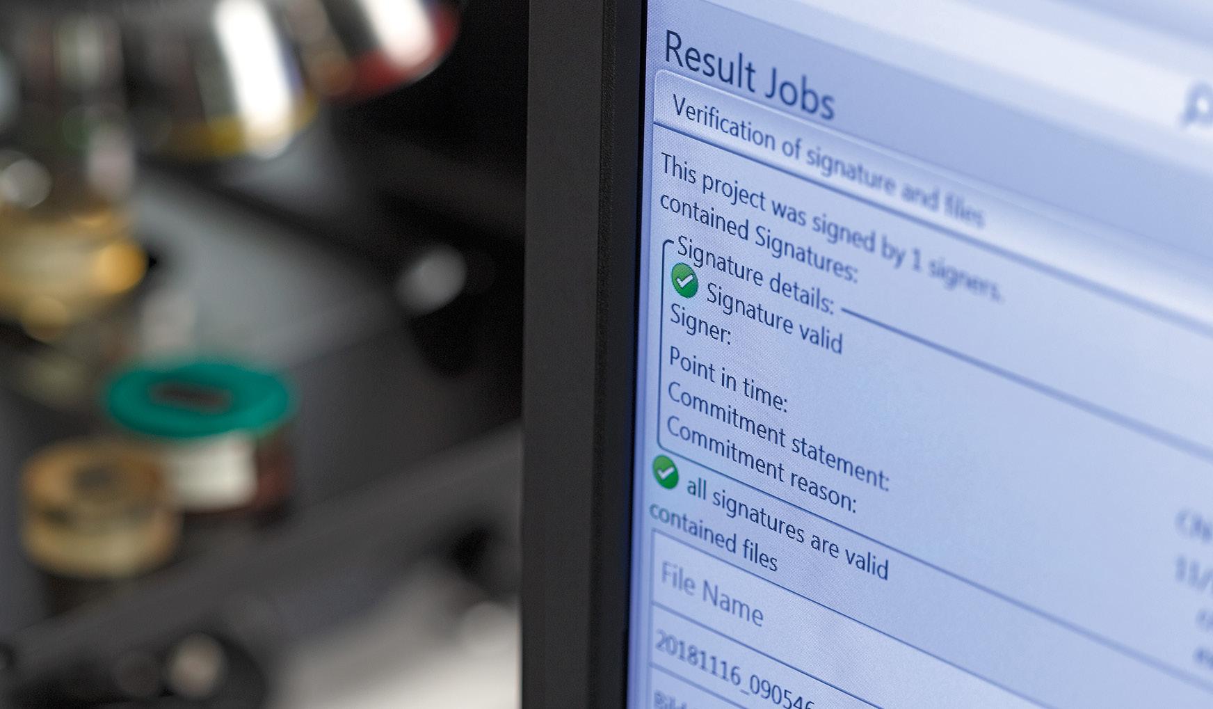
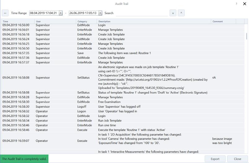
Audit Trail of all user activities
Use a variety of tools and functionalities in combination with the required qualification and validation activities to maintain GxP compliance for your images, tables, and reports:
• User management
• Audit trail
• Release procedure of workflows
• Electronic signatures, incl. countersign functionality
• Checksum protection of process-critical data
• Disaster recovery
• Combination of different clients via database
In Brief › The Advantages › The Applications › The System
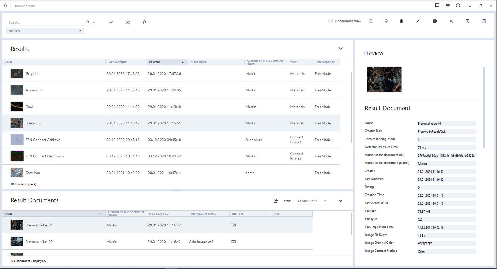
Easily browse and access all results, even those generated from multiple instruments operated via ZEN core in one central location.
• Access data collected from individual systems or from multi-modal workflows.
• Store your results and templates centrally across systems, laboratories and locations.
• Organize your data through document tagging.
• Browse your assets with powerful sorting and filtering features including search term history and quick filters.
• Upgrade to database and multi-user systems.
› The Advantages
› The Applications
› The System
As digitization continues to improve microscopic investigations, you’re faced with an ever-growing mass of images and data that needs to be managed, all the more so in multi-user laboratories. ZEN Data Storage enables you to separate image and data acquisition from post-acquisition works, making everyone in the lab work more efficiently in a number of ways:
• It’s easy for experts and non-experts alike to share instrument presets, workflows, data and reports.
• Access to all data from different microscope systems is a given, as is data from mobile and desktop devices – and from different locations too.
• You can access any ZEN Data Storage content with ZEN Data Explorer, a hybrid app for iOS and Android that lets you browse, view and annotate images and ZEN core job results.
• Existing image collections, including third-party images, can be uploaded automatically in the background or leverage the ZEN Data Explorer web interface.
• Use ZEN Data Explorer to access and explore even ZEN Connect projects.
• Group your images and data into collections, define read/write access and share them easily with your peers.
• With effortless correlation of data from different microscopes, you can perform multi-modal workflows and reap maximum information from your samples.
• Help your IT department implement security and backups by keeping all your data on one central server.
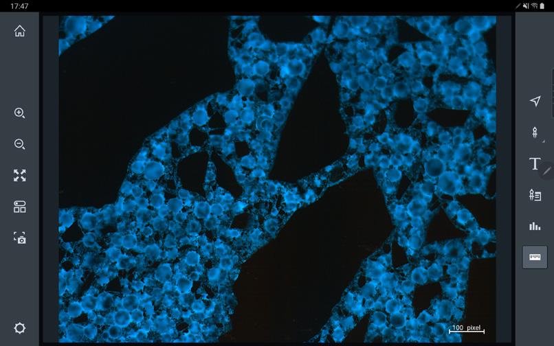
ZEN Data Explorer: The web-based app, included in the ZEN Data Storage server package, allows you to browse, view, and annotate images on smartphones and tablet devices.
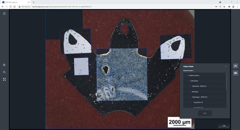
ZEN Data Explorer: Web view allows you to access data on ZEN Data Storage from your favorite browser. It also supports the display of ZEN Connect projects..
The System
Select Your Modules According to Your Requirements
General Workbenches
Functional Modules Workbenches
Job Mode
Basic Measurement
Measurement
Topo View
Database Interface
Reporting
Report Template Creator (MS Word® Add-In)
ZEN Connect Entry
Connection to APEER
APEER on-site Advanced
CAD Import
Connection to ConfoMap
Connection to Zaphire
Connection to GOM Inspect
Microscope Control
Online Measurement
TEM Lamella Preparation
Automated Imaging
Special Functions
Microscope Control
Camera Control
Panorama
General image acquisition and image analysis functionality via workbenches
Depending on the functionality and modules, the respective workbenches are available
Configurable mode to guide operators through a pre-defined workflow
Basic functions for interactive measurement
Extended functionality of interactive measurement
3D visualization of topography images and hightmap representation
Basic interface for storing data in databases
Creation of reports containing detailed information
Software to create report templates using MS Word®
Interactive acquisition and contextual display of images in single- and multi-instrument workflows
Connection to APEER, a cloud-based platform to share, run, and customize microscopy workflows
Download individual modules from APEER and execute them locally for specialized tasks
Import CAD data to create overlay images
Connection to ConfoMap for surface roughness analysis
Connection to Zaphire for automated measurements according to measurement templates
Connection to GOM Inspect for advanced topography analysis
Control of scanning electron microscopes and devices via software
Measurement of areas and non-area values in the live image
On ZEISS Crossbeam prepare lamella specimens for TEM investigation using automated workflows
On ZEISS FE-SEM correlatively acquire EM mosaic images via defined imaging protocols
Control of light microscopes and devices via software
Control of cameras from software
Manual acquisition of high resolution images; automated acquisition and stitching functionality on coded and non-coded stages Included
Manual Extended Focus
Motorized Extended Focus
Autofocus
Multi-channel Acquisition
Online Measurement
Tiles & Positions
Linkam (Hardware Control)
Functional Modules
Macro Environment
GxP
Image Analysis
ZEN Intellesis Segmentation
ZEN Intellesis Object Classification
ZEN Connect
ZEN Connect 2D Add-on
ZEN Data Storage Client
Third-party Import
Grain Size Analysis
Cast Iron Analysis
Multiphase Analysis
Comparative Diagrams
Layer Thickness Measurement
Non-Metallic Inclusion Analysis
Technical Cleanliness Analysis
Qual Data Export
Manually acquire multiple images at different focus positions and combine them to an image with a greater depth of field
Automatically acquire multiple images at different focus positions and combine them to create an image with a greater depth of field
Determine the focus position of your sample
Acquire multiple (fluorescence) image channels simultaneously
Measurement of areas and non-area values in the live image
Record exact and high-resolved images of large samples by automatically scanning pre-defined areas
Interface to control Linkam heating- and cooling stages
Description
Use Python programming language to generate customer specific macros
Ensure traceability and accountability of workflows (precondition for 21 CFR Part 11 compliance)
Creation of automatic measurement programs
Automated image segmentation based on machine learning algorithms
Automated object classification of segmented and analyzed images based on machine learning algorithms
Extend ZEN Connect functionality to correlative workspaces
Correlative Microscopy module for image acquisition and correlation on light and electron microscopes
Seamlessly connect to ZEN Data Storage Server for central storage of documents and templates
Import 3rd-party microscopy images and metadata into ZEN core
Determine grain sizes via different methods according to international standards
Analyze form, size and distribution of graphite particles in cast iron
Automated measurement of particle size and area content of multiphase samples, evaluation of porosity
Compare micrographs with standardized or customizable comparative charts (Wall Charts)
Automated or interactive thickness measurement of different layers
Create measurement files for Database like ZEISS PiWeb or qs-STAT Included Optional Special
Automated imaging, classification and reporting of non-metallic inclusions in steel
Automated identification and classification of particles compliant to cleanliness standards