Curiosity is always the first step when solving a problem.
Galileo Galilei
This is the origin of all innovations.
At Carl Zeiss innovations are a celebrated tradition. That’s why we work systematically and consistently on the development of innovative concepts.
The world of science and industry is changing constantly. Time and again, this change presents new challenges to all of us. It requires faster and more efficient development cycles, innovative and unique products and requires continuous optimization of processes in businesses and institutions.
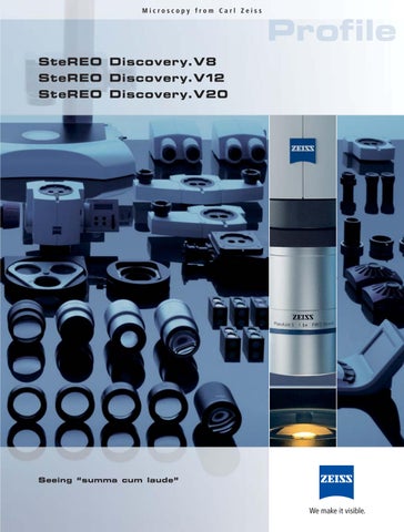
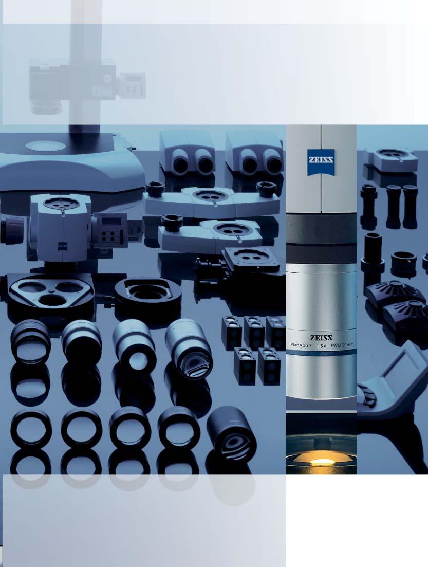
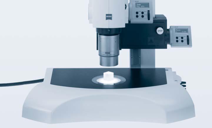
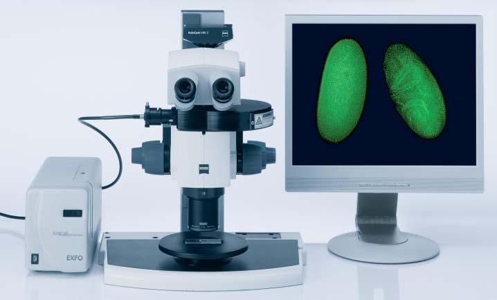
In this Profile, Carl Zeiss proudly presents the SteREO Discovery family of innovative stereomicroscopes. This is the sum of decades of experience in the conception, development and production of optical innovations.
In order to be able to offer you such advanced, forward thinking solutions, it’s not just calculus that is required –our enthusiasm and passion are vital. With precisely these qualities we will support, guide and assist you from the initial consultation through to realization - with all of our knowledge and experience.
So that you can rediscover ‘seeing’.
See so Much More
The pancratic magnification changer of the microscope body forms the core of a zoom stereomicroscope and, consequently, has a considerable influence on the optical performance of the entire system.
With the SteREO Discovery, Carl Zeiss has proven all its experience and competence, and has driven this system of common main objective (CMO) stereomicroscopes to the limits of what is optically feasible.
The SteREO Discovery.V12 and V20, with their maximum NAs of 0.144, are among the most powerful stereomicroscopes in the world.
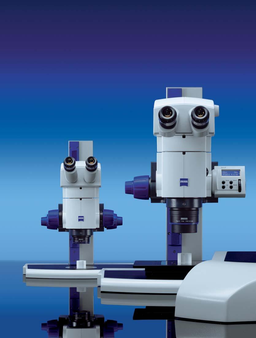
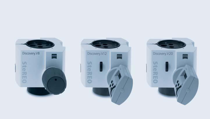
Three different microscope bodies are available:
SteREO Discovery.V8
- apochromatic zoom optics
- manual zoom 1x ...8x
- maximum NA of0.116
- engageable click stops
SteREO Discovery.V12
- apochromatic zoom optics
- motorized zoom 0.8x ...10x
- maximum NA of0.144
- electronic click stops
- double iris diaphragm
- selectable zoom speeds
- real time display ofmagnification, resolution,depth offield and the object field ofthe system
SteREO Discovery.V20
- apochromatic zoom optics
- motorized zoom 0.75x ...15x
- maximum NA of0.144
- electronic click stops
- double iris diaphragm
- selectable zoom speeds
- real time display ofmagnification, resolution,depth offield and the object field ofthe system
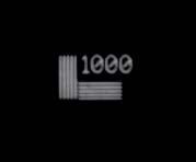
The first stereomicroscope to exceed the 1000 LP/mm resolution limit:
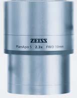
SteREO Discovery.V12 with PlanApo S 2.3x objective..
Double iris diaphragm to increase depth offield.Beneficial when observing and documenting three dimensional objects.
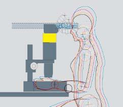
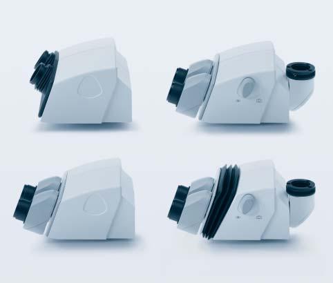
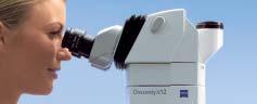
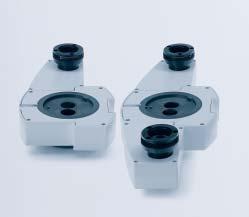
Observation tubes deliver two unreversed and upright images in stereo and contribute greatly to the ergonomic quality of the microscope. For example, with the ergotube it is possible to freely adjust the viewing angle within a defined range. Intermediate tubes help to optimize the viewing height of the stereomicroscope.
According to the results ofan international study,a viewing height ofapprox.500 mm with a viewing angle ofaround 20 degrees is best for avoiding tension and neck pain when using a microscope.
Binocular tube S 35°
- viewing angle of35 degrees,fixed
- adjustable interpupillary distance
55 - 75 mm
Binocular tube S 20°
- viewing angle of20 degrees,fixed
- adjustable observation positions (low and high) for two viewing heights
- adjustable interpupillary distance
55 - 75 mm
3 Binocular phototube S 20°
- viewing angle of20 degrees,fixed
- adjustable eyepiece positioning (low and high) for two viewing heights
- adjustable interpupillary distance
55 - 75 mm
- photo port 100/100,switchable
Binocular ergo-phototube S 5-45°
- viewing angle of5 ...45 degrees,adjustable
- adjustable eyepiece positioning (low and high) for two viewing heights
55 - 75 mm
- photo port 100/100,switchable
For simple equipment in reflected-light,use ofthe Intermediate tube S,40 mm is recommended to guarantee an ergonomic viewing height.
Center of Exemplary Performance
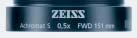
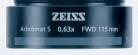
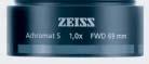
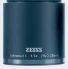
The objective is the eye of a microscope and is a decisive factor in determining the quality of the microscopic image. At Carl Zeiss objectives are a source of pride and a symbol of quality. For the Stereo Discovery objectives that range extends from the economical Achromat and powerful Plan Achromat through to the high end Plan Apochromat
Achromate S
For high contrast imaging ofthree dimensional structures
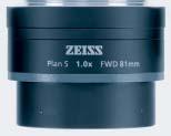
Plan-Achromat S (page 7)
Flat field corrected objectives for the observation and documentation of flat objects in particular; especially suitable for measurement tasks
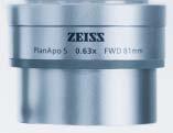
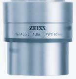
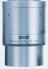
Plan-Apochromat S (page 7)
Offer an extremely high level of correction for flatness offield, resolving power and color fidelity
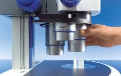
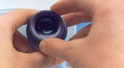
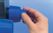
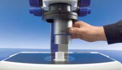
Change the objective simply by hand. The objective locks into place securely. In the SyCoP (System Control Panel),the encoded nosepiece ensures the automatic conversion ofall optical parameters, such as total magnification,object field, resolution and depth offield.
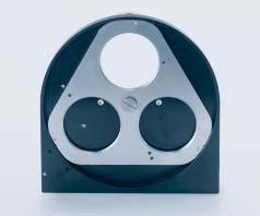
Objective nosepiece S,3x cod
The nosepiece can hold up to 3 objectives with different magnifications.It is recommended that parfocal objectives are used.This way,the observed position on the object remains in focus even after the objective has been changed.
Mounting surface of parfocal objectives
Large Fields Always in View
The intermediate image generated by the objective, zoom optics and tube lens is observed, and also highly magnified, using the eyepiece.
Eyepieces for people who wear spectacles make it possible to work safely, both with and without spectacles. Rubber rings protect the spectacles from being damaged. Eyecups also help.
All eyepieces can be focused and,therefore, allow individual adjustment for both eyes..
Eyepiece E-PL 10x/20 Br.foc. (not illustrated)
Economical widefield eyepiece (accepts reticles d = 26 mm)
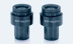
Eyepiece W-PL 10x/23 Br.foc. Powerful standard eyepiece with large,flat field of view (23 mm) (accepts reticles d = 26 mm)
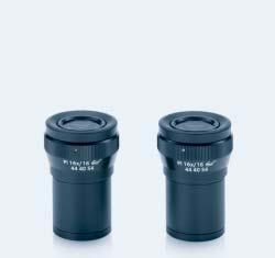
Eyepiece PL 16x/16 Br.foc.
For high magnifications with a large viewing angle of54° (accepts reticles d = 21 mm)
Eyepiece W 25x/10 foc. (not illustrated)
For maximum magnifications (accepts reticles d = 21 mm)
Reticles for measuring,counting and comparing (d = 26 or 21 mm)
Crossline micrometer 14:140Net micrometer 10 x 10/5; 10
Net micrometer 12.5 x 12.5/5; 10
The Backbone of a Microscope
Powerful modern stereomicroscopes such as the SteREO Discovery.V12 and V20 achieve resolutions of over 1,000 LP/mm. This places significantly greater demands on the construction of stand systems. Steady, stable and with minimal vibration –these are the properties of a stand required for precise and rapid focusing across the entire magnification range of the microscope.
A variety of interfaces makes it possible to add on components effectively for illumination, contrasting, and for positioning the object under the microscope.
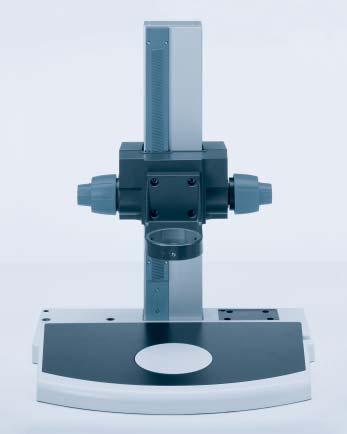
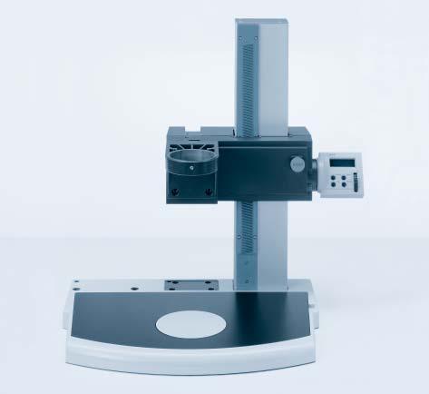
Coarse/fine drive with Profile S column on Stand base 450 to Profile S
This highly stable stand has a manual coaxial coarse/fine drive on both sides,and allows precise focusing.The specially coated,scratch resistant work plate is generously proportioned. It provides plenty ofspace and an excellent overview in the sample space.
Mount S with d = 76 mm support can be used at two different working heights.
- footprint: 450 x 300 mm
- insert plate: 410 x 250 mm
- round plate: d = 120 mm
- profile S column: h = 490 mm
- focus range: 340 mm
- load capacity: max.10 kg
- carrier for microscope: d = 76 mm
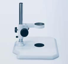
Stand N with 350 mm column*
The sandwich construction offers a compromise between stability and mobility.
For simple equipment:
- footprint: 440 x 370 mm
- round plate: d = 84 mm
- column d = 32 mm / h = 350 mm
- focus range: +/- 25 mm
- load capacity: max.5 kg
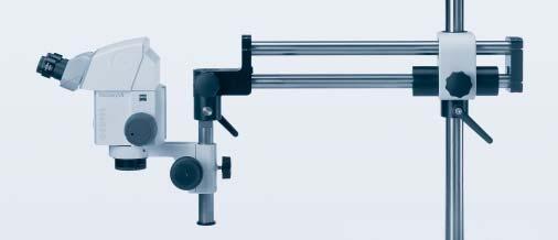
Horizontal armSDA Boom stand*
Suitable for examining larger objects.
- reach: up to 600 mm
- height ofcolumn: 600 mm
- focus range : +/- 25 mm
- load capacity: max 5 kg
* with Stemi mount with drive for column 32
Reliable,sensitive focusing ofcomplex equipment up to 10 kg: Coarse/fine drive with Profile S column.
Quicker to the Mark
Speed and reproducibility take their place alongside stability and precision. The motor focus is as precise and reliable as clockwork. New wear resistant materials make this possible.
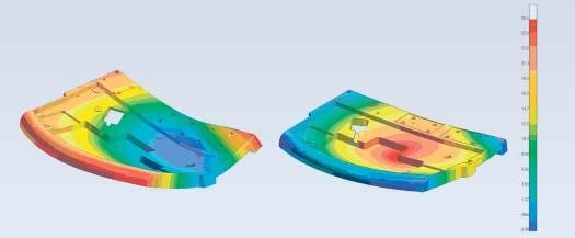
The ribbed die cast components ofconventional stand bases suffer significantly greater deformations...
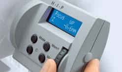
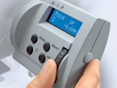
HIP (Human Interface Panel) replaces the knobs that were previously used for focusing. Always on display: the current Z position with a display accuracy of10 µm.
Quick and precise focusing with SyCoP. Aquick push ofthe joystick switches to the fine focus mode.
Focus motor with Profile S column on Stand base 450 to Profile S Focus manager,specimen protection and Z measurement are a standard part of the motor focus.
- footprint: 450 x 300 mm
- insert plate: 410 x 250 mm
- round plate insert: d = 120 mm
- profile S column: h = 490 mm
- focus range: 340 mm
- increment distance: 350 nm
- load capacity: max.17 kg
- support for microscope: d = 76 mm
...than the SteREO Discovery’s new Stand base 450 to Profile S,designed as a milled component.
The FEM slides show the possible deformation ofthe stand's base plate in the case ofassumed complex microscope equipment weighing 17 kg.
Scale factor: 500
Precise Positioning that Protects the Specimen
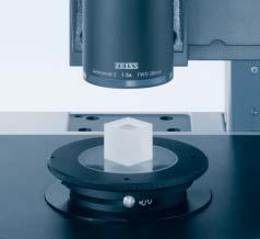
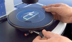
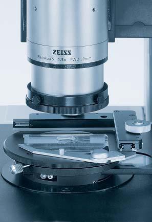
Specimen stages make it easier to move objects smoothly and evenly during observation. Depending on your requirements and applications, sliding, rotating, mechanical and ball-and-socket stages are available. They fit into the mount of the stand’s insert plate (d = 120 mm).
Gliding stage 110 x 110 S,d = 120 mm
For the sensitive positioning of even large samples in reflected and transmitted-light.Equipped with a glass plate 116 x 116 mm.
- positioning range: 110 x 110 mm
- stand mount: d = 120 mm
Alternative to glass plate:
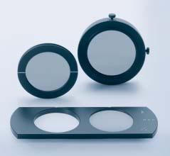
- mounting frame 116 x 116 / 84 mm for stage diaphragms - d = 40 mm opening
- d = 25 mm opening
- B/W plastic plate
Mechanical stage 100 x 100 S,d = 120 mm
(not illustrated) For the defined movement ofsamples in reflected and transmitted-light.Operation via horizontally arranged coaxial drive,left or right.
Equipped with a glass plate 116 x 116 mm
- travel range: 100 x 100 mm
- stand mount: d = 120 mm
Alternative to glass plate:
- mounting frame 116 x 116 mm / 84 mm for stage diaphragms - d = 40 mm opening
- d = 25 mm opening
- B/W plastic plate
- LED illumination mount for transmitted-light brightfield and polarizer S
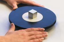
Rotating Pol stage for transmittedand reflected-light
For the precise rotation ofspecimens.
- stage diameter = 115 mm
- rotation range: 360°with graduation
- stand mount: d = 84 mm
Ifrequired,can be retrofitted with an object guide:
- adjustment range: 75 x 25 mm
Additional accessories for polarization in transmitted-light:
- Polarizer S,d = 84 mm
- full wave plate in slider
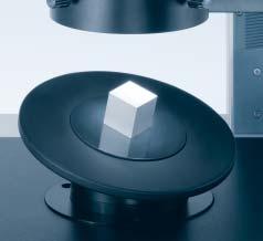
Gliding stage
For the positioning and rotating ofspecimens in reflected- and transmitted-light.
- stage diameter = 190 mm
- adjustment range: +/- 20 mm
- insert plate: d = 84 mm
- stand mount: d = 84 mm
Ball-and-socket stage
Because the stage can be tilted and rotated in all directions,three dimensional specimens can be examined efficiently from all sides. Preferably for applications in reflected-light. The exchangeable adhesive soft pad allows specimens to be affixed.
- stage diameter = 158 mm
- insert plate: d = 84 mm (adhesive soft pad)
- tilt range: +/- 30 degrees
- stand mount: d = 84 mm
Precisely on the Mark
There is an increasing demand for motorized stages in stereomicroscopy. They make it possible to scan large samples for documentation or analysis and to locate details on the specimen with speed, precision and in a reproducible way. When working with automated imaging techniques in particular, such as MosaiX or Mark&Find (AxioVision microscope software modules), these stages are already a prerequisite.
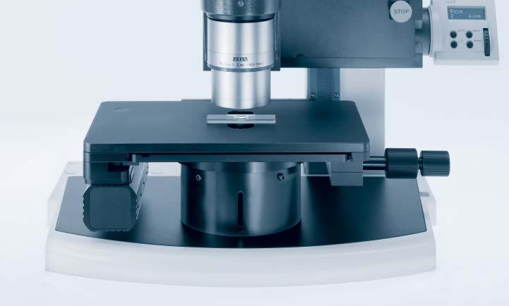
Mechanical stage 75 x 50 mot CAN (Illustrated without object guide)
Simple to operate – via the electronic coaxial drive arranged horizontally on the right ofthe stage or via PC.
- travel range: 75 x 50 mm
- speed: max.200 mm/sec
- increment distance: 0.1 µm
- accuracy ofreproduction: < 1µm
A selection ofmounting frames for specimen slides,petri dishes and reflected-light samples are available for different specimens.
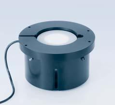
Stage carrier S,d = 120 mm
This makes it possible to mount motor stages securely and precisely on the extremely stable base plate.
- height: 85 mm
- stand mount: d = 120 mm
- LED illumination mount for transmitted light brightfield and Polarizer S
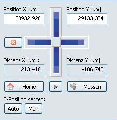
Precise location ofa sample detail via PC
Proven Classics
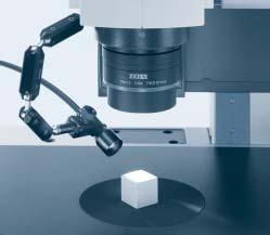
Cold-light illuminators have a long track record in stereomicroscopy. They illuminate the specimen with high intensity, and yet protect it from becoming too warm. Depending on the amount of light required, different cold-light sources are available with an extensive range of fiber optic components.
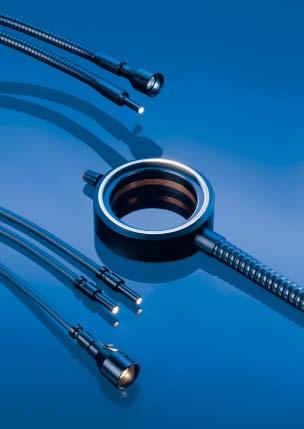
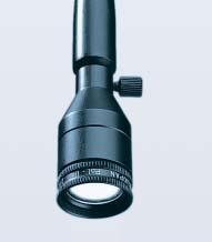
Single arm spot illuminator –
Variable oblique illumination with deliberate shadow cast effect
- one branch flexible or selfsupporting light guides with focusing attachment
- light guides with variable active diameter:
- d = 4,5 mm (fig.left)
- d = 8 mm (fig.above)
- d = 15 mm (fig.right) for large object fields
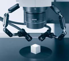
Dual arm spot illuminator –
Variable oblique illumination for the reduction ofdisruptive deep shadows
- flexible or selfsupporting light guides with active diameter:
d = 4.5 mm or d = 5.6 mm
- focusing attachments
Line light S,l = 50 mm
Converts the round cross section ofthe light guide into a narrow fiber optic slit.This produces an extremely flat angle ofillumination – the light spreads flatly over the specimen. The shadow cast effect makes even very small surface structures visible.Also suitable for high magnification objectives with smaller working distances.
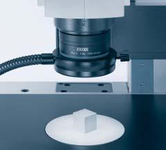
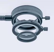
Slit ring illuminator for reflected-light brightfield –Ideal for the shadow free and homogeneous illumination ofobjects with large surface areas.
- with flexible light guides
- with active diameter:
d = 9 mm (fig.left)
- with active diameter
d = 15 mm (fig.right)
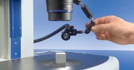
Extremely precise alignment ofthe illuminator is a must in order to achieve good 3D contrast in oblique reflected-light.Special clamps,illuminator carriers and articulated arms are available to ensure that the fixing is exact.Focusing attachments help to concentrate the light on the object field.
The better a specimen is illuminated, the more details it reveals to the observer. For example, fiber optic illumination components optimized for different applications generate very specific lighting effects on the surface of the specimen. The range extends from simple spot illuminators and ring lights through to specifically shaped glass fiber bundle geometries.
Ring illuminator for reflected-light darkfield
The light hits the surface ofthe specimen very flatly from all sides. As a result only the light dispersed from the structure ofthe specimen reaches the objective.Extremely fine structures are illuminated in their natural colors on a dark background.
- flexible light guide with active diameter: d = 9 mm
Move Your Specimen into a New Cold-Light
Powerful and effective:
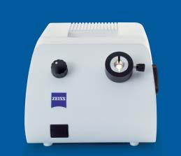
The ZEISS CL 1500 ECO cold-light source
- high light output,continuous control
- 15V/150W halogen lamp
- flicker free light for stable live images on the monitor
- quiet ventilation
- tailored standard accessories:
- flexible light guides with active d = 4.5 mm
- 2x goose-neck light guides with active d = 4.5 mm
- Slit ring illuminator with active d = 9 mm
All other light guides with an active diameter ofup to d = 9 mm can be used with the
Coaxial epi-illumination S
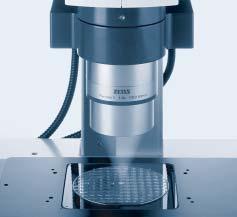
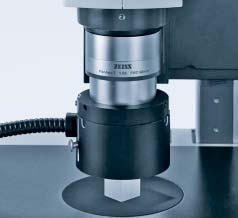
Particularly suitable for flat, specular specimens.Optimum image results are achieved using the PlanApo S 1.0x objective. The quarter wave plate should be used in the case ofvertical observation or documentation using the S/doc objective slide.
Diffusor S,telescopic,d = 66 mm
Indirect reflected-light illumination for the contrasting ofthree dimensional specimens with shiny surfaces. Glare is avoided due to the soft light. Recommended: sliding and/or ball-and-socket stage for improved alignment ofthe specimen.
ZEISS CL 1500 ECO.
Pure Daylight from Above
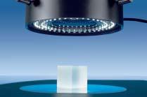
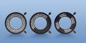
The VisiLED illumination system offers all the advantages of LEDs. Infrared free, offering the best daylight quality and electronically controllable – these are the reasons why this long lasting method of illumination is particularly recommended for illumination and contrasting tasks in stereomicroscopy.
Reflected-light brightfield with:
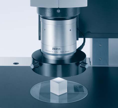
- VisiLED ring light S 80-55 BF (fig.above right) for objectives with a free working distance of55 to 135 mm.
- VisiLED ring light S 80-25 BF (fig.right) for objectives with a free working distance of25 – 50 mm.
Circular reflected-light darkfield with:
- VisiLED ring light S 40-10 DF (not illustrated)
- ALDF adapter for securing the ring light to the objective
VisiLED ring light/holder and distance rings are secured directly onto SteREO objectives (d = 66 mm).
The light from the
ripple and flicker free.It is also insensitive to fluctuations in the power supply.
Two operating units are available for controlling the VisiLED ring lights:
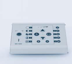
Multi Controller MC 1500
- for the control ofone or two ring lights
- brightness control with display
- segment control and rotation
- storage of4 illumination settings
- strobe mode and trigger operation
- thermo controller
- RS 232 interface
- foot switch optional
Multi Controller MC 750 (not illustrated)
- for the control ofone ring light
- adjustable transformer to set brightness
- thermo controller
VisiLED ring light is
Light and Shade
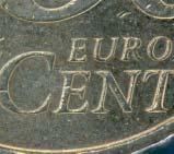
In addition to their high level of efficiency, the VisiLED ring lights also offer another substantial advantage. If required, they can be combined into segments and controlled individually. In reflected-light this makes variable oblique illumination possible. Shadows generated on the surface of the specimen in such a way reveal additional details.
Reflected-light brightfield, quarter circle
Reflected-light darkfield, full circle
Mixed-light
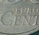
While the contours ofthe striking on this cent coin are highlighted in particular in darkfield,in mixedlight it is primarily the irregularities ofthe surface that are revealed more clearly.
All VisiLED ring lights can be controlled segment by segment.You not only choose between shadow free or oblique illumination – by rotating the segments you can also direct the illumination optimally onto the specimen.
In order to combine brightfield and darkfield in reflected-light,the VisiLED ring light S 40-10 DF is mounted onto the brightfield ring light using a special adapter.
With the MC 1500 Multi-Controller it is possible to work in brightfield,darkfield or mixed-light. Once illumination settings have been optimized, these can be stored and reproduced at the touch ofa button.
Thanks to the additional opportunities afforded by segmentation,virtually all contrasting possibilities are available in reflected-light.
Moveable for Optimum Contrast
The structures and details of transparent and semi-transparent objects can be made visible in transmitted-light.
By using a moveable mirror the direction of light can be changed in a targeted way. This allows the contrast to be geared towards the nature of the specimen in question.
The contours and outlines of specimens are also displayed clearly.
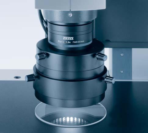
Transmitted-light equipment S
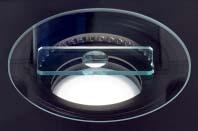
With this retrofittable module for stand base 450 it is possible to analyze samples in brightfield,darkfield and oblique transmittedlight.The large,rugged transmitted-light base offers plenty of room in the object space and makes it easier to screen batches of petri dishes or work with large samples
- footprint: 450 x 300 mm
- work surface: 410 x 250 mm
- glass plate: d = 120 mm
- illuminated object field: max.50 mm
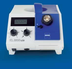
- cold-light source: ZEISS CL 1500 ECO or KL 1500 LCD or KL 2500 LCD
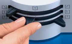
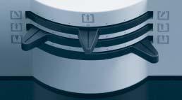
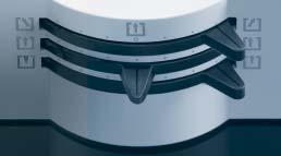
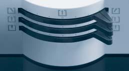
Ample possibilities for contrasting in transmittedlight.With 3 sliders it is possible to change the position ofthe mirror in relation to the specimen. Labeling makes it easier to reproduce the setting.
Top slider: sets the tilt ofthe mirror in relation to the specimen
Middle slider:moves the mirror in a north-south direction
Bottom slider:changes between two different reflectors for directional or diffused illumination
A Question of Adjustment
The continuous mirror adjustment with several degrees of freedommakes it possible to freely adjust the angle of illumination. This allows to individually optimize illumination and contrast for a very wide range of specimens.
Setting:Brightfield for transparent,high contrast specimens and also used to display contours and outlines
Setting:Oblique illumination makes low contrast structures visible in transparent and opaque specimens
Setting: One sided darkfield low contrast fine structures shine brightly against a very dark background
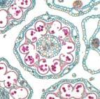
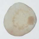
Daisy
Transmitted-light brightfield
PlanApo S 1.5x objective
Magnification: 150x*
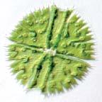
Desmid algae Micrasterias
Oblique illumination in transmittedlight PlanApo S 1.5x objective
Magnification: 150x*
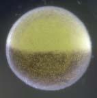
Frog embryo
Transmitted-light darkfield
PlanApo S 1.5x objective
Magnification: 150x*
See the Light
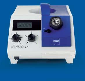
Depending on what is required and the tasks at hand,two different cold-light sources are available with LCD display and a broad range offiber optic illumination components.
KL 1500 LCD
-15V/150W light source
- for light guides with an active diameter ofup to 9 mm
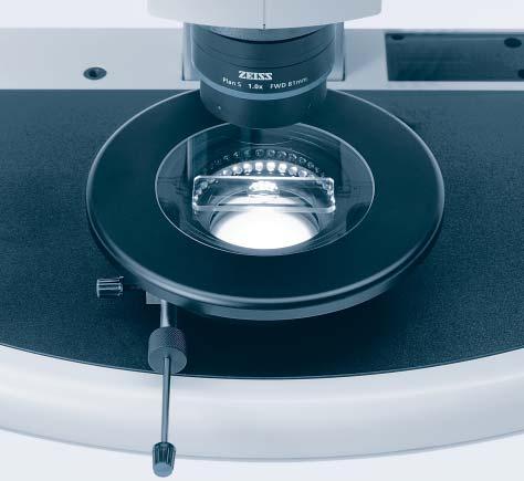
The VisiLED transillumination-contrast stage ACT (Advanced Contrast Transmitted) also offers the advantages of LED illumination for applications in transmitted-light. This dynamic and sophisticated illumination stage delivers brightfield, darkfield and oblique illumination with options to control the directionof illumination. This combination stage and illuminator is ideal for stereoscopic analysis of challenging, low contrast samples.
VisiLED transillumination-contrast stage ACT,d = 120 mm
This retrofittable transmitted-light solution allows analyses to be performed in brightfield and darkfield.Precise control ofthe variable slit diaphragm allows for generation ofreliefcontrast at high magnifications particulary.
- illuminated object field: max.50 mm
- stage diameter: 160 mm
- glass plate: 120 mm
-sliding stage adjustment range: +/- 10 mm
- insert plate mount: d = 120 mm
The LED is controlled using the MC 1500 Multi-Controller or via PC
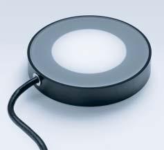
VisiLED transillumination BF,d = 84 mm
Retrofitted into the Stand base 450 to Profile S or the Stage carrier S for simple transmitted-light brightfield applications.
- illuminated object field: max.50 mm
- segment illumination for oblique illumination (using MC 1500)
- stand mount: d = 84 mm
The LED is controlled using the MC 750 or MC 1500 Multi-Controller.
Half Diaphragms –no Half Measures
Two horizontally arranged, moveable half diaphragms are positioned between an LED area light for brightfield and an LED ring light for darkfield. This variable slit diaphragm makes it possible to observe and document low contrast specimens in relief contrast at higher magnifications. It ensures stunning effects and renders considerably more information about the specimen.
Settings on VisiLED transillumination-contrast stage ACT
- brightfield area light “on”
- halfdiaphragms open
- darkfield ring light “off”
- brightfield area light “on”
- slit diaphragm positioned excentrically to the north, defined open
- darkfield ring light “off”
- brightfield area light “on”
- slit diaphragm positioned centrically,defined open
- slit diaphragm positioned excentrically to the south, defined open
- darkfield ring light “off”
Oblique illumination (“negative”reliefcontrast)
- brightfield area light “off”
- diaphragm closed
- darkfield ring light “on”
Transmitted-light darkfield (circular darkfield)
Bright Fluorescence Signals
Fluorescence stereomicroscopy means visualizing specific flurochromes and fluorescent proteins, working with large object fields and high resolution, three dimensional images.
With the “PentaFluar” S solution, the SteREO Discovery can be equipped for demanding fluorescence applications in no time at all.
Depending on the light source used,two different illuminators are available:
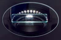
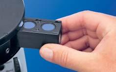
This intermediate tube works like a coaxial illuminator. Excitation takes place via two chromatic beam splitters through both observation channels.In this way,the visible object field is always fully illuminated when zooming.
- magazine for 5 filter blocks
- mechanical shutter
- iris for adjusting the diameter ofthe illuminated field
Embryos ofthe Drosophila melanogaster fruit fly,immunofluorescence, SteREO Discovery.V8 with Achromat S 1.5x objective, visual magnification 120x with 10x eyepieces
Specimen: Sameer Phalke,Institute for Genetics,Halle University, Germany
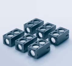
The reflector turret holds up to 5 different filter blocks.Each filter block has an excitation filter and two emission filters permanently built in:
- Filter block 38 GFP BP
- Filter block 43 Cy3/Rhod/RFP
- Filter block 45 Texas Red/FRFP
- Filter block 46 YFP
- Filter block 47 CFP
- Filter block 50 Cy5
- Filter block 57 GFP LP
- Filter block 58 Cascade Yellow
Polarization Brings it to Light
Polarizing equipment makes it possible to analyze birefringent materials using the stereomicroscope. Simple measurements can then be performed, for example the determination of the relative birefringence. The use of polarizers also helps reduce distracting reflections.
For transmitted-light analyses in polarized light,the Rotating Pol stage is used with the Polarizer S and can be equipped, when necessary,with an specimen guide 28 x 75 mm and a Full wave plate in a slider (first order red).
The Polarizer S,d = 84 mm can also be mounted directly into the Transmitted-light equipment S for simple analyses between crossed polars in transmitted-light.
- for inserting into the rotating stage
Polarizer S,d = 84 mm (not illustrated)
- for inserting into transmitted-light equipment and stages
Analyzer S, rotatable,d = 66 mm
- for clamping to objectives
Full wave plate in slider
- as slider for Rotating Pol stage
For improved illumination ofshiny surfaces,rotating Polarization filter can be screwed onto the focusing attachments (for light guides with an active diameter ofd = 4.5 mm).
Analyzer S fitted to the objective then allows disturbing reflections to be minimized.
Additional accessories for analyses in polarized reflectedlight include:
- Polarization filter set S VisiLED (not illustrated)
- Polarization filter set S, d = 66 mm for slit ring illuminators
Polarizer S
SyCoP – State of the Art
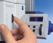
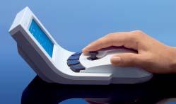
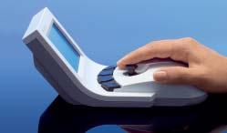
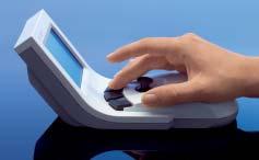
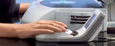
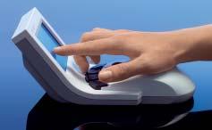
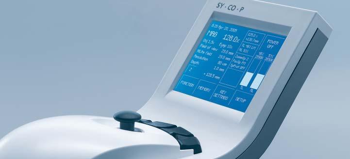
SyCoP (System Control Panel) is a new operating element, patented by Carl Zeiss, for controlling complex stereomicroscopes and illumination systems. It combines push buttons, a joystick and a touchscreen to create a handy, mobile control unit. All the essential functions of the stereomicroscope are integrated in one place, enabling safe and efficient one handed control of the system so that you never have to look away from the specimen.
The joystickfor zooming and focusing.
The keysfor illuminating and contrasting.
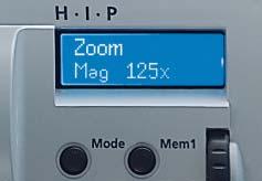
HIP for zooming:
HIP offers a choice between three different speed profiles and provides a real time display ofmagnification,diameter ofthe object field,resolution and depth offield. Additionally,preset magnifications can be stored and recalled via the two memory keys.
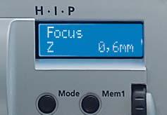
HIP for focusing:
This HIP also offers three different speed profiles and provides a real time display of the current Z position.Additionally,preset focus can be stored and recalled via the two memory keys.
HIP (Human Interface Panel) replaces the conventional knobs for focusing and zooming on the SteREO Discovery.V12 and V20.
Complete attention can be given to the specimen... ...thanks to SyCoP.
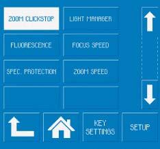
The touchscreenfor switching to additional functions and for information.
The wheel responds dynamically to the movement ofthe thumb: slowly and sensitively with small movements and more quickly with larger movements.
SyCoP Delivers Information
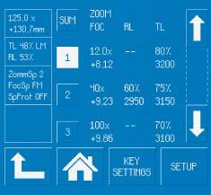
A further innovation in stereomicroscopy. SyCoP delivers information. In addition to its function as a control unit, the SyCoP also displays real time data on all the important optical parameters of the current microscope setting.
In the FUNCTION menu it is possible, for example,to set ClickStops,activate electronic specimen protection,select speeds for zooming and focusing and activate the light or focus manager.
Under SETUP,up to five instrument settings per user can be programmed and called up by touching the screen.
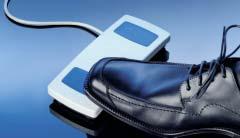
The EMS electronics module not only allows the microscope to be operated via SyCoP but also via AxioVision software or foot pedals.
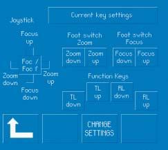
Under SETUP,important and frequently used functions ofthe stereomicroscope can be assigned individually to the keys ofthe SyCoP or to the foot switch.
The main window of SyCoP displays information on the important, current optical data relating to the stereomicroscope,such as:
- total magnification
- visible object field
- maximum possible resolution
- depth offield
Additional settings relating to the basic instrument and illumination are displayed in the status window.Buttons on the touchscreen lead to the respective sub-windows.
Protect Valuable Information
Whether it’s for research or routine tasks in biology, medicine or industry – the requirements for the reliable and rapid documentation of microscopic images are becoming ever more demanding.
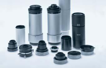
A series ofadapters for the 60N interface on the microscopes allows easy coupling to the following documentation systems:
- AxioCam digital microscope cameras
- video cameras
- digital compact cameras
- analog and digital mirror reflex cameras
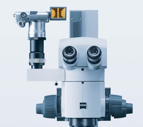
SteREO Discovery.V8 with a Canon PowerShot on the Intermediate phototube S,left 100/100. Digital consumer compact/mirror reflex cameras are connected via the 60N interface.
Intermediate phototube S, left 100/100
For the adaptation ofa camera, with switching between 100% observation or 100% photo
Intermediate phototube S with two 50:50 ports
For the adaptation oftwo cameras, with permanent splitting for simultaneous stereoscopic observation and documentation
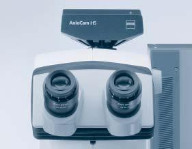
With AxioCam, Carl Zeiss has created a series of digital microscope cameras in order to address the ever expanding range of tasks performed in laboratories and research institutes. The accompanying AxioVision software ensures that the processes of microscopic image analysis can be completed simply, quickly and accurately.
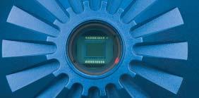
A feature ofAxioCam cameras: Encapsulated CCD sensor with C-Mount adapter
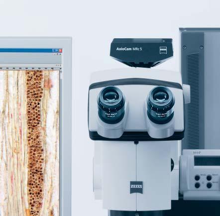
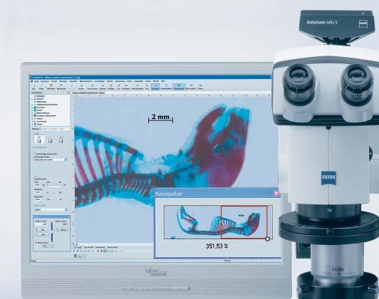
SteREO Discovery.V12 with binocular ergo-phototube S 5-45°and AxioCam MRc5
Compact,powerful and fast.
A camera for virtually all applications in stereomicroscopy:
- resolution: 5 mega pixels in color
- sensor size: 2/3”
- FireWire/IEEE 1394 interface
- connection: C-Mount
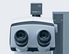
AxioCam IC
Two genuine microscope cameras for biomedical and industrial applications provide pin sharp,true color images. AxioCam ICc3
The high-speed camera for the digital documentation of dynamic processes in B/W.
- basic resolution: 330 K pixels
- image rate: up to 198 images/second (5x5 Binning)
- sensor size: 1/2”
- FireWire 1394a
- connection: C-Mount (see www.zeiss.de/micro for more cameras)
AxioVision is The Software
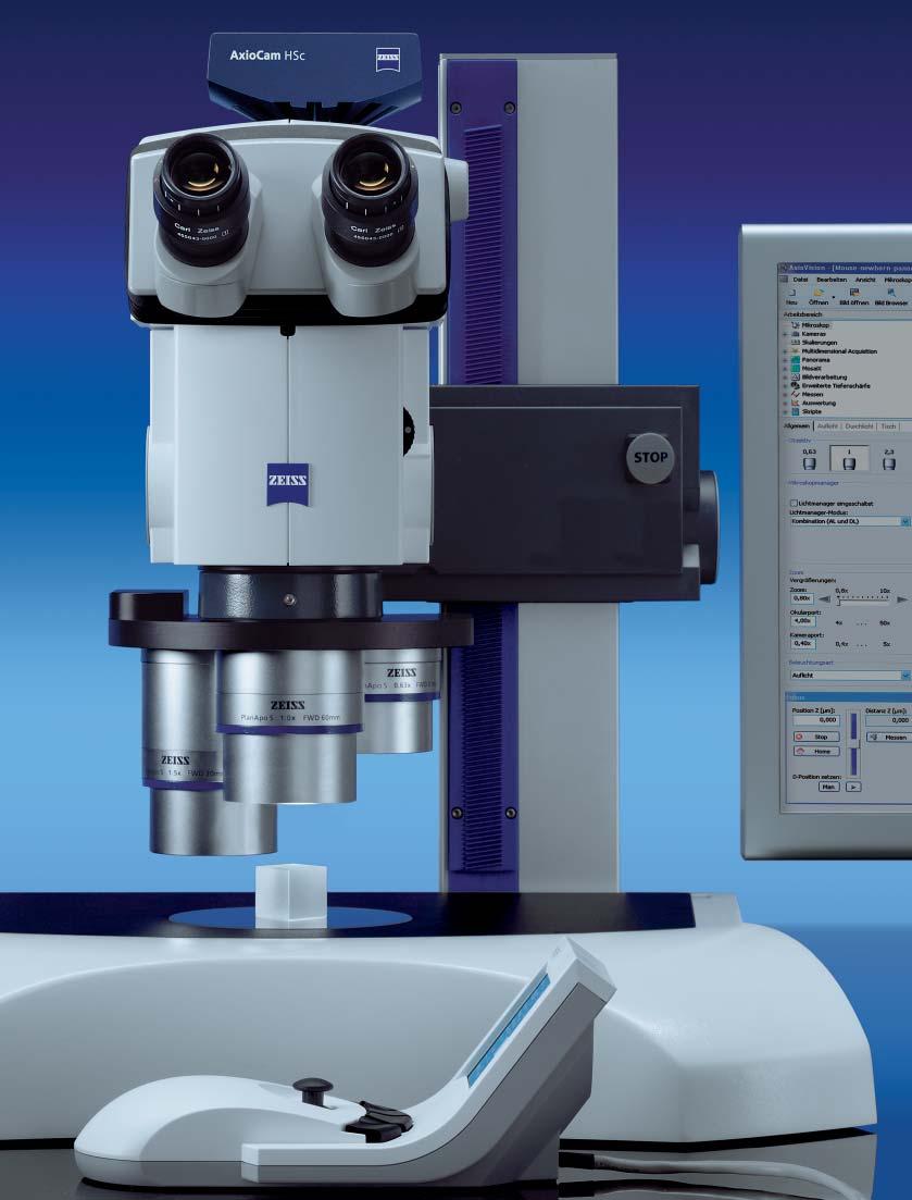
AxioVision – a complete suite of software for microscopy and image analysis. Geared to current requirements in stereomicroscopy thanks to its unique modular structure and offering attractive options for the future development of this field, AxioVision integrates microscope control, image acquisition, image processing, image management and archiving to form a complete system.
Extended Focusmodule
In situations where the height ofthe sample exceeds the depth offield ofthe stereomicroscope. This module allows information collected in a Z-stack to be combined in to a single image.
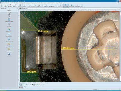
Interactive Measurement module
For the measurement ofmorphological parameters on interactively defined contours.
(see www.zeiss.de/axiovision for more modules)
Electronic component
Reflected-light brightfield
SteREO Discovery.V12 with PlanApo S 0.63x AxioCam MRc5
Specimen: Carl Zeiss MicroImaging GmbH
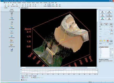
Topography module
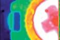
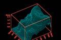
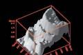
Height maps can now be generated from Z stacks and displayed in three dimensions.The topographic image created in this way contains all the important information. Other displays are also possible:
Color coded height image
Isometric grid projection Shadow projection
Perpendicular Perfection
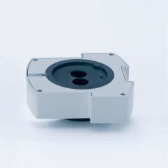
The S/doc objective slider is recommended for professional documentation as well as for more precise measurement in the microscopic image. Depending on the position of the photo port, you can choose to shift the objective centrically under the right or left observation channel.
The now perpendicular, axial view of the object results in freedom from parallax and optimum imaging quality.
Simple
Transmitted-light
Specimen:
Dr.Kenji Imai,M.D.,PhD.
GSF National Research Center for Environment and Health, Neuherberg,Germany
switching from stereoscopic observation to a parallax free macro view ofthe object by the camera
Objective slide S/doc - switched to left channel for documentation - central position for stereoscopic viewing- switched to right channel for documentation
Skeleton specimen ofa newborn wild-type mouse
brightfield SteREO Discovery.V12 with PlanApo S 0.63x AxioCam MRc5
Panorama module. Using this module,high resolution overview images can be generated perfectly from single images,without the need for a motorized stage.
SteREO Discovery.V8
SteREO Discovery.V12
ObjectivesEyepieces
SteREO Discovery.V20 NameFWDMagnificationObject
Operating concept Telescope or CMO (Common Main Objective)
Pancratic zoom range
Magnification range
SteREO Discovery.V81x ...8 xmanual zoom body
SteREO Discovery.V120,8x …10 xmotorized zoom body
SteREO Discovery.V200,75x ...15 xmotorized zoom body
SteREO Discovery.V810x …80xmax. 346 Lp/mmBasic version* and max. resolution 3x …460xmax. 801 Lp/mmwith exchangeable optics for stereo observation
Binocular tube S 20°Binocular ergo-phototube S 5-45°
Interpupillary distance at 2 viewing heights, adjustable from 55 mm to 75 mm
Intermediate tubes Intermediate phototubes S 100/100 and S 50/50 Drawing intermediate tube S Y intermediate tube S (for 2D macroscope setting)Intermediate tube S, fixed 40 mm (ergo component)
Reflected light polarizer for ring lights and spotsFiber optic cold-light
Microscope cameras AxioCam IC, MR, HR, HS
Adaptation for digital compact and mirror reflex cameras
AxioVision microscopy and image analysis software
*Basic version with 1x objective and 10x eyepieces
Behind the Scenes
position in μm
On the SteREO Discovery.V12 and V20, mechanical zoom control curves have been replaced by ones that are generated electronically. This level of precision yields, considerably sharper microscopic images across the entire zoom range. The result is more relaxed viewing in 3D and improved contrast at high magnifications.
The motorized zoom body of the SteREO Discovery.V12 and V20 deliver images that are twice as sharp as traditional mechanical zoom bodies.
Magnification
Depth of field curve, within these parameters the images are in focus
Typical defocus curve of a single zoomchannel with mechanical zoom curve
Typical defocus curve of a single zoom channel with electronically generated zoom curve
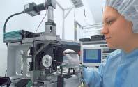
Before assembly begins,each lens is exactly calibrated against a reference “null lens set”and the values are saved in a data pool. This feeds the database used by a computer to select optimally matched lenses for the zoom system. By doing this,an optimally coordinated lens family is developed for every stereomicroscope.
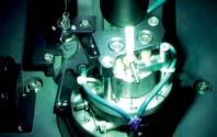
The rotating reflex ofa lens ofthe zoom optics. As soon as it is in the circle …
… the moveable micro clapper on the computer controlled glue leveling machine automatically adjusts the final fine alignment.
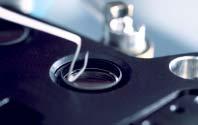
The lens which has been positioned carefully is then fixed immediately.The machine lays highly calibrated, uninterrupted glue beads through a strong 0.5 mm cannula and hardens it using UV light.
In the zoom body adjusting device,the precise procedures ofall moveable optical elements are programmed. To do this,approxamately 7,000 reference points are analyzed by computer.In doing so,each stereomicroscope recieves its own completely individual zoom control curve.