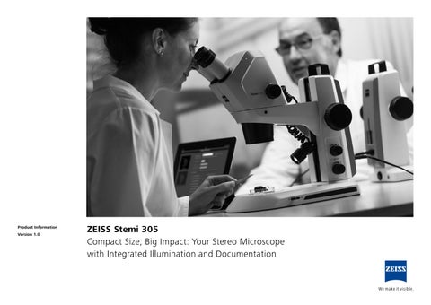
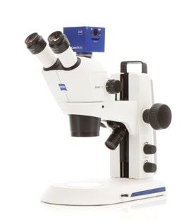
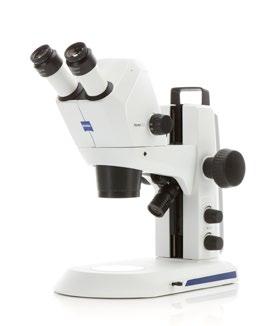
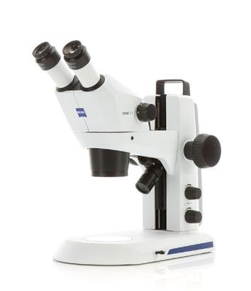
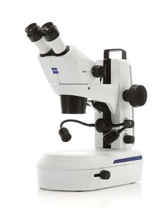
All-in-one design. Easy to Use. Easy Documentation, too.
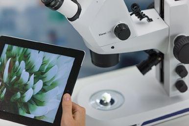
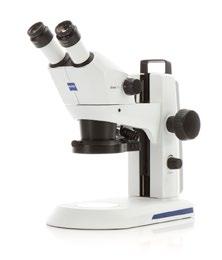
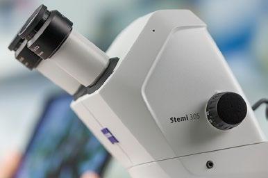
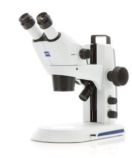
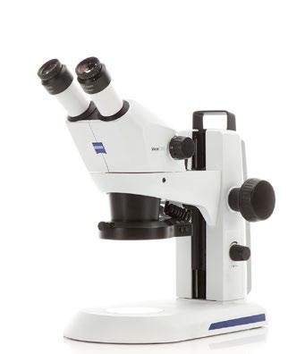
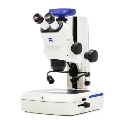
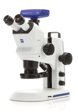
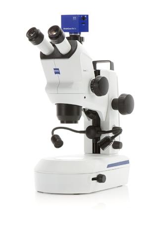
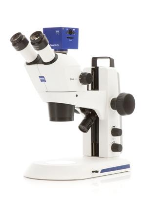
Stemi 305 is your compact stereo microscope with a 5:1 zoom – equally at home in the biology classroom, research lab or on the industrial shop floor. Choose between predefined microscope sets to get the optimal illumination equipment for your applications. Stemi 305 lets you observe samples as they really are: three-dimensional and crisp in contrast – no preparation needed. Enjoy all the advantages of an easy-to-use microscope with integrated LED illumination for reflected and transmitted light – plus fast, easy-to-use documentation. Just snap your images with the integrated 1.2 Megapixel Wi-Fi camera and share them using Labscope, the iPad imaging app. Or choose the conventional phototube for access to all ZEISS Axiocam cameras and free ZEN lite imaging software. This Greenough microscope will give you crisp 3D impressions, versatile object illumination and easily acquired images to share, whenever you want.
› The Applications
› The System
› Technology and Details
› Service
Simpler. More Intelligent. More Integrated.
More than Design. All-in-One Device.
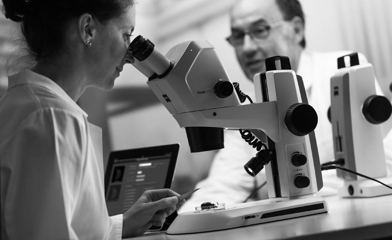
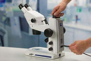
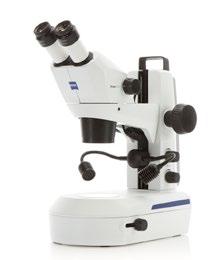
It‘s compact and it‘s fun. Yet Stemi 305 integrates everything you need into a single Greenough stereo microscope. Just plug it in and switch it on – that’s the installation done, with no extra accessory boxes or cables to clutter up your space. Then all you have to do is illuminate your object, focus and start snapping images. Use Stemi 305 cam with on-board Wi-Fi camera to easily save your results, share them and collaborate on projects with friends, colleagues and classmates. Store Stemi 305 away when work is done, then take it out and get started again in minutes.
Illumination. Crisp for Any Application.
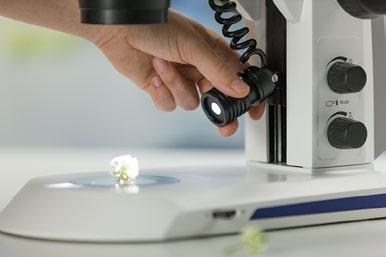
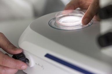
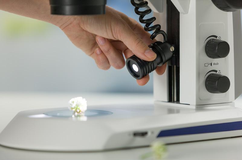
Despite its ease of use, Stemi 305’s integrated illumination is variable to contrast every specimen at its best. Simply press a button to select and combine up to two reflected light contrasts and transmitted light. While each zoom body is equipped with near-vertical light to illuminate deepenings in the sample, the second reflected light and transmitted light units are interchangeable. This enables predefined microscope sets for education, laboratory or industrial use to optimize contrast in your application range. White LEDs in your Stemi 305 generate the brightlight of daylight color so each image appears crisp and clear. What’s more, long-life LEDs are noiseless, maintenance-free – and real energy savers. › In Brief › The Advantages
Documentation. Integrated and Wireless.
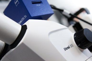
Documentation is important for lab work, essential for industrial inspection. In the classroom your ability to acquire and share images is a key resource for vivid, exciting science courses. Stemi 305 comes with two options for documentation. Choose Stemi 305 trino with a conventional phototube for access to any ZEISS Axiocam microscope camera and free ZEN lite imaging software. Or go for Stemi 305 cam with its integrated camera and Wi-Fi router. Create your own virtual classroom by using Labscope, the iPad imaging app, to stream live images from several microscopes to all connected iPads. It’s easy to share, compare and discuss the work of all the students on your own wireless network. Fun, too.
› In Brief › The Advantages › The Applications
The System
Technology and Details
Service
Your Insight into the Technology Behind It
ZEISS Stemi 305 for Laboratory
Versatile Illumination for Sample Preparation
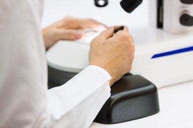
Workspace is always limited in a laboratory where you are observing, preparing or dissecting model organisms and other bio specimens. You may be looking at oocytes or embryos, larvae or adult animals, or at plant components such as roots and leaves. For all these reasons you need variable contrasting methods in transmitted light, but also reflected light. Use the mirror-based transmitted light unit of stand K LAB to observe and manipulate even uncolored transparent specimens. With stand K LAB reflected and transmitted light can be selected easily or combined. To document your results choose Stemi 305 cam or Stemi 305 trino with stand K LAB, double spot K and ergonomic hand rest.
Compact, versatile and well prepared for laboratory work. The tiltable and shiftable mirror features brightfield, one-sided darkfield and oblique light – plus polarization contrast as an option. Rotate to frosted and plain mirror side, then decide between crisp and diffuse brightfield contrast.
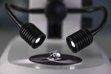
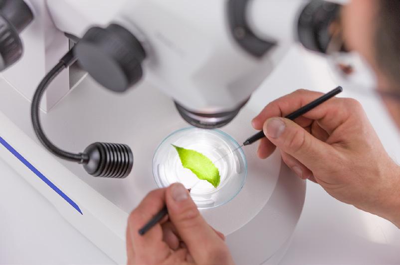
For sample preparations in reflected light, the double spot illumination with self-carrying goosenecks is optimal. It creates half-shadow effects that lead to a good 3D impression without overly dark shadows. During dissections the specimen stays illuminated even if the manipulating hand covers one of the spots.
extended work add the
For
ergonomic hand rest to keep your hands relaxed, even during long preparations.
The Advantages
The Applications
Your Insight into the Technology Behind It
ZEISS Stemi 305 for Industry
Segmentable Ringlight for Quick Inspections
Technology and Details
Service
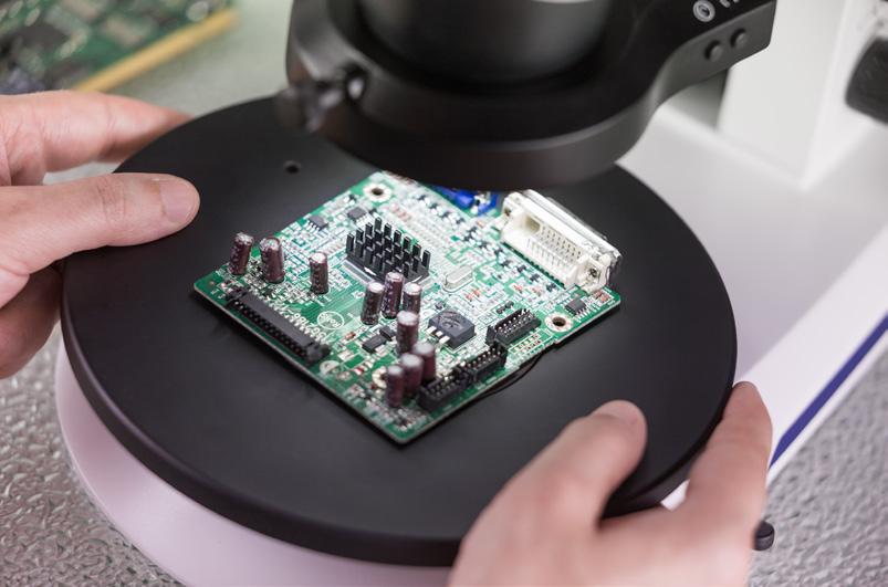
Stereo microscopes are essential to everyday routines in your production line or quality department: inspecting, assembling or repairing electronic or optoelectronic components, small mechanical parts, sensors or measuring devices. And if you work in electronic industries your microscopes operate in electrostatic protected areas (EPAs). Choose Stemi 305 MAT for visual inspection or small parts assembly. It provides stand K MAT with reflected light LED controls and anti-static surface resistance to enable use in EPAs. It also includes two reflected light illuminations: the integrated vertical illuminator to look at holes, threads and cavities – and the segmentable ringlight K LED. Simply press the dimming button at the side of the focus column to switch quickly between vertical spot, ring illumination and mixed light. To document or archive your results use Stemi 305 trino with stand K MAT and segmentable ringlight K.
Compact, easy to use and suited for electrostatic protected areas.
The shadow-free ringlight features four different segment modes: full-, half- and quarter-circle and 2-opposite quartercircle. Set the segment illumination, then turn the light direction manually in 90° steps to quickly inspect for scratches, defects or residues without moving your specimen. Or use auto rotating mode to get a spatial impression of the object surface just by changing shadows.
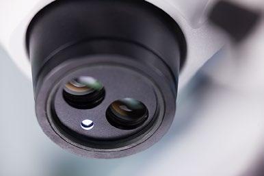
Stemi 305 is equipped with an integrated near-vertical LED spot to illuminate holes and indentations – even through front optics.
Your Insight into the Technology Behind It
Document Your Results. Archive and Share.
Enjoy the flexibility of Stemi 305 trino with access to ZEISS Axiocam microscope cameras and ZEN imaging software. The fixed 50/50 division between left eye and camera path is optimal for live demonstrations: your students or customers can follow your microscopic work live on the monitor. Combine Stemi 305 trino with Axiocam ERc 5s and get the advantage of various interfaces: HDMI for direct monitor access, USB to use with Windows PC and free ZEN lite imaging software, or LAN to connect to a digital network and control the camera with the iPad imaging app Labscope.
Stemi 305 cam is your compact and easy-to-use solution for educational tasks. The fast, color Wi-Fi camera and WLAN router are already integrated into the microscope body. In “Access point” mode each Stemi 305 cam creates its own WLAN: up to six iPads can connect directly to the integrated camera and display its live image using the iPad imaging app Labscope. To create a high quality digital classroom, enable “Add to existing WLAN” mode and connect several Stemi 305 cams to the same digital network. Your students can then share their microscope images and collaborate on projects with colleagues and classmates. Let them learn the fun way.
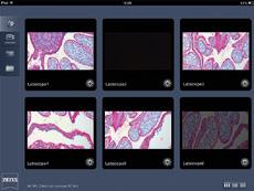
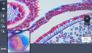
The iPad imaging app Labscope displays the live images of all Stemi 305 microscopes in your network to each connected iPad. With one touch you can look at each student's results. It's easy to snap an image, add annotations and measurements, and save it – or export it directly to your server. Once connected to the internet you can share your images, reports or videos with others via email, social media or cloud services. With Labscope, you are saving your images in the ZEN compatible .czi file format including all metadata –or you can select the space-saving .jpg format. Download Labscope free from the Apple App Store.
ZEISS Stemi 305 trino
ZEISS Stemi 305 cam
ZEISS Labscope, the iPad Imaging App
› In Brief
› The Advantages
› The Applications
› The System
› Technology and Details
› Service
Tailored Precisely to Your Applications
Typical Applications, Typical Samples Task
Industry
• PCB Electronics
• Entertainment Electronics
• Micro Technology
Use Stemi 305 for assembly, visual inspection, and repair of various 3D shaped industrial work pieces.
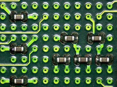
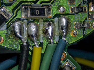
Visual inspection for damage to circuit boards, e.g. oxidation, stress corrosion, inaccurate drill holes.
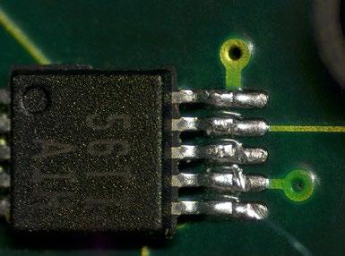
Inspection of the quality of soldered connections, e.g. for improper wiring or for damaged or missing components.
Manufacturing, inspection and repair of thick film or hybrid circuits.
• Car Industry Inspection of injection nozzles, air bags ABS systems.
Inspection of large parts, motor or chassis components.
ZEISS Stemi 305 Offers
• Compact Stemi 305 MAT microscope set includes integrated vertical illumination to illuminate holes and deepenings, and a shadow free ringlight.
• ESD safe by antistatic surface treatment, usable in electrostatic protected areas.
• The ringlight is segmentable for distinct shadow effects. Quickly change the light direction to find scratches and defects without moving the specimen.
• To investigate structures of flat surfaces set double arm gooseneck to a low position to create grazing light. To reduce reflections from shiny parts add optional polarization equipment.
• For large part inspection use cost effective boom stand A in combination with front lens 0.5×. Benefit from the integrated vertical illumination of Stemi 305.
• To document with highly resolved images use Stemi 305 trino with Axiocam microscope camera.
• Get the lowest cost imaging from Stemi 305 cam with integrated camera and iPad solution. The images can be archived easily to your local server via wireless LAN.
• Dental Laboratory
Finish all-ceramic crowns accurately and reliably, identify and remove casting beads precisely in the framework.
• Use Stemi 305 flexible tilting arm stand U to share your microscope with two or three work places. The integrated vertical illumination of Stemi 305 is shadow free and homogeneous – and always correctly adjusted to the object field.
› In Brief
› The Advantages
› The Applications
› The System
› Technology and Details
› Service
ZEISS Stemi 305 at Work
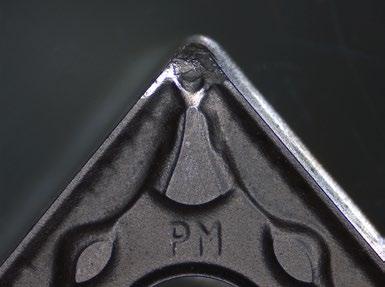
Indexable insert; ringlight full circle, zoom 0.8×
• LED illuminators to stands K: spot, double spot gooseneck, segmentable ringlight, flat or mirror-based transmitted light stands
• Controller K – for standalone use integrated camera, near vertical spot or ringlight K
• Fiberoptic cold light sources CL4500 LED and CL1500 Hal with spot, annular ring, linear, vertical, diffuse and area illuminators, fiber optic transmitted light unit
• Polarization equipment for filters for spots, ringlights and transmitted light units
Illumination Techniques
Reflected and transmitted light: brightfield; darkfield; polarization; oblique light
4 Stands
• Space saving table top stand K
• Stand K EDU with reflected light (=RL) LED electronics and flat transmitted light unit
• Stand K LAB with RL LED electronics and mirror-based transmitted light unit
• Stand K MAT with RL LED electronics and ESD features (antistatic)
• Large table top stand N
• Boom stands A and SDA, tilting arm stand U
5 Accessories
Eyepiece reticles, gliding, ball/socket and rotating stages, ergo hand rest for stand K LAB
6 Software
• ZEN lite imaging software
• Labscope/Matscope iPad imaging apps
7 Recommended Cameras
• Axiocam ERc 5s
• Axiocam 105 color
• Axiocam ICc 1
• Axiocam ICc 5
› In Brief
› The Advantages
› The Applications
› The System
› Technology and Details
› Service
Your Flexible Choice of Components
Microscope Sets for:
Education
• Binocular tube
• Integrated near vertical illumination
• LED spot, zoomable and height-adjustable, for oblique and grazing light illumination with strong shadow
• Flat transmitted light base for brightfield and darkfield illumination
• Optional: polarization equipment for spot and transmitted light
• Order number: 435063-9010-100
• Binocular tube
• Integrated near vertical illumination
• Double arm gooseneck, self-carrying, for variable oblique light illumination with distinct shadow effect
• Tiltable mirror base for brightfield, darkfield and oblique light illumination
• Optional: ergonomic hand rest, polarization equipment for spots and transmitted light
• Order number: 435063-9020-100
• Binocular tube
• Integrated near vertical illumination
• LED segmentable ring light for shadow free ring illumination and oblique light segment illumination: half circle, quarter circle, two-spot
• Rotating illuminating segments
• ESD properties: antistatic coating of microscope body and stand
• Order number: 435063-9030-100
› In Brief
› The Advantages
› The Applications
› The System
› Technology and Details
› Service
System Overview
› In Brief
› The Advantages
› The Applications
› The System
› Technology and Details
› Service
System Overview
› In Brief
› The Advantages
› The Applications
› The System
› Technology and Details
› Service
System Overview
The Advantages
Technical Specifications
ZEISS Stemi 508
Your Apochromatic Stereo Microscope with 8:1 Zoom for Excellent Image Contrast and Color Accuracy
Your Apochromatic Stereo Microscope with 8:1 Zoom for Excellent Image Contrast and Color Accuracy
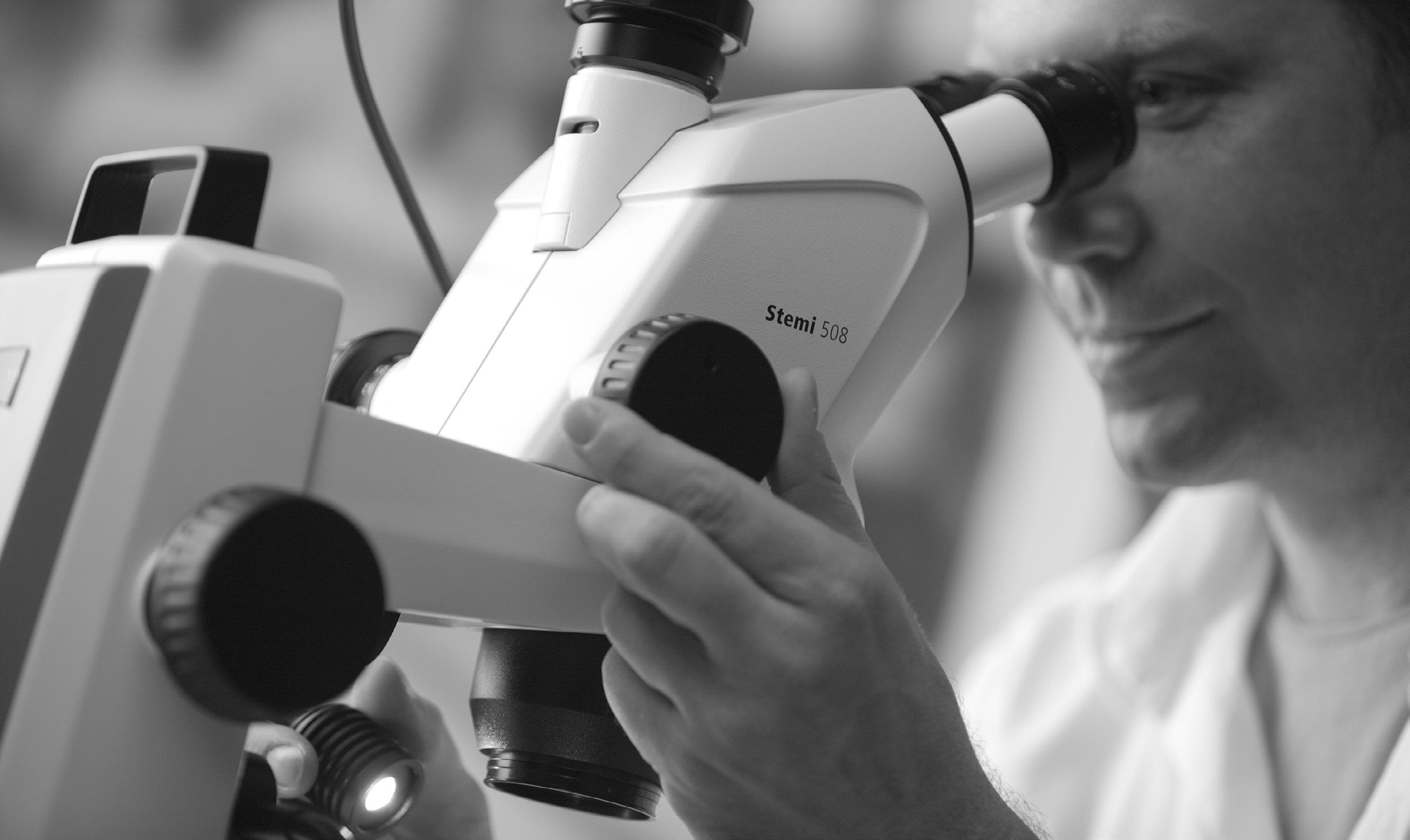
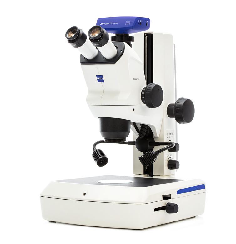
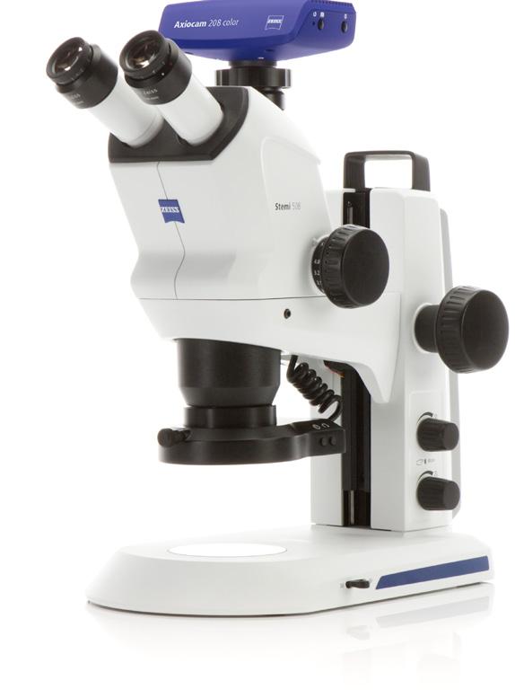
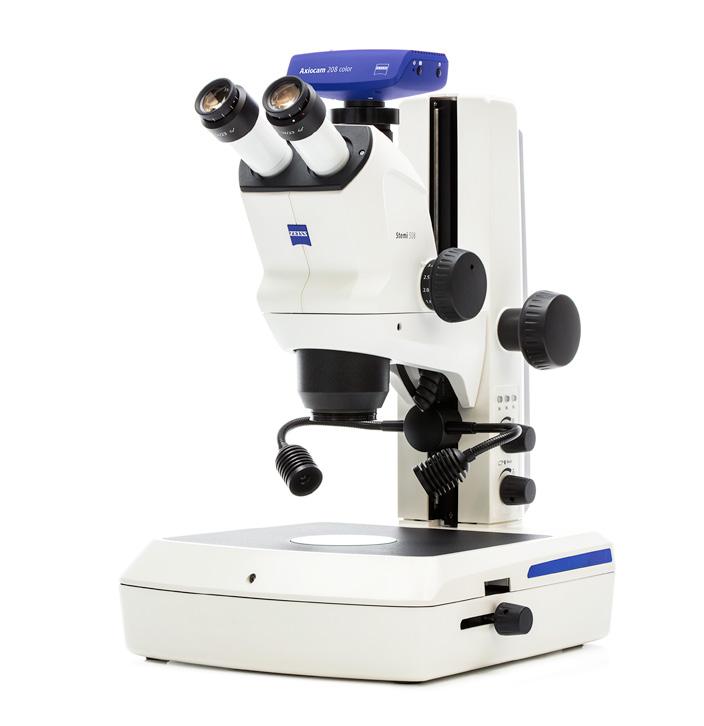
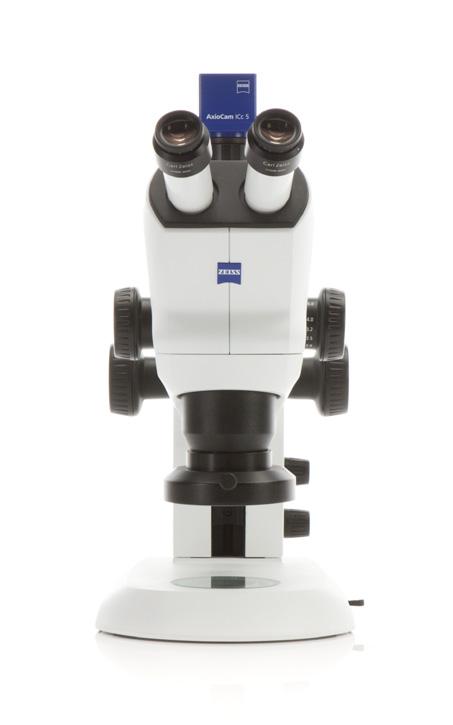
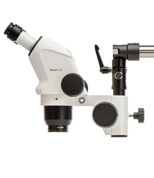
Stemi 508 is compact, reliable and equipped with optics and mechanics designed for heavy workloads. With the large 36 mm object field you always keep the overview of your sample. The 8:1 zoom then allows to bring details up to 50× magnification. You even have larger samples? Add interchangeable optics and observe an area of up to 122 mm, making Stemi 508 a top per former in its class. Stemi 508 offers better ergonomics than any other Greenough-type stereo microscope: The low viewing angle of 35° lets you keep a relaxed posture even after hours of work.
With Stemi 508 you observe and document your samples exactly as they are: rich in detail, sharp in focus and free from distortion or color fringes. Stemi 508 is your robust all-rounder for everyday lab work and industrial inspections: accurate, ergonomic – and always easy to use.
› In Brief
› The Advantages
› The Applications
› The System
› Technology and Details
› Service
Simpler. More Intelligent. More Integrated.
Large 8:1 Zoom with Apochromatic Correction
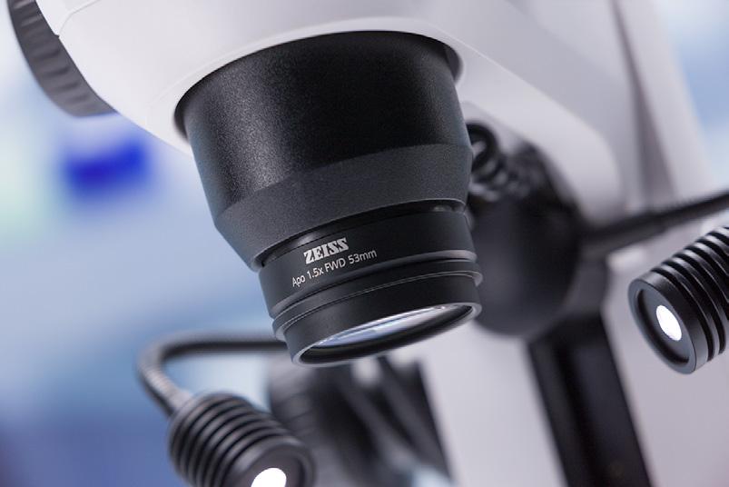
Stemi 508's apochromatic zoom optics and efficient stray light suppression give you a crisp threedimensional image. You visualize your objects distortion-free and without color fringes, in fields of view up to 122 mm. The large 8:1 zoom lets you observe even minute structures in high contrast. Select from a range of apochromatic front optics and eyepieces to reach any magnification between 2× and 250×. Double your resolution or enjoy large working distances up to 287 mm without compromising optical quality – it's your choice.
Precise Mechanics for Heavy Workloads
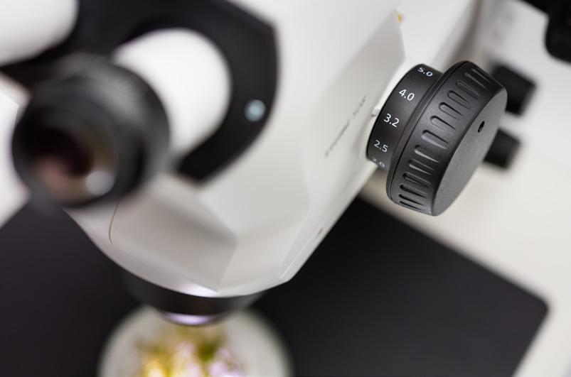
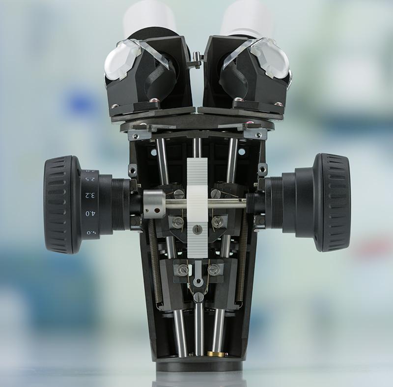
Stemi 508 is designed for heavy workloads, using durable and reliable mechanics. Its precise zoom adjustment ensures a well-balanced 3D impression: enjoy relaxed stereo viewing without eyestrain. Whether you zoom continuously variable or in reproducible mode activating click stops –the image stays sharply focused over the whole magnification range. You often work on the microscope for long hours and appreciate a comfortable posture. The low viewing angle of 35° makes Stemi 508 more ergonomic than other Greenough-type stereo microscopes.
Created for All Your Applications
Choose from a wide range of different stands and accessories to get exactly what you need for your application. From compact stands to flexible and stable boom stands, from basic transmitted light to polarization contrast. Whether your object needs special illumination or simply the brightest light – you have various fiberoptic light guides and LED illuminators to choose from.
Position your specimen precisely by adding a gliding, tilting or rotating polarization stage. Via interchangeable camera adapters, Stemi 508 can be combined with any ZEISS Axiocam camera and most DSLR cameras or camcorders. A c-mount adapter 0.5x for Axiocam cameras up to chip size 1/2" is always included.
In Brief
The Advantages
Your Insight into the Technology Behind It
Greenough Stereoscopic Design
Technology and Details
Service
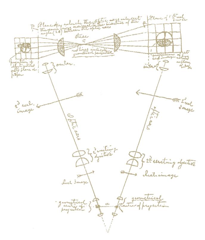
The basic idea behind a stereoscopic microscope is simple. It was formulated in 1896 by the biologist Horatio S. Greenough, who wanted to see small biologic samples magnified, but with the same quality as with unaided eyes. In other words: in three dimensions and with all the depth information he needed to understand the irregular shape of his specimen intuitively. He reckoned, one could build a microscope with two separate beam paths facing the object from two directions - exactly as human eyes do when observing a small object at a distance of 250 mm. The brain would fuse the two images together and produce a spatial image of the object with a high degree of depth perception. This thinking led to the first factory-produced stereo microscope being developed by ZEISS.
Stemi 508 is a Greenough-type stereo microscope, utilizing long working distances for easy specimen handling and large fields of view. It is com pact, rugged and simple to use – and so typically employed for heavy workloads in routine microscopy, such as printed circuit board inspection or dissecting biological specimens.
Hand drawing by Horatio S. Greenough (1896), leading to the world's first industrially-manufactured stereo microscope.
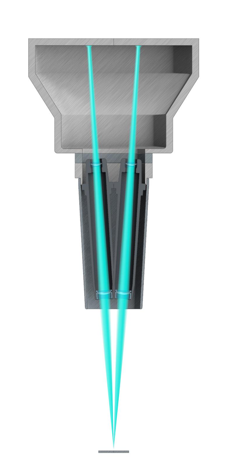
Schematic beam path of a Greenough type stereo microscope.
ZEISS Stemi 508 features a Greenough optical system with powerful 8:1 zoom. The twin body tubes are inclined by an angle of 11° – for an impressive stereoscopic image impression.
› In Brief
› The Advantages
› The Applications
› The System
› Technology and Details
› Service
Your Insight into the Technology Behind It
Apochromatic Optics for Images Free of Color-fringes
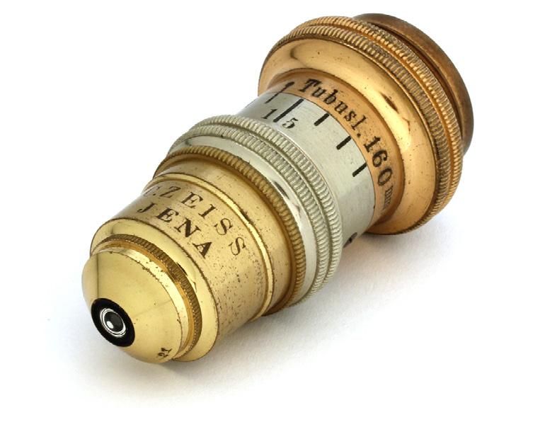
Producing microscopes with pre-calculated optical characteristics based on Abbe's calculation was already a huge step for ZEISS. But, especially working with large apertures, Abbe recognized aberrations he called "chromatic differences of spherical aberrations" caused by the quality of the optical glasses. As a result, different colors of light focus at different distances from the objective. Observed through a microscope, objects appear to have colored fringes! It was already known that chromatic abberations can be reduced by combining glasses with differing dispersion – eg achromatic doublets made of crown and flint glass – but to further increase the degree of chromatic correction,
a larger number of lenses and optical glasses with special dispersive properties were needed. Only with the development of new optical glasses by Otto Schott, the production of apochromatic objectives by Carl Zeiss (based on the calculation of Ernst Abbe) could be commercialized. Apochromatic objectives correct the lens aberrations for three colors of the visual spectrum (red, green and blue) by bringing the three wavelengths into focus in the same plane. Apochromatic objectives therefore produce images which are sharper and more brilliant. Therefore these objectives are favourable, whenever image quality is of importance doing evaluations and documentation.
1886
First apochromatic microscope objective, color corrected for three wavelengths based on the calculations of Ernst Abbe. The foundation for this achievement was in part formed by the concerted attempts of Abbe and Schott to improve optical glasses.
Chromatic aberration
Chromatic aberration
Chromatic aberration f
Lenses with apochromatic correction
Lenses with apochromatic correction
Lenses with apochromatic correction
The Advantages › The Applications › The System › Technology and Details › Service
Your Insight into the Technology Behind It
White LEDs Replace Halogen Light for Microscopy Lighting
LEDs – light emitting diodes – are semiconductor diodes that emit visible light when a forward electric current is applied. The color of the light is nearly monochromatic and determined by the semiconductor material. To produce white light, blue LED chips are combined with different phosphors that convert a fraction of the blue light to longer wavelengths. The resulting LEDs show a broad, continuous emission spectrum. Given their excellent properties, white LEDs have largely replaced halogen lamps as standard illumination in stereo microscopy: White LEDs can be perfectly integrated into microscope stands since they are compact, maintenance-free and noiseless, and they don't generate vibration either. Since it doesn’t contain any infrared or ultraviolet parts,
white LED light contrasts your objects in a gentle way. Since LEDs rarely ever fail, their lifetime is defined by “lumen maintenance”, which is the operating time until the initial light output has degraded to 70 %. At ZEISS we specify 25,000 hours equivalent over 12 years (a 5 day week, 8 hours per day). Modern white LEDs feature high brightness and daylight quality (CCT 5000 – 6000 K), they are energy saving (their luminous efficiency already reaches fluorescent tubes) and convince with very short switch-on times. Even in the most intense fiber optic cold light sources, innovative high power LED packages have replaced 150 W and 250 W halogen reflector bulbs – as in our ZEISS CL 6000 LED and CL 9000 LED cold light sources.
distribution of a phosphor-based white LED: blue LED light combines with a broadband
Typical design of white LEDs: a blue LED chip is coated with yellow phosphors that partly convert the blue light to longer wavelengths.
Spectral
phosphor spectrum.
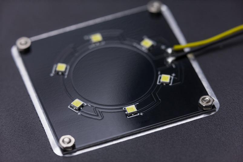
The flat transmitted light unit in stand K EDU is powered by six white High Brightness LEDs. The LEDs are directly mounted on a printed circuit board. It's black color gives the dark background needed for transmitted light darkfield contrast.
› In Brief
› The Advantages
› The Applications
› The System
› Technology and Details › Service
Expand Your Possibilities
Stand M – Generous Sample Space. Large Focusing Range. Including LED Illumination.
Stemi 508 is a high quality stereo microscope that's highly suitable for the most demanding tasks in laboratory work as well as industrial failure analysis. And a sophisticated microscope needs a coequal stand: Stand M offers generous specimen space and a large focusing column. On the other hand, Stand M is compact enough to be used in small lab rooms - all the more, since LED electronics are integrated and thus no external light sources necessary. Stand M LED controls up to two reflected light LED illuminators and transmitted light in parallel. Mix their illumination to optimally contrast your specimens, store the settings by a button press – and quickly switch between your light scenarios by using the memory section of stand M LED.
To optimize Stemi 508 to your applications' needs, you can choose from a range of changeable LED illuminators. All of them use intense white LEDs – long life, maintenance-free and of daylight quality.
ZEISS Stemi 508 with stand M provides a large sample space. Easily handle large specimen or several Petri dishes in parallel. Cope even with big industrial workpieces.
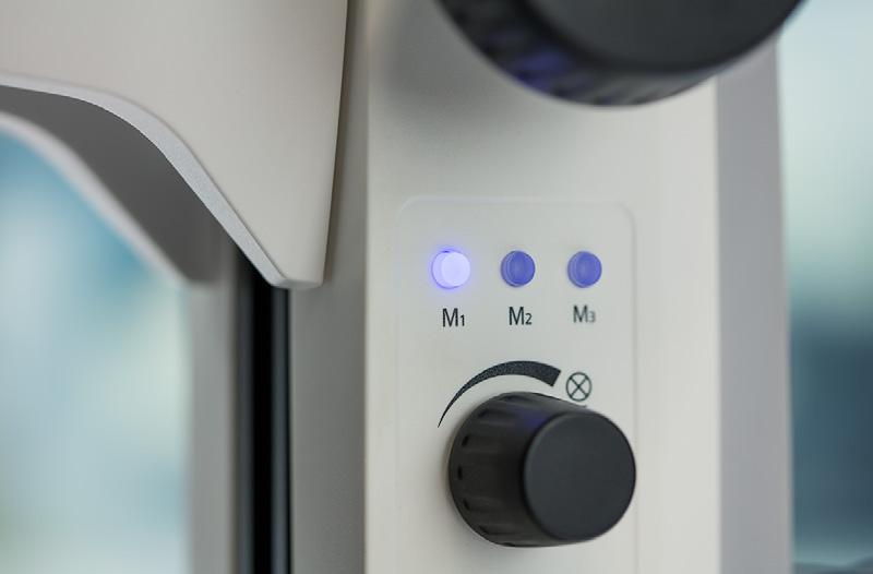
The memory section of stand M LED allows storing and quick recall of up to three customer specific illumination conditions. Press an "M" key long to store, press it short to recall your individual light scenario.
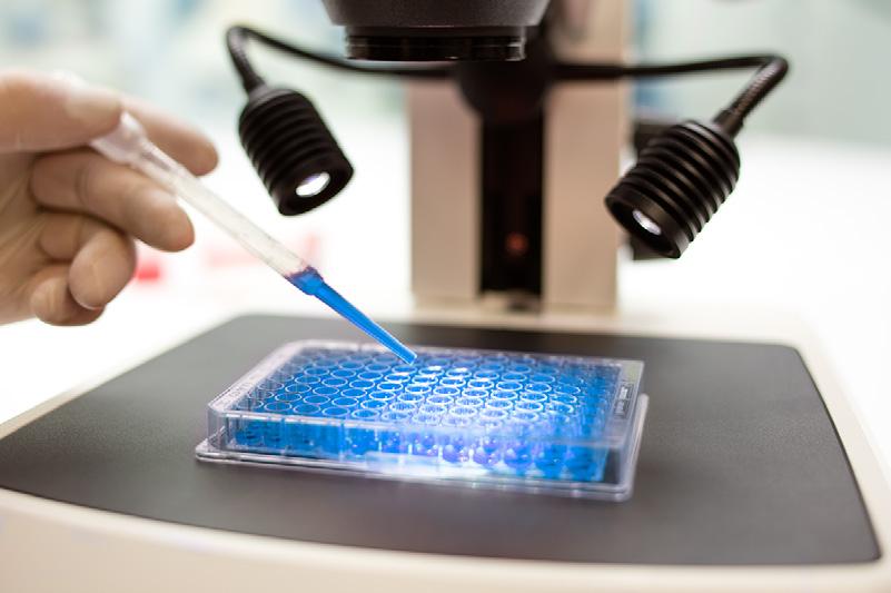
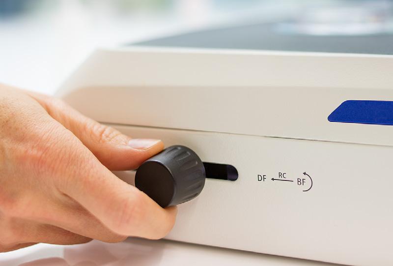
Stand M LED can easily be equipped with transmitted light: Choose between mirror-based transillumination unit M LED or the flat brightfield-darkfield transilluminator, that is integrated in the stand base without adding height.
› In Brief
› The Advantages › The Applications › The System › Technology and Details › Service
Expand Your Possibilities
Stand K – Compact. Easy to Use. All-in-One Design.
Workspace is limited in your lab or quality department? Do you usually stow away your systems and set them up again? Or you often work with untrained or multiple users? – In all these ways, the stand K line is made for you. With its small footprint and integrated LED illumination, a K stand turns your Stemi 508 into a compact all-in-one device that's easy-to-use and quick to pack up and reinstall wherever it's needed. To ensure the integrated LED illumination is optimized to your application range, choose from different stand versions.
Stand K EDU with flat transmitted light unit and carrying handle for easy transport is your compact choice for classroom environments. Add a single spot illuminator K LED to study your opaque specimen in strong 3D contrast.
Stand K LAB's compact transillumination base with tiltable mirror features brightfield, one-sided darkfield and oblique light. Add the double spot illuminator K LED for reflected light dissections.
Stand K MAT is equipped with reflected light LED controls and ESD features (antistatic coating), and thus optimizes your Stemi 508 for PCB inspection and small parts assembly in electronics industry. Add the segmentable ringlight K LED as illumination – and a gliding or tilting stage, if precise positioning of your specimen is needed.
In Brief
The Advantages
The Applications
The System
Technology and Details
Service
Expand Your Possibilities
Document and archive your results.
And share the images throughout your digital network.
It’s full of possibilities.
HDMI port:
Direct access to your monitor without computer. Camera settings via remote control / OSD. Snap your images on an SD card.
USB port:
LAN port:
Easy access to your digital networks. Camera settings and imaging with the free ZEISS imaging app Labscope. Efficient control of digital classrooms with Labscope Teacher.
Easy access to your Windows PC. Camera settings and imaging with the free ZEISS imaging software ZEN lite, ZEN starter or Labscope for Windows.
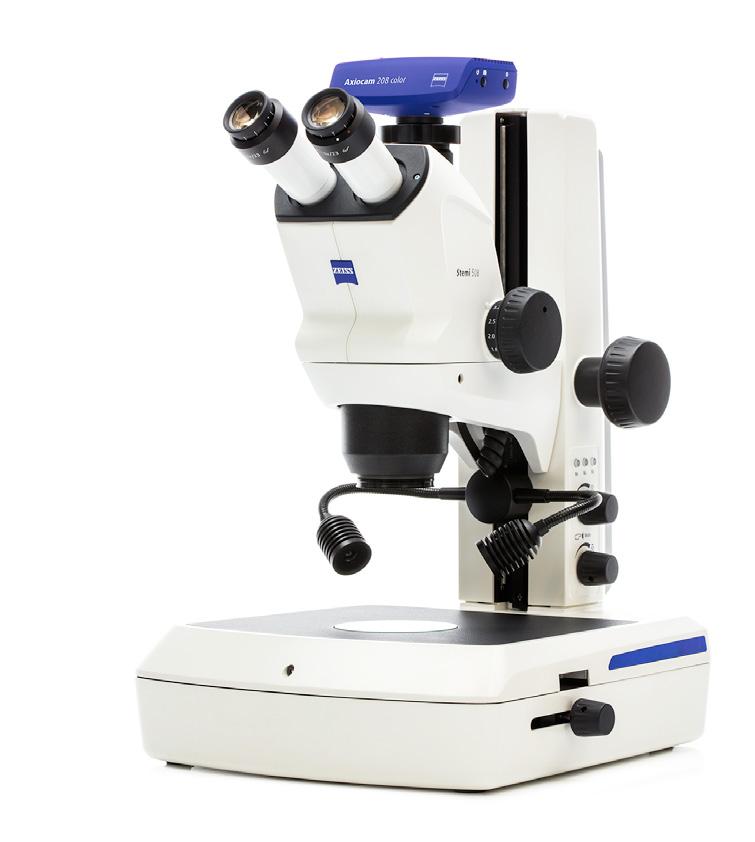
Use Stemi 508 with Axiocam ERc 5s or Axiocam 208 color and take advantage of its various interfaces to display the live window and snap your images.
Connect several Stemi 508s and other ZEISS network-compatible microscopes – and transform your lab into a Wi-Fi enabled network. Simultaneously observe the live images of all connected microscopes on your iPads, iPhones or Windows PCs. With the imaging software Labscope you snap images, record videos and measure your microscopic samples easier than ever – and finally archive all results on your local server.
Tailored Precisely to Your Applications
Typical Applications, Typical Samples Task
PCB Inspection
Failure Search and Analysis
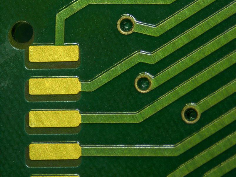
Check PCBs in the quality lab. Look for improper quality of contacts, engraving or wiring, metal or processing residues, and faulty solder joints. Document your results.
Search for reasons of faulty circuits and report your failure analysis.
ZEISS Stemi 508 Offers
• Compact stand K MAT with segmentable ringlight is the right solution for limited space. Document your results with Axiocam 105 with 5 Megapixel resolution.
• Use the 10 zoom click stops of Stemi 508 to reproduce your magnification easily.
• To inspect and document shiny metal parts without strong reflections, create diffuse light using fiber optic diffusor S, diffuse face light S or the fluorescent tube ringlight. Use a ball-andsocket stage to tilt the specimen in a defined way.
Diamond Industry
Medical Devices
Evaluate quality of diamonds by visual inspection, find inclusions and impurities.
Assemble small high-precision components, e.g. in hearing aids or cardiac pulse stimulators.
Sensor Manufacturing Centre and align optical sensors or optical fibers.
Clocks and Watches
Printing and Engraving
Minted Coin and Medal Manufacturing
Forensics
Assemble small part components of watches, e.g. minute wheels, springs, cocks, bridges, pinions, bell and ratchet wheel screws, dial, mainspring, bolt and shoulder screws.
Evaluate the surface quality of paper or engravings under grazing light.
Inspect for scratches on proof quality silver coins.
• Enjoy the large zoom range, high resolution, impressive image contrast and excellent color correction of Stemi 508.
Art Restoration
Analyze, restore, clean and conserve paintings, sculptures and other works of art, analyzing layer by layer and identifying the material.
• Zoom from the whole watch in the overview up to the 8 times magnified detail.
• During a long day, enjoy relaxed viewing with Stemi 508 and precise mechanics made for extensive use.
• Stemi 508 offers fiber optic line light S for intensive grazing light with strong shadow effects. To see scratches on polished surfaces, use fiber optic darkfield ringlight. Use gliding stage S or stand base 300 with mechanical xy stage to screen the specimen in comfort.
• The apochromatic color correction of Stemi 508 with interchangeable front optics is essential for fiber analysis.
• Achieve optimal contrast with the high brightness LED light of CL 6000 LED and the range of fiber optic light guides.
• Use flexible floor stand S or stable ball bearing stand SDA, along with shadow-free fiber optic ringlight. Benefit from Stemi 508's excellent image contrast and large field of vision.
In Brief
The Advantages
The Applications
The System
Technology and Details
Service
ZEISS Stemi 508 at Work
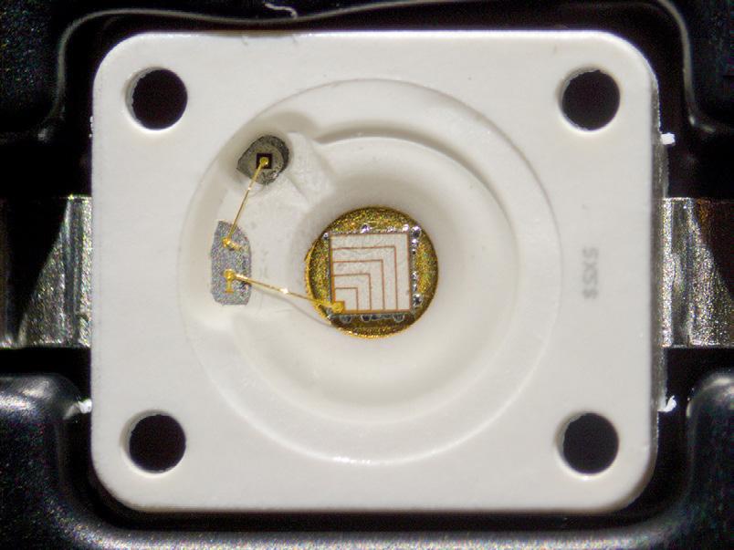
High brightness LED, chip, bond wires and packaging Spot K LED, oblique reflected light, zoom 1.25×
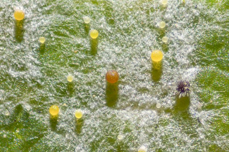
Powdery mildew ( sawadaea) on Norway maple, cleistothecia
Spot K LED, reflected light darkfield, zoom 2.0×
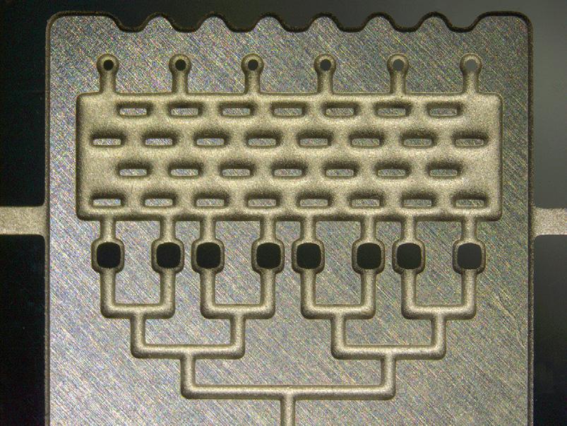
Microfluid device
Segmentable ringlight K LED, half circle mode, zoom 0.8×
Hazelnut (corylus)
Transilluminator M LED, brightfield, zoom 5.0×
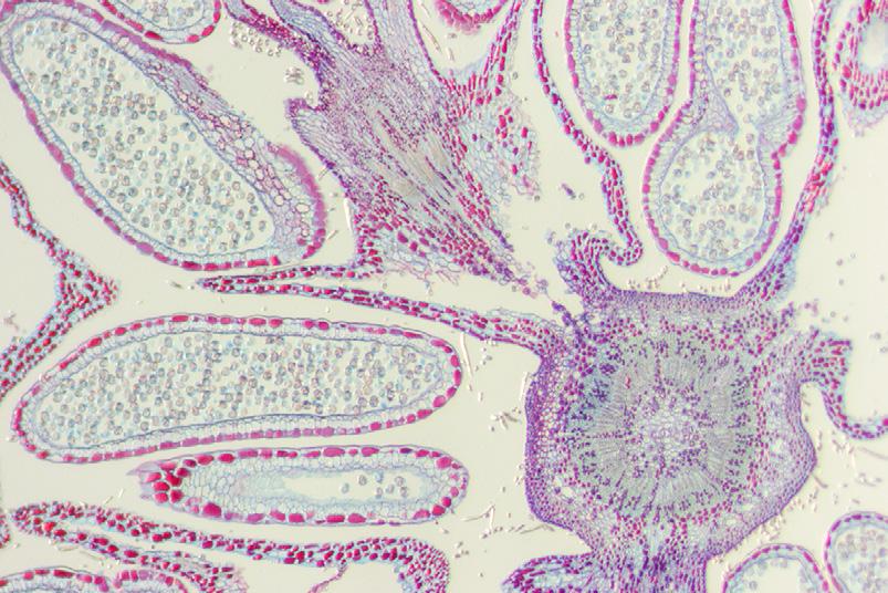
Royal fern, sori and sporangia
Spot K LED, oblique light, zoom 0.63×, front optics 5 Apo 0.63×
› In Brief
› The Advantages
› The Applications
› The System
› Technology and Details
› Service
Your Flexible Choice of Components
1 Microscopes
• Stemi 508 (binocular)
• Stemi 508 doc (phototube 0/100 %, right path)
• Stemi 508 trino (phototube 50/50 %, right path)
All zoom bodies incl. eyepieces 10×/23 Br. foc and all phototubes with camera adapter 0.5× for c-mount cameras (changeable via 60N interface.)