Accelerate Your Time to Insight
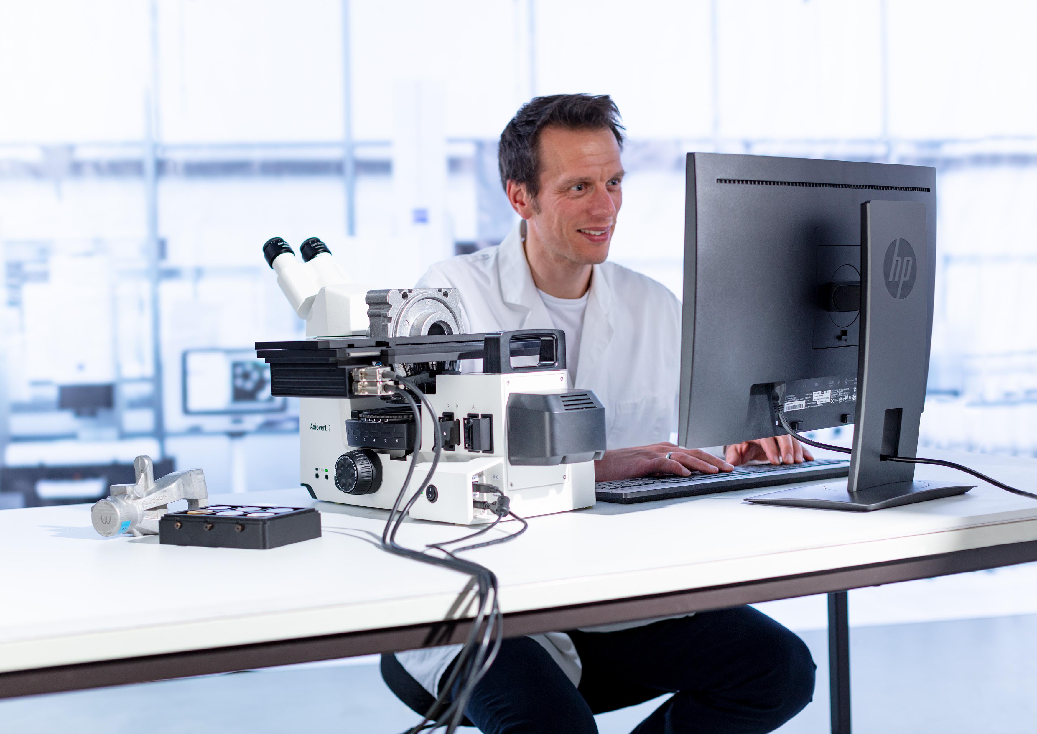
Your Inverted Microscope System for the Materials Lab and Smart Documentation
ZEISS Axiovert System

Your Inverted Microscope System for the Materials Lab and Smart Documentation
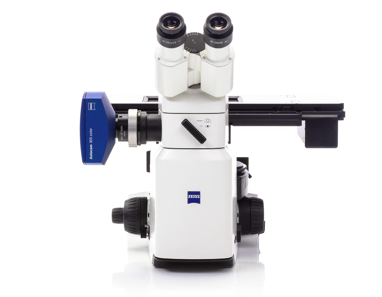
If you’re looking for a compact microscope that effortlessly produces high quality images of even large and heavy samples, then look no further than ZEISS Axiovert.
This inverted microscope is a joy to use in the materials lab for routine and research tasks. With an ergonomic design and smart technology, let the system take care of selecting optimal settings for a consistently well illuminated and sharp image. And if you want to increase productivity even further, opt for additional automation features with a motorized Z-focus and stage.
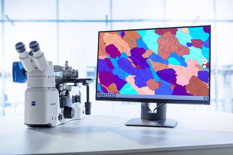
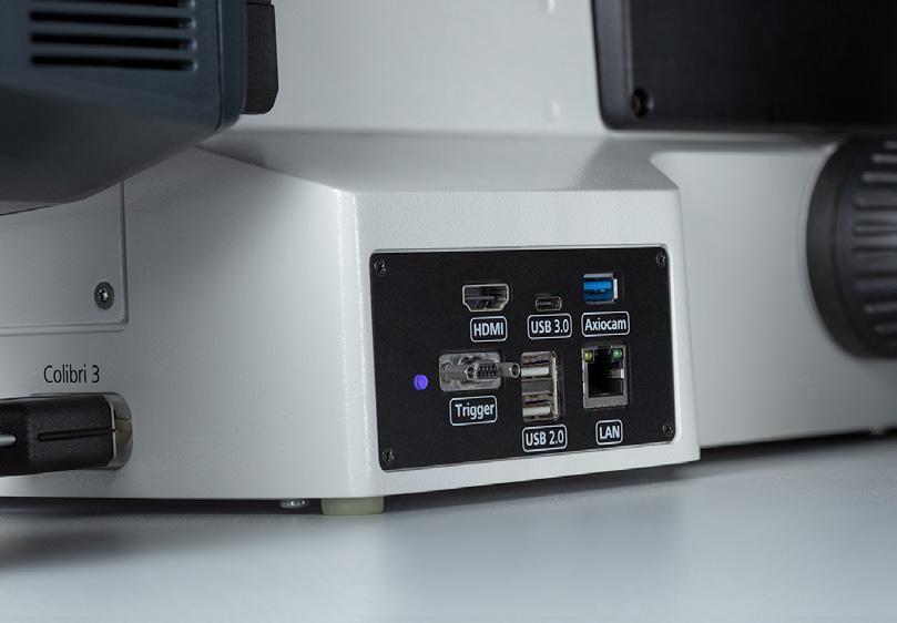
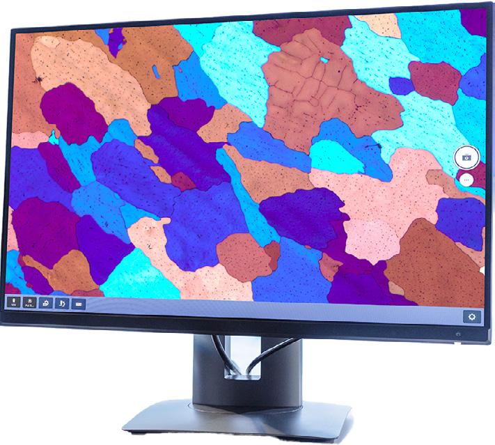
With Axiovert 5, you don't even need a PC to view and document images – just connect with a monitor and save directly to a USB device.
Select the right system for your needs with a choice between the manual Axiovert 5 with smart microscopy for fast and reliable results or the motorized Axiovert 7 for higher demands on workflow automation.
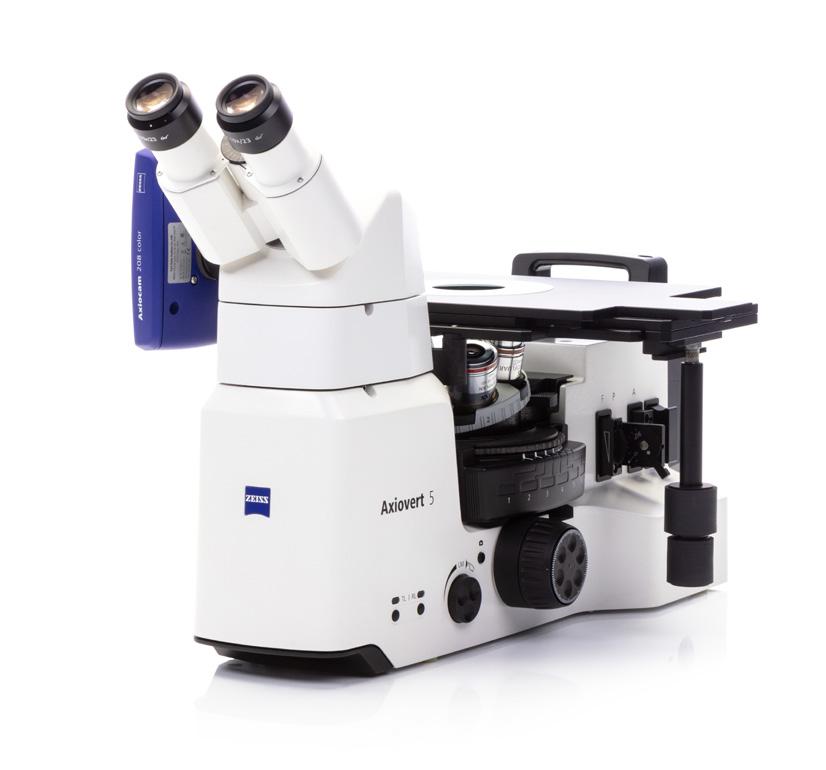
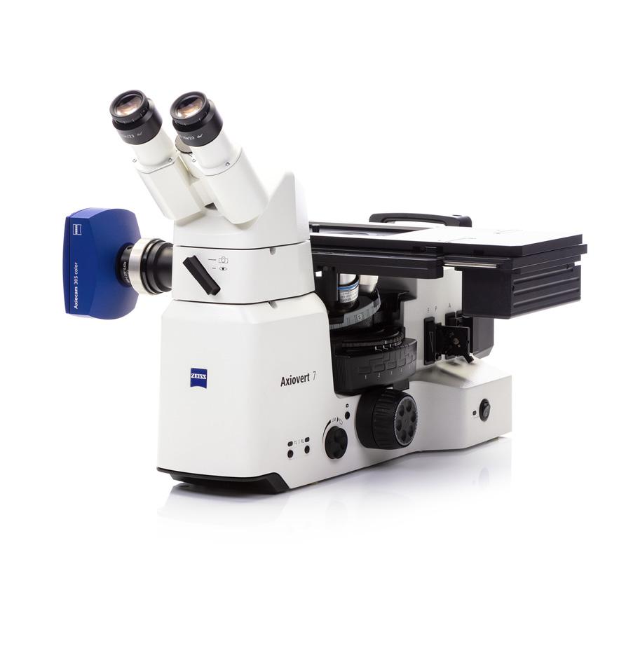
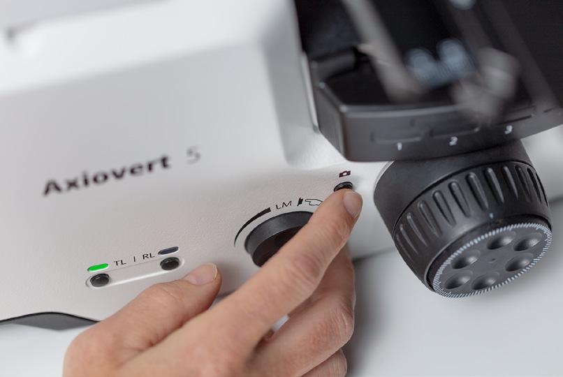
Axiovert is a smart microscope, delivering excellent results with fast time to image. Simply focus and press a single button to save a high-quality image of your material sample.
Change samples with no need for refocusing to save time and effort. Just place the sample on the stage, focus once and stay in focus for further samples, even when changing magnification.
Axiovert makes your routine tasks easier and more convenient. With a wide range of classic and advanced contrast methods, you can examine even large and heavy samples with stability.
Extend your throughput with additional automation and future proof your requirements with optional accessories and software.
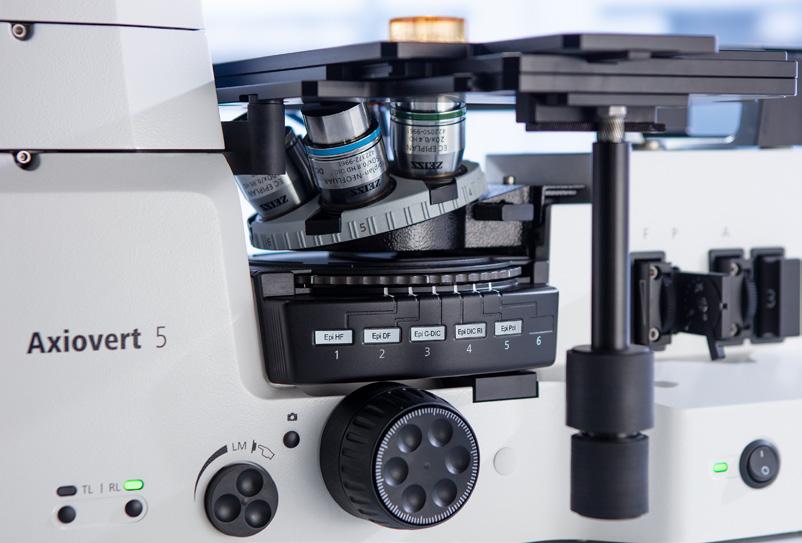
Axiovert is designed to make everyday operations as comfortable and convenient as possible. Controls such as the focus drive, stage drive, light manager and image capture, are ergonomically arranged on the microscope for relaxed and efficient handling.
The light manager provides uniform brightness at all magnifications, eliminating manual adjustments to the lamp intensity when you’re changing objectives.
Let the system take care of optimal settings for image acquisition whilst you focus on your results.
› In Brief
› The Advantages
› The Applications
› The System
› Technology and Details
› Service
Always stay focused on your sample, thanks to smart microscopy. Camera settings such as white balance, exposure time, and image enhancement functions are done automatically. Without needing additional imaging software or even a computer, you can:
• Snap images and record videos directly
• Use a mouse (and optionally, a keyboard) to control your camera via On Screen Display (OSD)
• Save settings
• Store images with all microscope and camera metadata as well as scaling information
• Predefine the name or rename your image
• Browse and view your snapped images with the file browser
• Connect to Wi-Fi or a network, independently from the ZEISS Axiocam you’re using
• Save your data directly at the stand via a USB device.
Smart functionality for digital documentation for routine applications.
Efficiency gain: Eyes and hands stay on the microscope.
Brief
The Advantages
The Applications
The System
Technology and Details
Service
Adapt ZEISS Axiovert 5 to Your Lab Space and Task.
If lab space is limited, you can use Axiovert 5 in standalone mode without the PC and control the microscope via the OSD menu. For your daily checks, use our free imaging app, ZEISS Labscope. Simply download it to your tablet or Windows PC and you‘re ready for image acquisition. Or for more sophisticated experiments, ZEISS ZEN core imaging software is recommended.
Imaging and Documentation without a PC
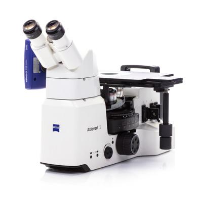
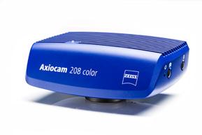
Axiovert 5 operates independently of a computer system
ZEISS Labscope for Connected Routine Imaging
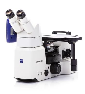
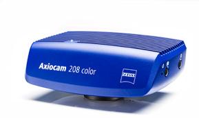
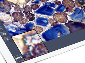
Operating Axiovert 5 with Labscope imaging software is ideal for standard imaging
ZEISS ZEN core for Advanced Applications
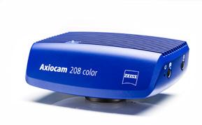
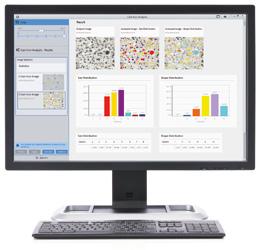
Use the ZEISS imaging software suite ZEN core to perform advanced imaging and analysis tasks with Axiovert 5 and Axiovert 7
In Brief
The Advantages
The Applications
The System
Technology and Details
Service
With Axiovert 7 you will benefit from higher productivity, repeatable processes based on predefined parameters, and better comparability of results thanks to the motorization of the Z-axis and a motorized XY stage.
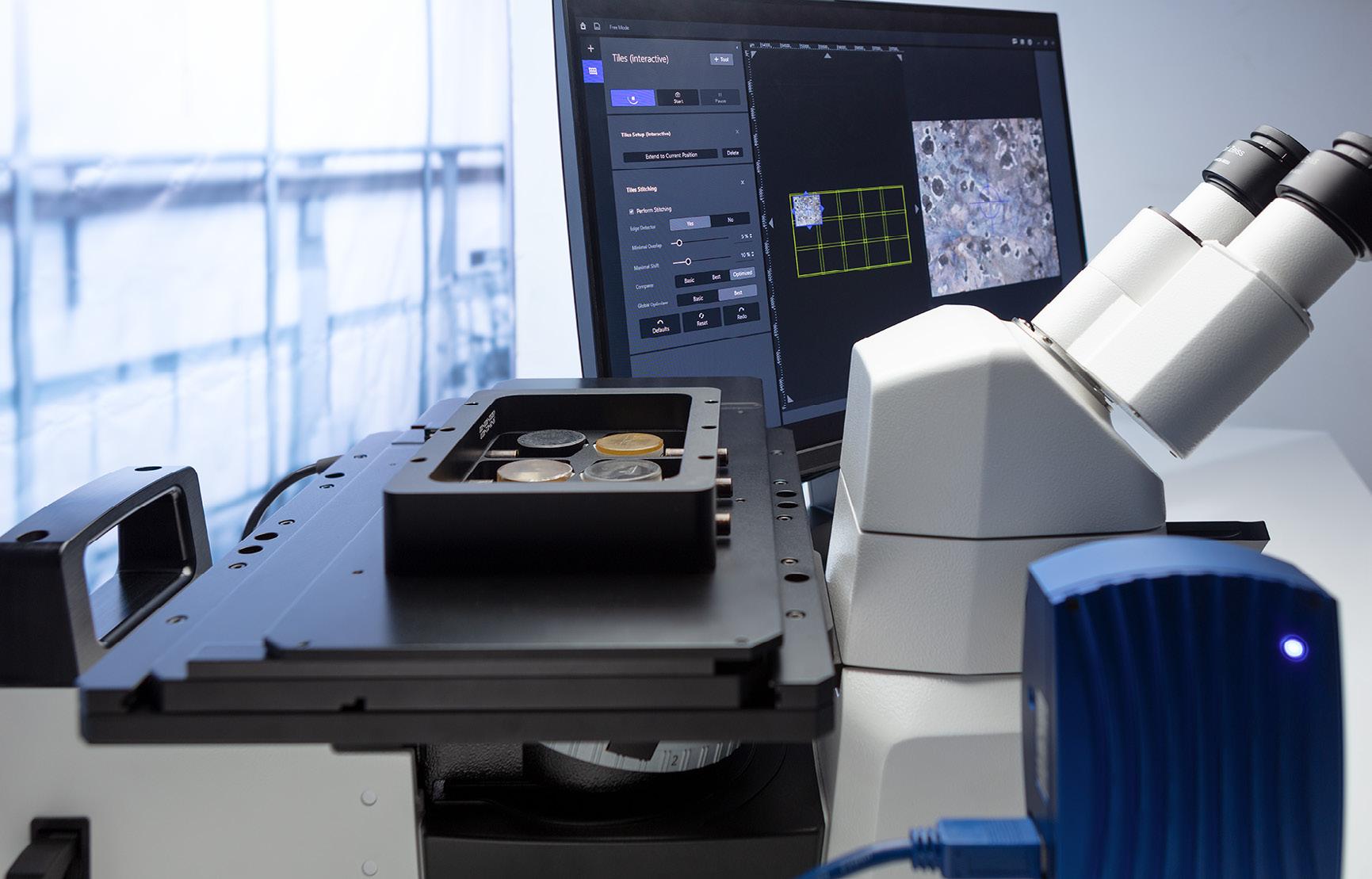
Take advantage of:
• Autofocus functionality – find the focus position automatically
• Parfocality function – remains in focus when the objective is changed
• Extended depth of field - automatically acquire multiple images at different focus positions (Z-stack) and combine them to create an image with enhanced depth of field
• Panorama images – create composite images of larger sample areas in just a few clicks
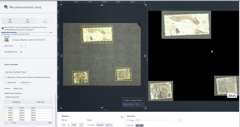
• Tiles and positions – record exact, highly resolved images of multiple field of views by automatically scanning predefined areas
• Guided acquisition – take an overview scan, detect regions of interest (ROI) automatically, start detailed scan of these ROI
• Correlative microscopy – examine samples with different light and electron microscopes. Relocate ROIs automatically using ZEN Connect
The coded components of Axiovert not only make your work easier and more comfortable, but also ensure that erroneous operation and the associated falsification of the examination results can be largely ruled out. With Axiovert, you’re assured reliable and reproducible results.
The coded components of Axiovert detects changes to objectives or contrast techniques, then adjusts dependent parameters – such as light intensity and scaling –automatically. This allows multi-faceted routine workflows to be processed more quickly and easily. Using process parameters that you or others have stored, anyone can reproduce an exact workflow at any time and achieve comparable results, independent of individual users’ operating habits or preferences.
› In Brief
› The Advantages
› The Applications
› The System
› Technology and Details
› Service
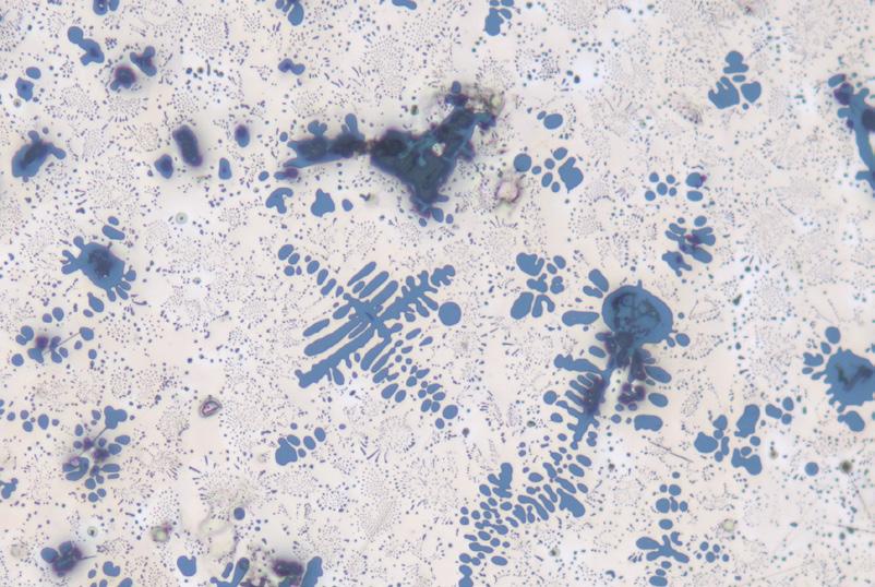
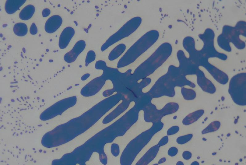
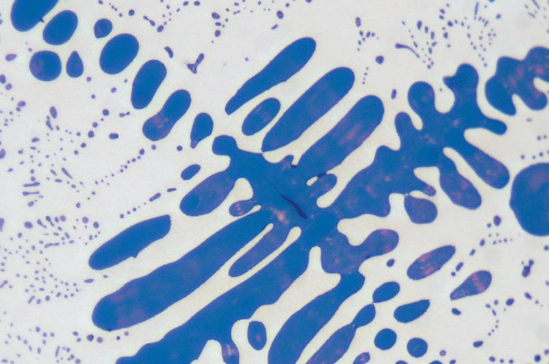
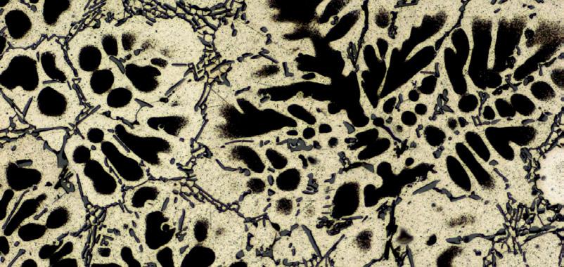
Use the contrast method of reflected light brightfield to analyze the micro structures of etched surfaces. Recognizing grain boundaries, you can draw conclusions on grain sizes, phases and structural constituents. See colors and pigments. Detect impurities and structural constituents, such as graphite in cast iron, prior to etching.
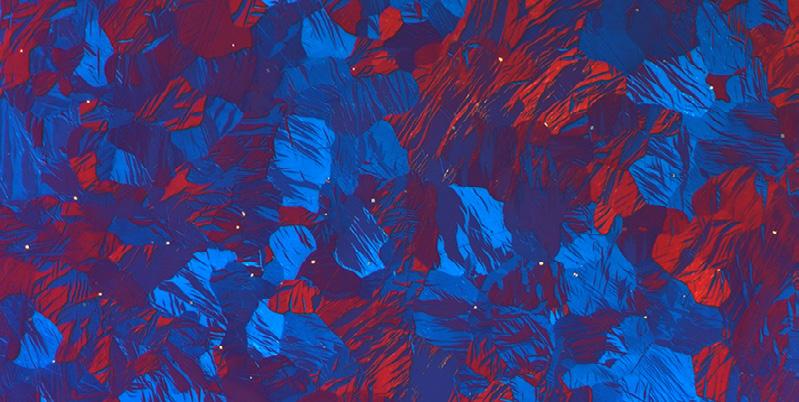
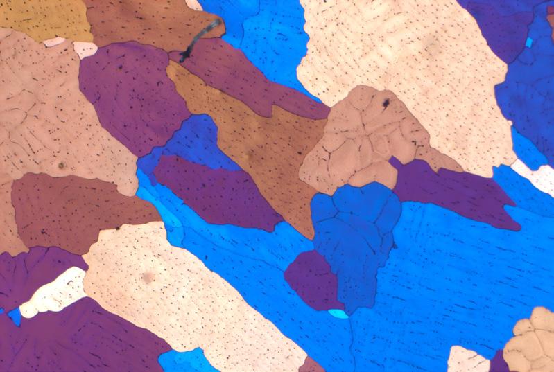
Use the polarization contrast to analyze the structure of anisotropic materials, such as magnesium, aluminum, bronze and brass. In polarized light, the individual grains of crystal lattice will show their characteristic color.
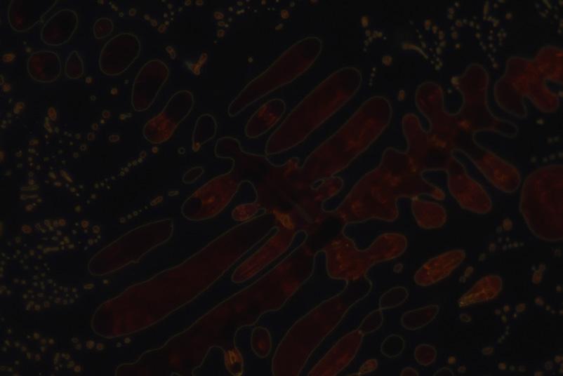
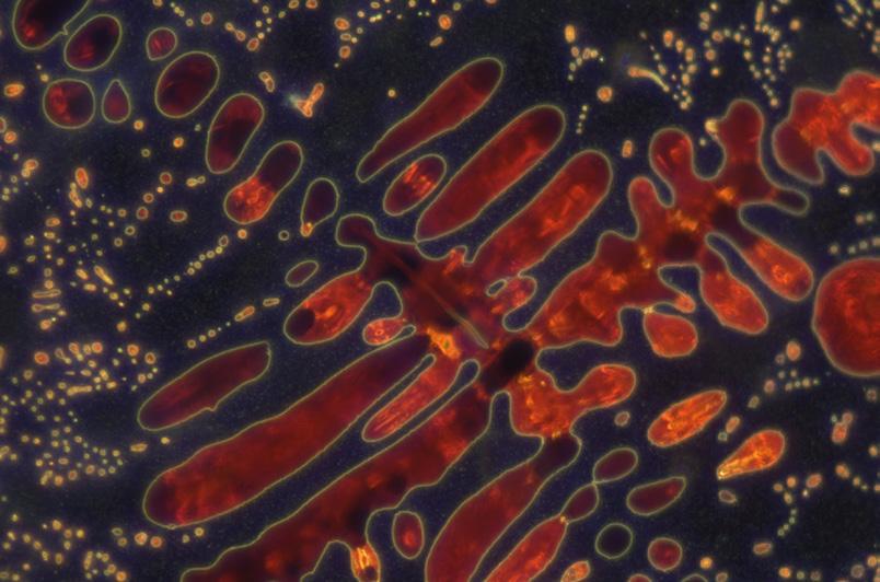
In reflected light darkfield, mechanical surface faults such as fracture sites, pores and inclusions show up just as well as cracks, scratches and cavities. You can assess the surface quality of processed work pieces precisely, and make out grain boundaries on etched cuts just as easily.
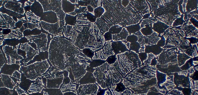
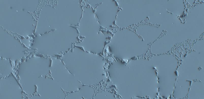
The Differential Interference Contrast (DIC) lets you detect tiny structural differences in height with particular sensitivity. Differences in height, whether in the form of natural differences or artifacts produced by preparation, take on a 3D effect as relief-like structures.
In Brief
The Advantages
The Applications
The System
Technology and Details
Service
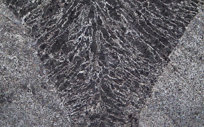
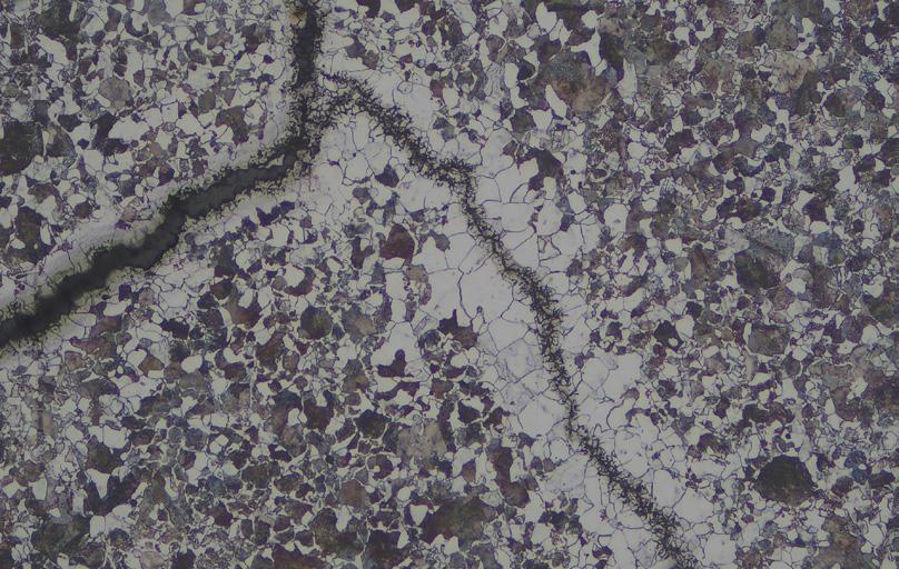
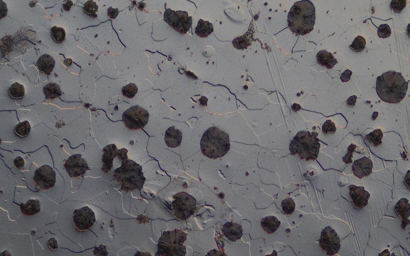
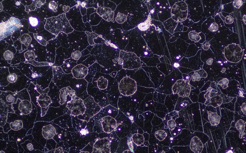
• Determine grain sizes, structure, distribution and phases
• Quickly carry out on-site analysis
• Study modes of material failure: fatigue, corrosion, creep deformation, stress cracks or fractures
• Clearly visualize all sample features – Axiovert supports your examination with all essential contrast methods including darkfield and differential interference contrast (DIC)
• Get your work done quickly and easily – the Smart Microscopy concept assures simple operation and rapid image acquisition
• See your samples as they really are – document your sample just as you see it through the eye piece
• Provide reproducible results – encoded components assure that you always get the right light intensity and scaling
In Brief
The Advantages
The Applications
The System
Technology and Details
Service
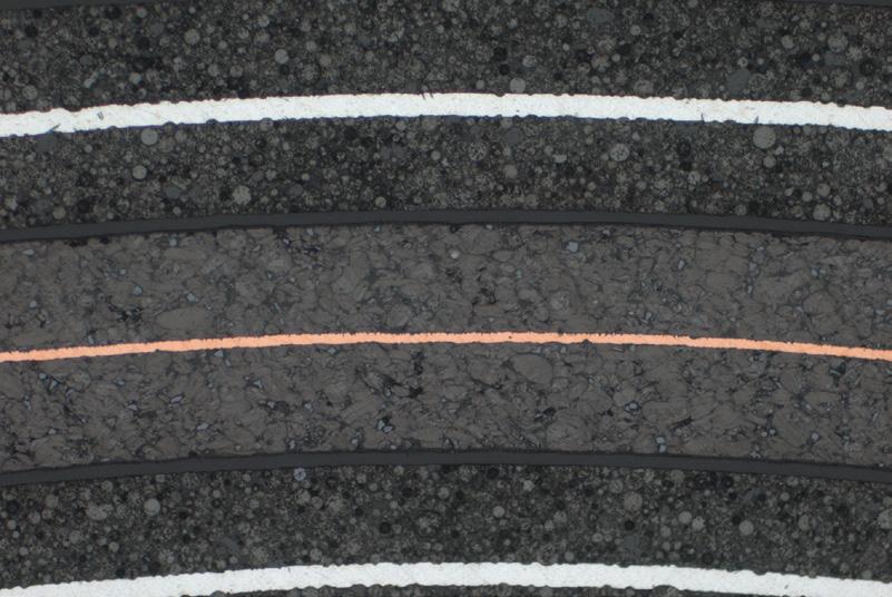
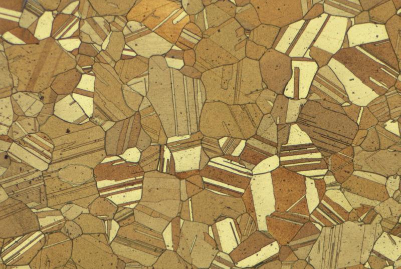
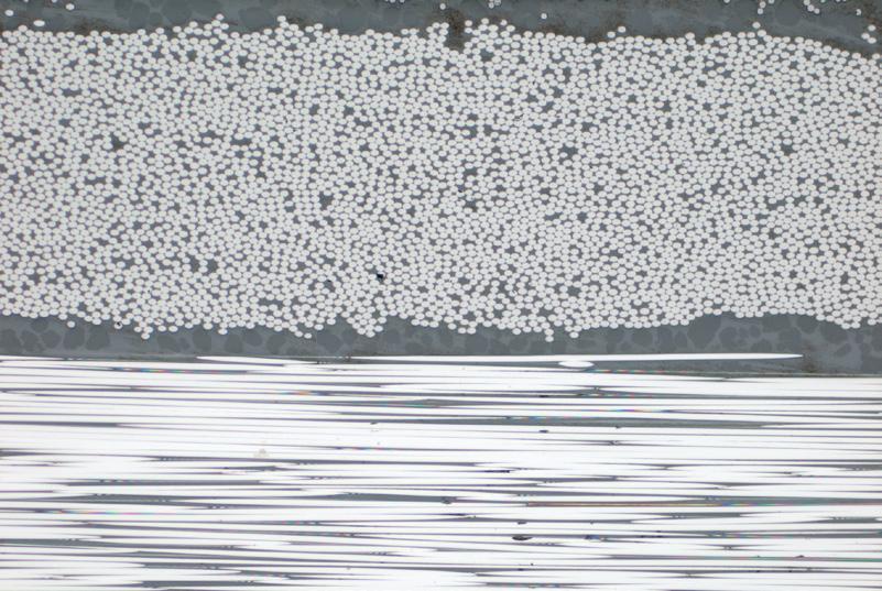
• Analysis of structure (e.g. phases, grain sizes, texture, precipitates) and structural defects (e.g. inclusions, porosities, voids, cracks)
• Measurement of layer thicknesses and geometric properties
• Analysis of anisotropic samples in polarization contrast (e.g. grain size of aluminum alloys by Barker etching, zinc alloys, graphite, titanium alloys, magnetic materials)
• Clearly visualize all sample features – Axiovert supports your examination with all essential contrast methods including darkfield and differential interference contrast (DIC)
• Improve your statistics with large area scanning of your sample with motorized XY stage
• Get more meaningful results from your images with ZEN core microscopy analysis software.
• Provide reproducible results – encoded components assure that you always get the right light intensity and scaling
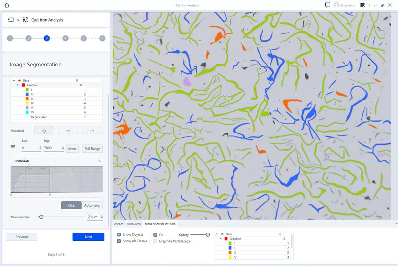
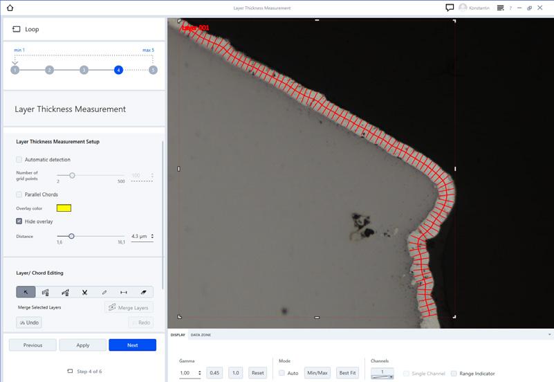
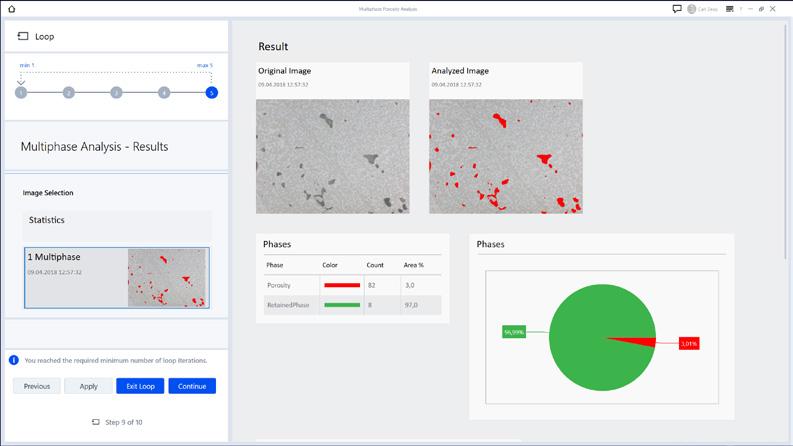
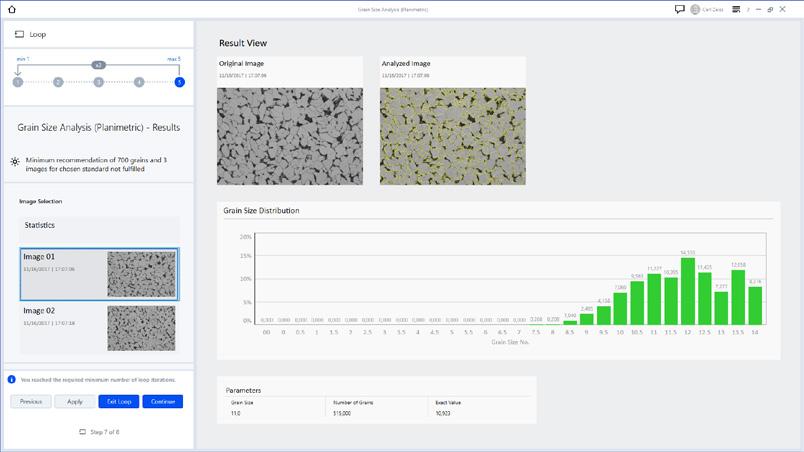
Typical tasks and applications
• Imaging and analysis of microstructure of metal materials
• Quantitative microstructure analysis
• Evaluation according to international standards
• Grain size analysis
• Multiphase analysis
Get these benefits from ZEISS Axiovert
• Reveal microstructural information using different contrast methods
• Use brightfield contrast to get information about the overall number, size and shape of features within a material
• Enhance grain boundaries and particle edges with darkfield contrast to reveal sharper features and clearer definition of interfaces
• With circular differential interference contrast (C-DIC) your sample surface appears as a 3D relief so you can easily detect polishing marks
• Acquire large areas easily with a motorized stage
• Encoded components assure that you always get the right light intensity and scaling to provide reproducible results
› In Brief › The Advantages
› The Applications
› The System
› Technology and Details
› Service
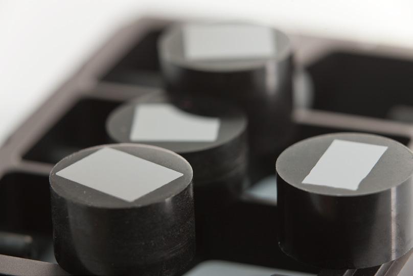
The type and amount of non-metallic inclusions (NMI) in steels strongly affect the mechanical and physical properties of these steels.
Metallographic analysis of NMI is governed by industry standards that are supported by the modular and customizable ZEN core software which guides the user quickly and easily through the workflow, generating a report and inclusion gallery compliant with the standards.
Axiovert 7 with ZEISS ZEN module Non-Metallic Inclusion Analysis confirms that manufacturing processes, grade and quality of the product meet strict specifications for impurities or defects that can cause a component to fail or impact its tensile strength, toughness and fatigue.
Powerful inspection views and automated deformation axis detection features make analysis easy, intuitive and repeatable. With additional GxP functionality, ZEN core users are able to offer their customers full traceability and data integrity in NMI analyses, meaning that grade certification is auditable, particularly advantageous for customers in regulated industries
Supported Standards
• ASTM E45
• ISO 4967
• JIS G0555
• GB/T 10561
• EN 10247
• SEP 1571
• DIN 50602
The Applications
The System
Technology and Details
Service
ZEISS Axiovert Systems for Material Applications
The Axiovert systems offer instrument variants for routine tasks and advanced research applications. Each configuration has been optimized for specific applications with all relevant contrast techniques available to support your needs.
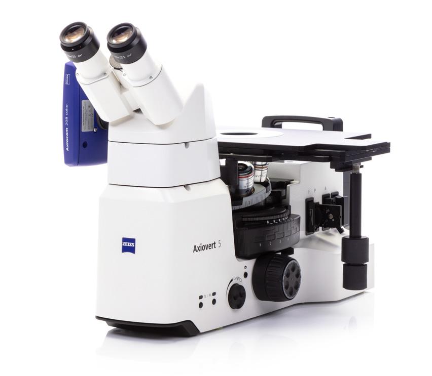
ZEISS Axiovert 5
Manual microscope with coded components for reproducible and reliable results of samples acquired with reflected light such as metallographic cuts and large specimens.
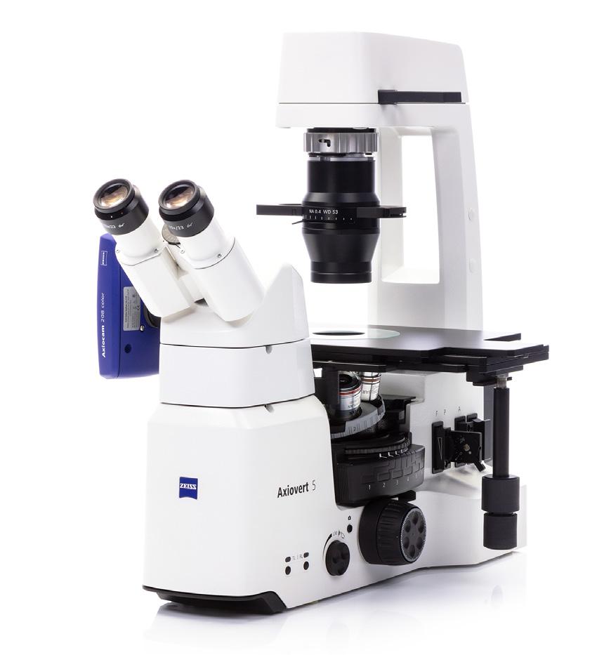
ZEISS Axiovert 5 for Reflected and Transmitted Light
Add additional transmitted light contrast capability to Axiovert. Broaden your application space and examine transparent or thin cut samples.
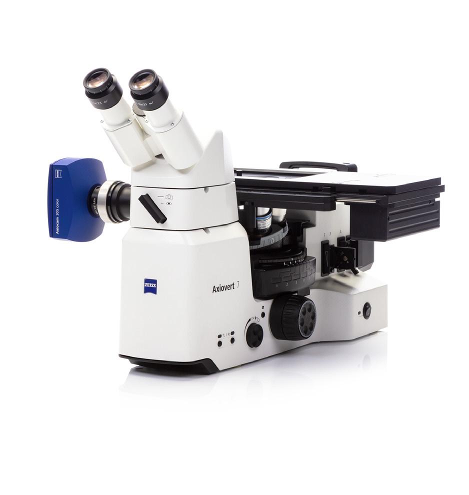
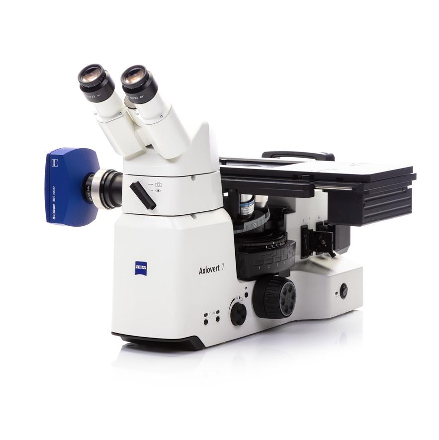
ZEISS Axiovert 7
Motorized microscope with coded components for applications with demands on workflow automation and advanced imaging tasks.
› In Brief
› The Advantages
› The Applications
› The System
› Technology and Details
› Service
1 Microscope
• Axiovert 5
• Axiovert 7
2 Recommended Classes of Objectives
• EC-Epiplan
• EC-Epiplan-Neofluar
3 Recommended Cameras
• Axiocam 105
• Axiocam 208
• Axiocam 305
• Axiocam 705
• Axiocam 712
4 Stages
• Mechanical Stage
• Gliding Stage
• Scanning Stage
Operating Modes
• Stand-Alone Operation (without PC) via On Screen Display (OSD)
• Via Labscope
• Via ZEN core on PC
Auxiliary microscope, d=30
444830-9902-000
Eyepieces with graticules (by choice)
Eyepieces (by choice)
Camera adapter 60N-C 2/3" 0.5x (for Axiocam 105 color R2, Axiocam 208 color and Axiocam 305)
426112-0000-000
Camera adapter 60N-C 2/3" 0.63x (for Axiocam 202 mono and Axiocam 305) 426113-0000-000
Camera Adapter 60N-C 2/3" 0.63x; rotating +/- 2°
426113-9010-000
or: Adapter 60N - T2 1.0x 426103-0000-000 in combination with:
Camera adapter 60N-C 1" 1.0x 426114-0000-000 Axiocam 208 color 426570-9001-000 Axiocam 305 mono R2 426560-9041-000 Axiocam 305 color R2 426560-9031-000
105 color R2 426570-9010-000
(further cameras by choice) Axiocam 202 mono 426570-9011-000
Camera Adapter T2-C 1" 1.0x; adjustable 426105-0000-000
Camera Adapter T2-C 1" 1.0x 426104-0000-000 or:
Binocular tube 45°/23 425523-9000-000
Slider with mirror 100 % 425146-9051-000
Slider with beam splitter 50 % 425146-9061-000
Slider with beam splitter 20/80 % 425146-9070-000
Binocular phototube, left port 45°/23 (50:50) 425523-9020-000
Intermediate phototube H = 50 mm, left port 425146-9031-000
Binocular phototube, 45°/23 (50:50) 425523-9030-000
LD condenser 0.4 for slider 424228-9020-000 (further condenser by choice)
Binocular ergotube 30-60°/23 425523-9040-000
Note: Ergo intermediate pieces and Intermediate phototube can be combined up to a maximum of 100 mm
Ergo intermediate piece H = 25 mm 425146-9020-000
Ergo intermediate piece H = 50 mm 425146-9040-000
Microscope stand Axiovert 5 RL TL SCB 431030-9170-000
Microscope stand Axiovert 7 RL TL 431030-9180-000
Compensator pol Lambda sub. +/– 10° 453706-0000-000
Compensator Lambda 453707-0000-000
Analyzer slider A 12x35 mm, 90° rotatable 453667-9901-000
Objectives (by choice)
follwing external light sources can be used with Axiovert
diaphragm 423655-9020-000
Illuminator RL LED 10 W (included with the stand) 423004-9030-000
For stand-alone operation (without PC) of microscope stand functions with ZEISS Axiocam (202, 208, 305) via On Screen Display
For usage with On Screen Display (OSD) is required:
– Cable, High-Speed-HDMI-Cable, Premium, black, 2m
– Keyboard, USB, Design “Business Slim”, Language English US
– Keyboard, USB, Design “Business Slim”, Language German
– Mouse, Optical USB-Scroll Mouse
– Wi-Fi adapter package (by choice) – TFT Monitor (by choice)
(USB 3.0 flash drive , Type -A cable and a camera cable is included)
Only for stands:
Microscope stand Axiovert 5 RL SCB
431030-9070-000 Smart control box (SCB)
Microscope stand Axiovert 5 RL TL SCB
431030-9170-000
Microscope stand Axiovert 5 RL SCB 431030-9070-000
Microscope stand Axiovert 7 RL 431030-9080-000
For reflected light:
Reflector module bright field ACR P&C 424928-9901-000
Reflector module dark field ACR P&C 424922-9901-000
Reflector module DIC/Pol ACR P&C 424939-9000-000
Reflector module DIC/Pol red Lambda
Reflector module C-DIC/TIC ACR P&C 424941-9000-000
protection set 434308-9010-000 (included with the stand)
Polarizer slider A 6x30 mm, 90° rotatable 427710-9000-000
Stop slider A 14x40 mm with aperture luminous field diaphragm 423730-9151-000
DIC slider (by choice)
› In Brief
› The Advantages
› The Applications
› The System
› Technology and Details
› Service
Mounting frame for steel specimen
432322-9000-000
Trackball XY; CAN 432903-9000-000
Stage insert, metal; D=72 mm, d=10 mm
451326-9901-000
Joystick XY; CAN 432903-9011-000
Mounting frame K; low profile, for reflected light, d=72 mm
432334-9001-000
Stage insert, metal; D=72 mm, d=20 mm
451327-9901-000
Stage insert, metal; D=72 mm, d=23 mm
451328-9000-000
Stage insert, metal; D=72 mm Drop Shape
451328-9010-000
Electronic Coaxial Drive; CAN 432904-9901-000
Scanning stage 130x85 mot P; CAN 432031-9902-000
Glass insert 115 mm, d=10 mm
451228-4001-000
Glass insert 115 mm, d=27 mm
451228-4002-000
Glass insert 115 mm, d=50 mm
451228-4006-000
Mechanical stage 40x40 R/L, reflected light
432043-9010-000 (receptacle for stage inserts D=115 mm)
Converter CAN - USB Rev.2 (without fig.)
432909-9901-000
Stage insert, metal; D=115 mm, d=10 mm
432328-9020-000
Stage insert, metal; D=115 mm, d=20 mm
432328-9040-000
Stage insert, metal; D=115 mm, d=25.4 mm
432328-9050-000
Stage insert, metal; D=115 mm, d=30 mm
432328-9060-000
Gliding stage Z with stage inserts d=24 and d=48
471722-9902-000
In Brief
The Advantages
The Applications
The System
Technology and Details
Service
Air Conditioning and Quality
Temperature range for operation with indicated performance (24 h per day, regardless of whether the microscope is in operation or switched off)
Relative Humidity < 80 % at 40 °C
Atmospheric Pressure / Altitude 800 to 1060
Pollution Degree 2
Mains Connection
Nominal AC Voltage
Nominal Frequency 50/60 Hz
Max. Current 1.4 A
Rating for Microscope Stand 24 VDC, 5 A
Protection Class IP20 (IEC 60529)
Overvoltage Category II
System Features
Optical System Infinte, ICS
Nosepiece 6× nosepiece, encoded
Reflector Turret 6× reflector turret, encoded
Focus manual coarse / fine focus; 13 mm focus range with adjustable focus stop motorized focus drive (resolution 78 nm) 13 mm focus range with adjustable focus stop
Reflected Light Illumination (included) White 10 W LED, average lifetime >60000 hrs
Contrast Methods (Reflected Light / Transmitted Light)
Differential Interference Contrast (DIC)
Circular Differential Interference Contrast
Interference Contrast (TIC)
Snap Button on Stand
Ergonomically positioned on both sides of the stand; allows to snap images, record videos, start workflows
Light Manager Set, save and recall the optimal image brightness according to specific combination of nosepiece position and reflector turret position.
useable
not useable
› In Brief › The Advantages
› The Applications
› The System
› Technology and Details › Service
Your microscope system from ZEISS is one of your most important tools. For over 170 years, the ZEISS brand and our experience have stood for reliable equipment with a long life in the field of microscopy. You can count on superior service and support - before and after installation. Our skilled ZEISS service team makes sure that your microscope is always ready for use.
• Lab Planning & Construction Site Management
• Site Inspection & Environmental Analysis
• GMP-Qualification IQ/OQ
• Installation & Handover
• IT Integration Support
• Startup Training
• Predictive Service Remote Monitoring
• Inspection & Preventive Maintenance
• Software Maintenance Agreements
• Operation & Application Training
• Expert Phone & Remote Support
• Protect Service Agreements
• Metrological Calibration
• Instrument Relocation
• Consumables
• Repairs
• Decommissioning
• Trade In
• Customized Engineering
• Upgrades & Modernization
• Customized Workflows via APEER