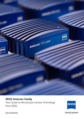
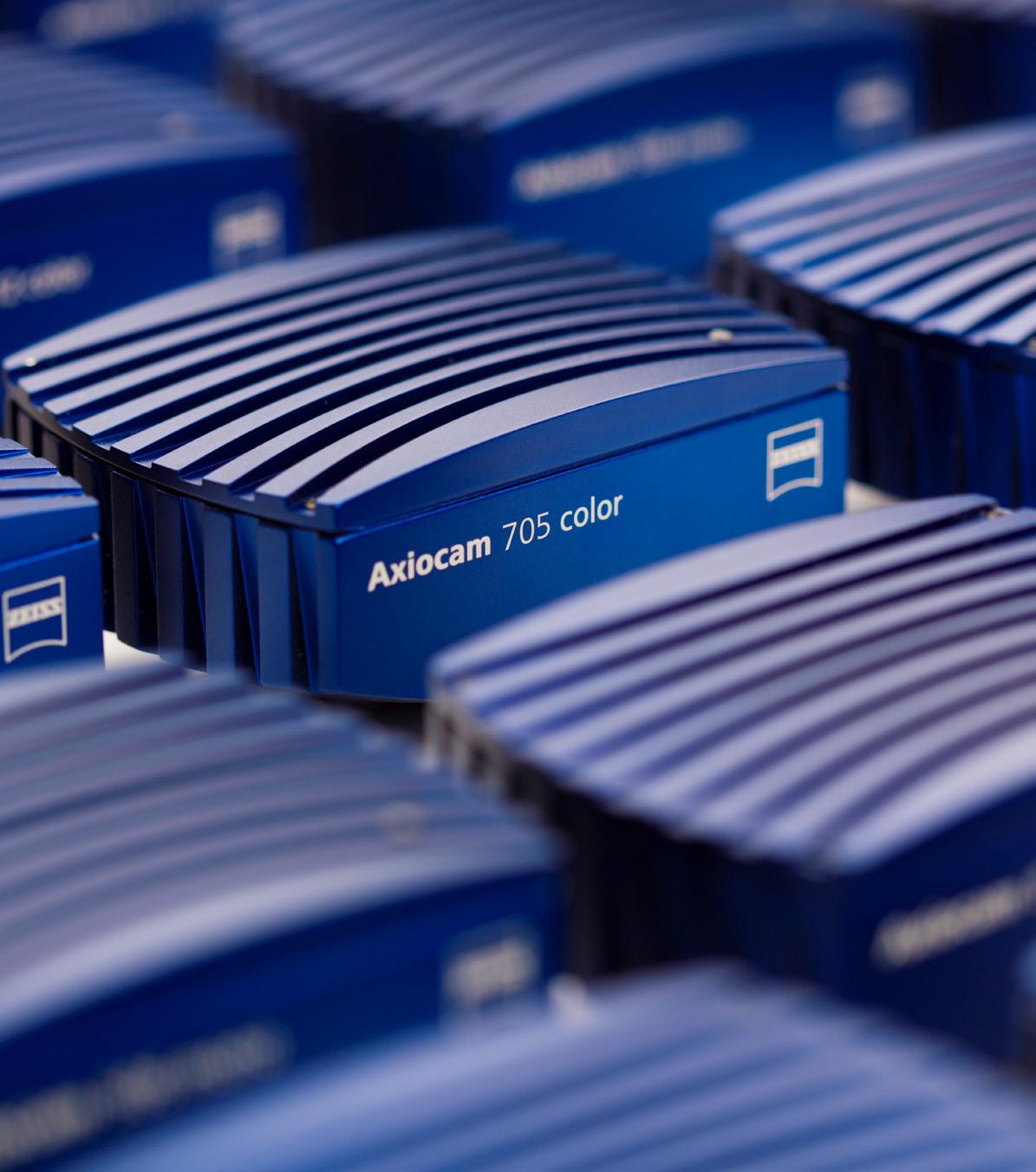
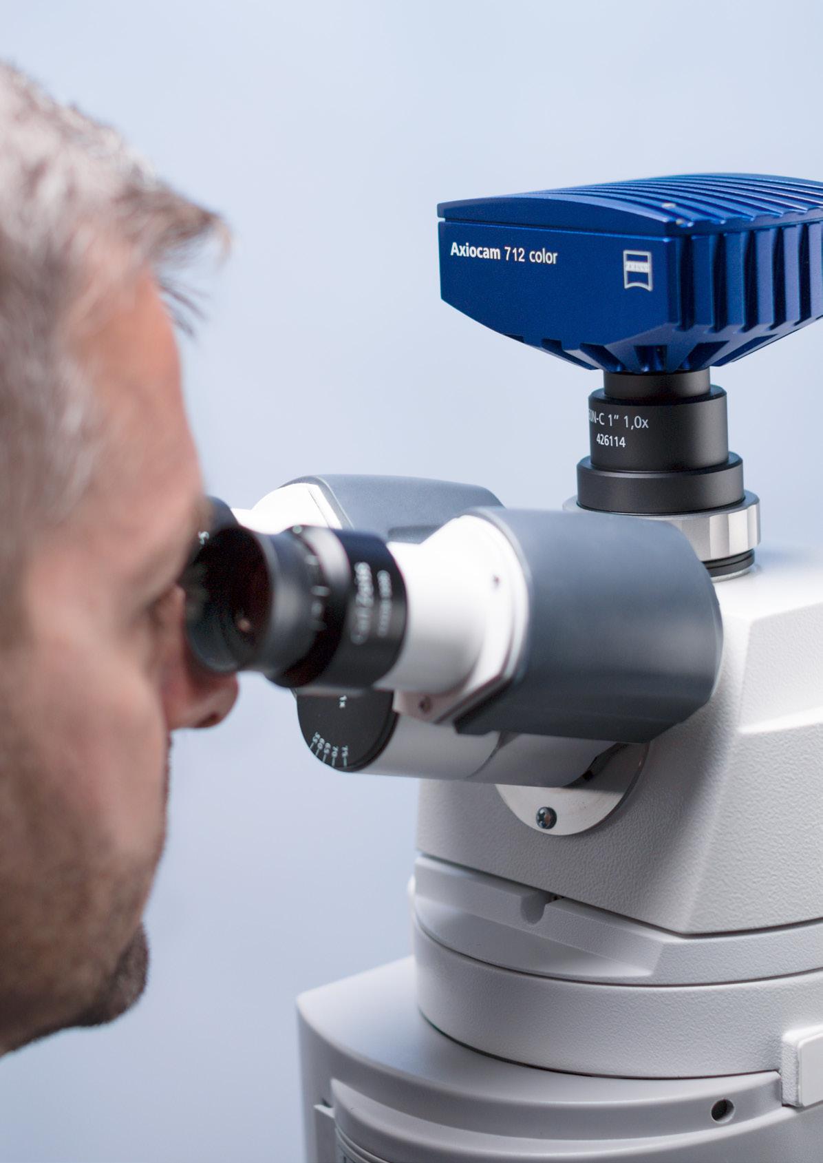
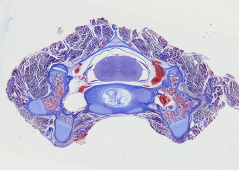
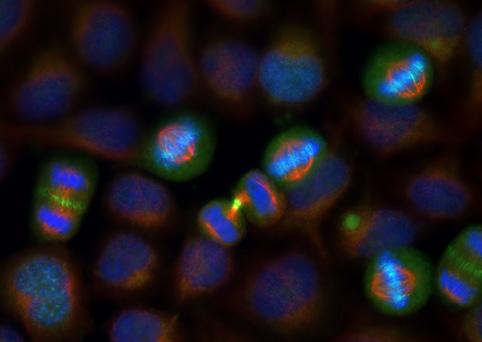
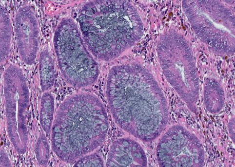
Camera technology as diverse as your imaging and documentation tasks.
Recent years have seen rapid developments in the processing and documentation of microscopic images. Technology has evolved from video cameras connected by frame-grabbing control cards to today’s purely digital cameras operated via USB, FireWire or quick Ethernet interfaces. Whichever camera technology you are using for microscopy, you will always need high contrast resolution, good sensitivity and dynamic performance, and a high readout speed. Often, short exposure times and the option of recording a quick series of images will be just as important as exact color reproduction.
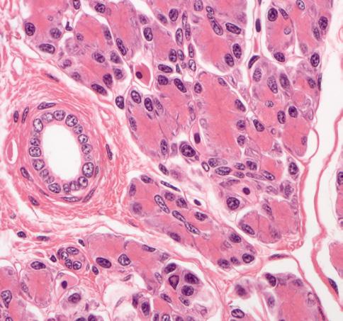
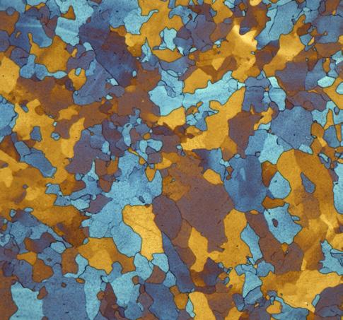
All things considered, there is simply no such thing as the perfect microscope camera – it just depends on the applications at hand. This guide aims to give you an overview of the whole portfolio of ZEISS Axiocams. These dedicated microscope cameras range from compact color cameras for routine documentation to fast, sensitive monochrome cameras for gentle live cell imaging. Explore typical applications and use the performance matrix to decide which Axiocam is the right one for you. Use the camera terminology chapter to learn about fundamental principles and the words which are used to describe them.
Welcome to the fascinating world of microscope cameras. With this compendium, you’ll get an overview about the whole portfolio of ZEISS Axiocams. Discover many exciting applications and use it as your guide to selecting the best camera for your imaging and documentation tasks.
Cameras for Teaching or Routine Applications
These cameras meet the needs for easy operation and efficiency. You benefit from live images with exactly the right resolution and crisp contrast. Page 8
High End Color Cameras
These cameras all deliver outstanding true color images in high resolution. Their high dynamic range and high frame rates meet the needs of even the most demanding pathology or histology imaging. For polarized light applications a special camera provides meaningful color-coded visualization.
Integrated Network Cameras
These cameras can be connected to your WiFi – giving you freedom of sharing your images with colleagues. Already integrated into the microscope stand, these cameras are always well adjusted.
Page 50
Software
Each Axiocam comes with a bundle of free software for basic imaging tasks or can be combined with several high end modules of ZEN imaging software tailored to your applications.
Page 54
High End Fluorescence Cameras
These sensitive monochrome cameras are dedicated to capture even faint signals from your living samples. Each Axiocam contributes a unique combination of resolution, sensitivity and speed to your most demanding live cell imaging experiments.
Knowledge Base
Learn about fundamental terms of camera technology and their meaning. See how sensor type, resolution, frame rate and sensitivity are interconnected and influence your results.
Cameras for Teaching or Routine Applications
Enjoy efficient, easy operation.
These cameras meet the needs for easy operation and efficiency. You benefit from live images with exactly the right resolution and crisp contrast.
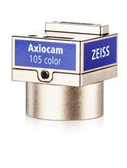
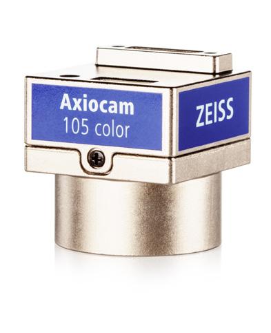
ZEISS Axiocam 105 color Your 5 Megapixel Microscope Camera for Documentation in Routine Labs
Axiocam 105 color is your small, no-frills microscope camera. With its compact design, it makes quick and efficient work of your daily documentation needs.
With its USB 3.0 connection, experience a high speed data transfer rate for handling your high resolution 5 megapixel color images. Offering an exposure time range of 30 µs to 1 s and a live frame rate of up to 30 images per second, the camera allows you to be well prepared to cover multiple tasks. Document your results quickly and conveniently. With its attractive price-performance ratio, you can also expand the capability of your fluorescence imaging system with color imaging. Axiocam 105 color is ideal as a secondary camera on fluorescence microscopes that are traditionally equipped with monochrome cameras. The camera’s small form factor also lends itself well to environments with limited space.
Recommended for
• Applications with bright samples
• Documentation
• Education / Teaching
• Routine tasks
• Materials testing
• Quality assurance / Quality control
• 5 megapixel CMOS sensor
• 30 images per second at full 5 megapixel color resolution
• 8 bit digitization
• High resolution with 2.2 µm pixel
• Easy to use super-speed USB 3.0 connection
• Color and black & white imaging modes
• Fast and efficient operation with ZEN imaging software
Simpler. More Intelligent. More Integrated.
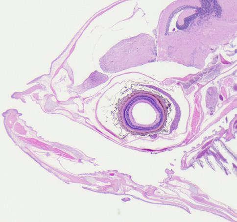
Fish, HE staining, brightfield, acquired with ZEISS Stemi 305
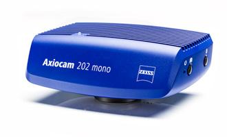
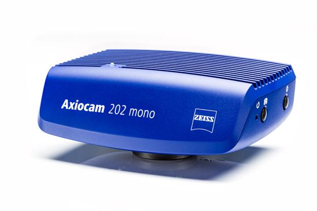
ZEISS Axiocam 202 mono
Your 2 Megapixel Stand-alone Microscope Camera for
Routine Fluorescence Documentation
Axiocam 202 mono is your 2 megapixel monochrome microscope camera with automatic functions for routine fluorescence applications. With this CMOS sensor camera you can easily acquire monochrome images in stand-alone mode with no need of a PC. Since the camera automatically adjusts the exposure time you only need to press the snap button to capture and store your fluorescence images on a USB flash drive. If needed you can adjust parameters in the OSD (on screen display) menu before you acquire the image. In combination with the smart microscopes Axiolab 5 or Axioscope 5 you can even capture multichannel fluorescence images by simply pressing one button.
Recommended for
• Applications with bright fluorescence samples
• Documentation
• Education / Teaching
• Fluorescence imaging applications with live and fixed cells
• Documentation of fluorescent cell cultures
• Routine tasks in cell laboratories
• 2 megapixel CMOS chip sensor with image diagonal of 13 mm and large pixel size for high sensitivity in fluorescence documentation
• Choose between 12 bit or 8 bit digitization
• Store images directly on USB flash drive in stand-alone mode
• Single button multichannel fluorescence acquisition when combined with Axiolab 5 or Axioscope 5 stands in standalone mode (with no PC)
• Automatic exposure and gain adjustment for easy fluorescence image capture
• Connect directly to a monitor by a HDMI cable for live image display for search and focussing and review of acquired images
• Perform secure image data transfer to TWAIN-compatible 3 rd party software solutions with the TWAIN driver.
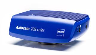
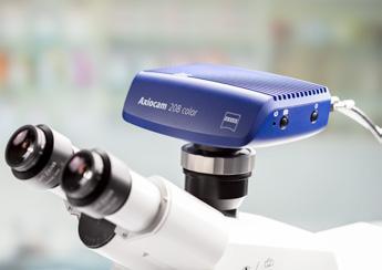
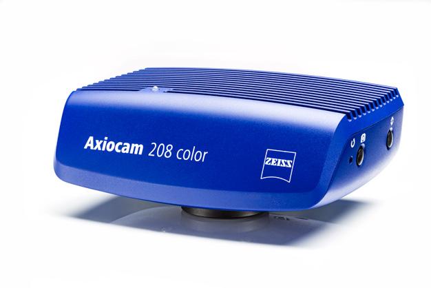
Your Fast, 4K Microscope Camera for Smart Digital Documentation
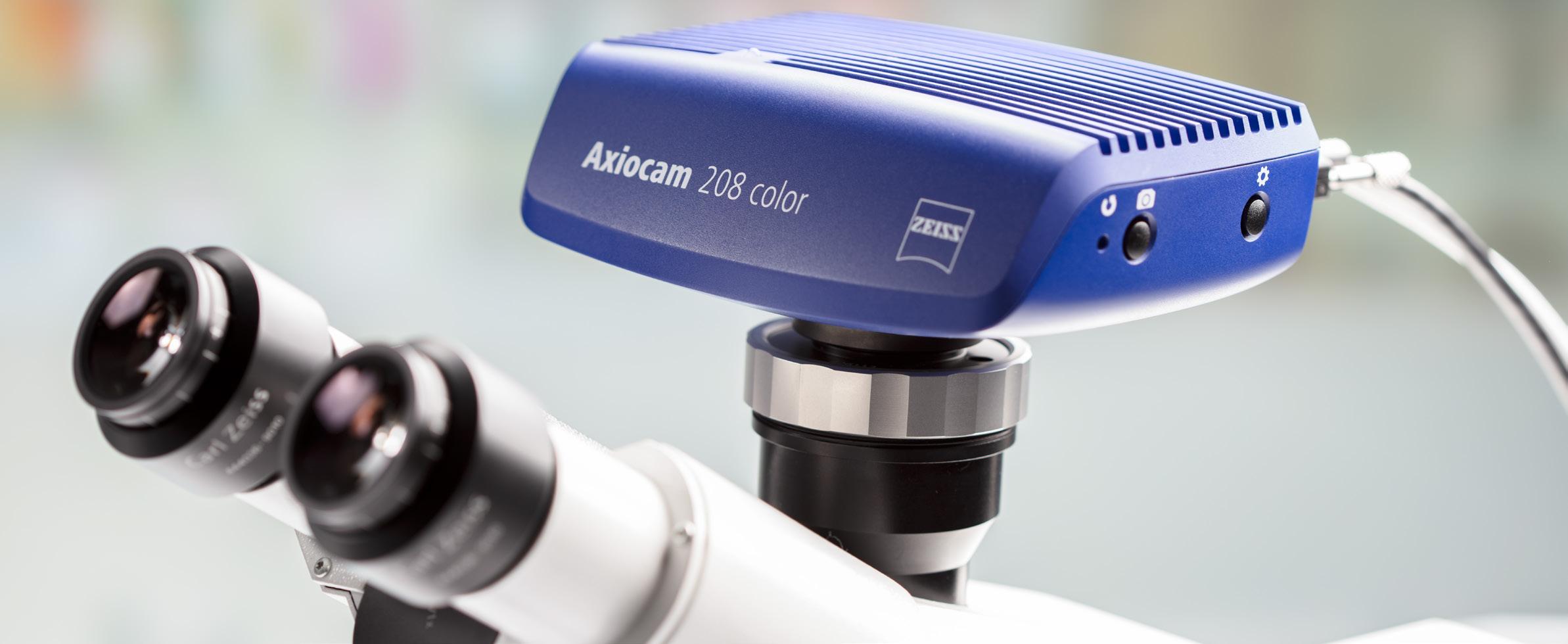
Axiocam 208 color is your smart versatile 8 megapixel color microscope camera suitable for education, documentation and routine applications. This CMOS camera delivers crisp, detail rich live images with high color fidelity at full 4k resolution in outstanding 30 fps. Choose between three modes of operation:
1. In stand-alone mode, you don’t need a PC to acquire microscope images. The camera automatically adjusts brightness and white balance and offers live image enhancement functions like sharpening, denoising and HDR. Digital documentation of your specimen has never been easier.
2. Alternatively, connect the CMOS camera via USB or to a network and control it wirelessly with the easy-to-use imaging app Labscope. Since you can connect multiple cameras to the network, Axiocam 208 color is the ideal solution for digital classroom applications and for connected laboratories, too.
3. In addition, you can use the powerful imaging software ZEN with your Axiocam 208 color.
Recommended for
• Documentation
• Education / Teaching
• Routine tasks
• Materials research
• Quality assurance / Quality control
• Fast high resolution live image for co-observation
• Full 4K resolution in outstanding 30 fps
• Brilliant color rendering
• Live image enhancement functions like sharpening, denoising and HDR
• Use in stand-alone mode and save images on USB flash drive, use Labscope imaging app or ZEN imaging software
• Easy and effortless digital documentation – especially suitable for education, digital classroom and routine documentation
• Ethernet or USB 3.0 as digital data interface
• Use the optional WiFi stick and Labscope imaging app to control and transfer data wirelessly
• Document your samples as you see it in the eyepieces
• Stand-alone operation with camera control by intuitive On Screen Display via mouse and keyboard without a PC
• Connect directly to a monitor by a HDMI cable for live image display for search and focussing and review of acquired images
• Perform secure image data transfer to TWAIN-compatible 3 rd party software solutions with the TWAIN driver.
Simpler. More Intelligent. More Integrated.
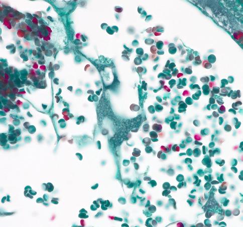
Trichrome stained blood vessels in transmitted light brightfield, acquired with ZEISS Axiolab 5, objective: Plan- APOCHROMAT 40× 1.4
Red bone marrow in transmitted light brightfield, acquired with ZEISS Axiolab 5, objective: Plan- APOCHROMAT 40× / 1.4
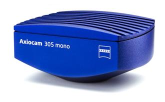
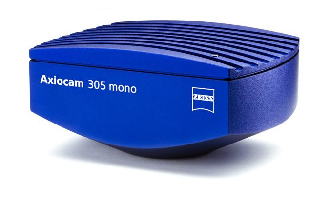
ZEISS Axiocam 305 mono
Your Fast 5 Megapixel Microscope Camera for Routine Fluorescence Applications
Axiocam 305 mono your 5 megapixel camera from ZEISS for fluorescence imaging for your routine lab and enables a range of applications for live cell observation. The state-of-the-art CMOS Global Shutter technology lets you follow and capture samples accurately. Thanks to its high dynamic range, you can acquire images with various high contrasts and intensities in a single image. A dark homogenous background helps you see even the finest structural details. And it’s a really fast camera, acquiring up to 36 frames per second at full 5 megapixel resolution. Highly sensitive sensor technology and sophisticated camera engineering means your Axiocam 305 mono will deliver reproducible results every time. The sensor is temperature-stabilized, resulting in reproducible quality and reduced background noise. Easy to use ZEN imaging software fully supports the robust camera performance by an intuitive user interface through a simple and fast USB 3.0 connection.
Recommended for
• Fluorescence imaging applications with live and fixed cells
• Documentation of fluorescent cell cultures
• Routine tasks in cell laboratories
• Materials research in near infrared wavelengths
• Time lapse recording
• Multi channel imaging without the need for hardware trigger synchronization
• 5 megapixel CMOS global shutter sensor
• 11.1 mm image diagonal
• Fast readout with 36 images per second in full color resolution
• 12 bit digitization finer gradation in signal
• Small 3.45 micron pixels for better sampling at low magnifications
• Global shutter architecture for distortion-free images
• Active thermal stabilization of the sensor for extremely reproducible image quality
• Easy to use super-speed USB 3.0 connection
• Fast and efficient operation with ZEN imaging software
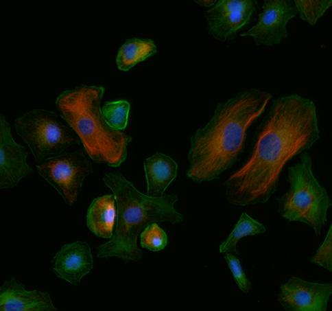
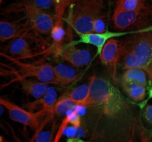
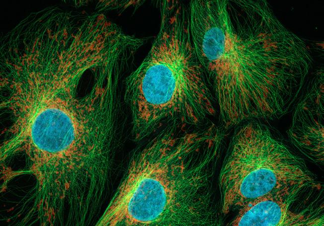
Astrocytes. Green: GFP, red: tubulin – Alexa 568, blue: Hoechst 33342, acquired with ZEISS Axio Imager.D2, objective: Plan APOCHROMAT
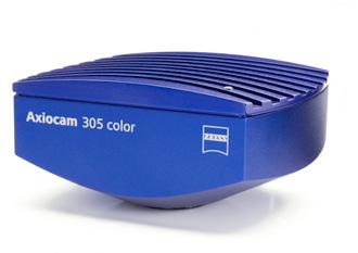
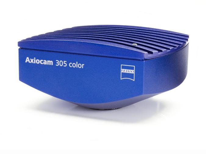
ZEISS Axiocam 305 color
Your Fast 5 Megapixel Microscope Camera for
Routine and Research Labs
Axiocam 305 color is your 5 megapixel camera for high resolution imaging at fast speeds. With state-of-the-art CMOS global shutter technology, you can follow and capture samples distortion-free and with great accuracy. Thanks to this highly sensitive sensor technology and precise camera engineering, your Axiocam 305 color allows the capture of quality color images for a wide range of applications. Acquire great color images with crisp contrast or use the optional black & white mode to document basic fluorescence. With this fast camera offering up to 36 frames per second at full resolution, achieve efficient searching, fast focusing and ergonomic handling at your digital microscope workplace. Cover more of your area of interest with its 2/3″ sensor format and produce great color images on your compound, stereo, or zoom microscope.
Though a simple and fast USB 3.0 connection, control the camera and experience robust performance with easy to use ZEN imaging software and its intuitive user interface.
Recommended for
• Applications with bright samples
• Documentation
• Routine tasks
• Materials research
• Quality assurance / Quality control
• Fast high resolution live image for co-observation
• Fast image acquisition and time-lapse recording
More Intelligent. More Integrated.
• 5 megapixel CMOS global shutter sensor
• 11.1 mm image diagonal
• Fast readout with 36 images per second in full color resolution
• 12 bit digitization for finer gradation in signal
• Small 3.45 micron pixels for better sampling at low magnifications
• Global shutter architecture for distortion-free images
• Active thermal stabilization of the sensor for extremely reproducible image quality
• Easy to use super-speed USB 3.0 connection
• Color and black & white imaging modes
• Fast and efficient operation with ZEN imaging software
Simpler.
Liver of Amphiuma in brightfield, HE-staining, acquired with ZEISS Axio Imager, objective: EC Plan-NEOFLUAR 20× / 0.50
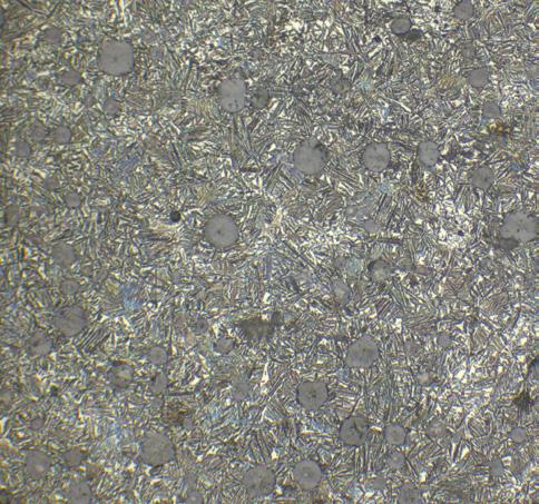
Pure iron in brightfield, reflected light, acquired with ZEISS Axio Observer, objective: EC Epiplan-APOCHROMAT 50× / 0.9
High End Color Cameras
Get True Color Images in High Resolution
These cameras all deliver outstanding true color images in high resolution. Their high dynamic range and high frame rates meet the needs of even the most demanding pathology or histology imaging. Large sensor areas offer best coverage of your microscope field of view.
Your Fast 5 Megapixel Microscope Camera for True Color Image Acquisition in High Resolution
This flexible 5 megapixel scientific color camera strikes the perfect balance of speed and resolution. Delivering more than 60 frames per second at full 5 megapixel resolution, this camera captures even the most dynamic processes without compromising image resolution. Subsampling or sub-region readout accelerate your acquisition speed to hundreds of frames per second.
Recommended for
• High-resolution microscopy
• High-framerate imaging
• Research
• Documentation
• Industrial applications
• Materials research
• Quality control
• Medical microscopy
• Pathology
• Cytology
• 5 megapixel cooled color CMOS sensor with 11 mm diagonal
• 62 frames per second in full 5 megapixel resolution
• Best-in-class color rendition
• Color and monochrome imaging modes
• Exclusive noise inhibition technology for low-light imaging
• Dynamic range of 1:25,000 in high-dynamic range (HDR) mode
• Combined analogue and digital pixel binning
• Small 3.45 µm pixels for high-resolution imaging
• Hardware triggering
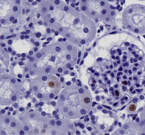
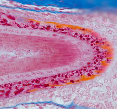
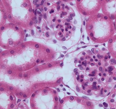
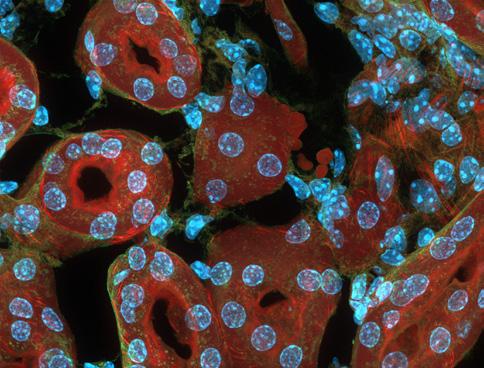
Rat kidney section, objective: Plan- APOCHROMAT
Simpler. More Intelligent. More Integrated.
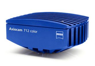
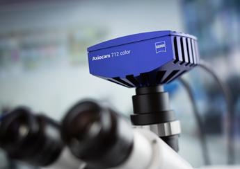
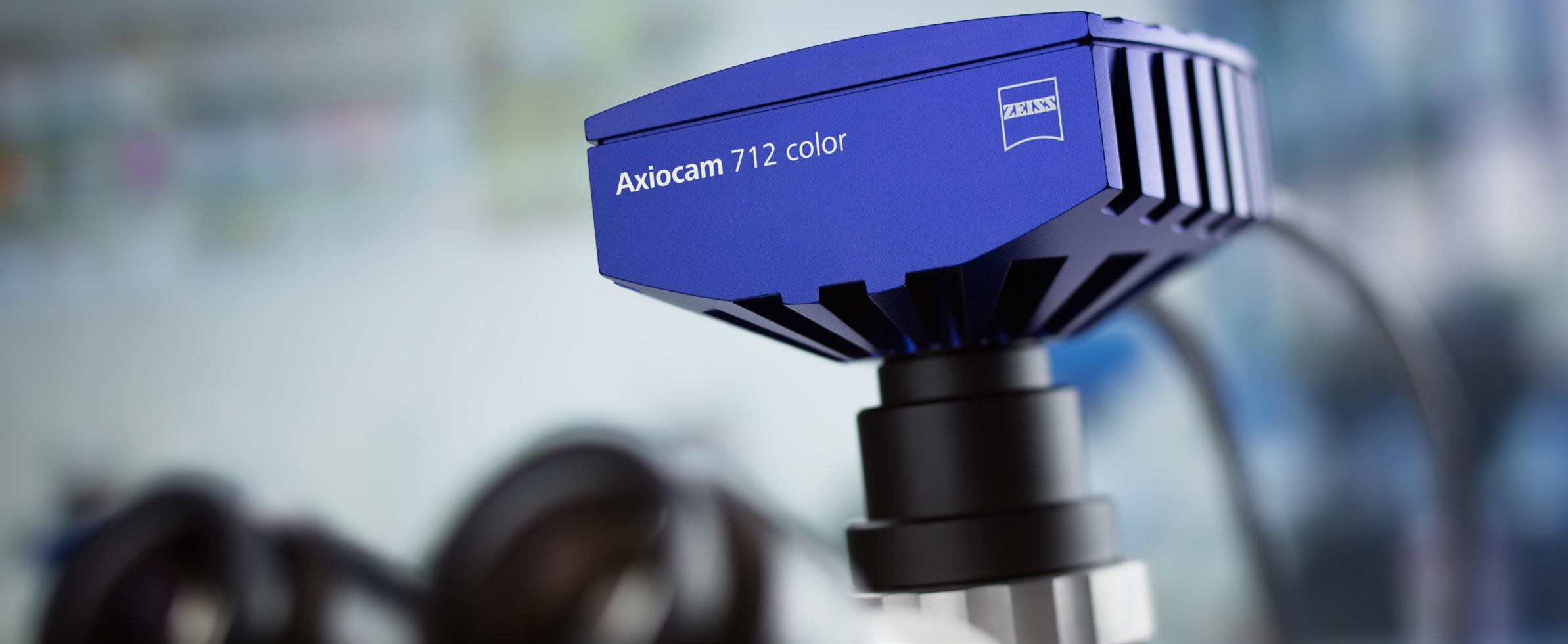
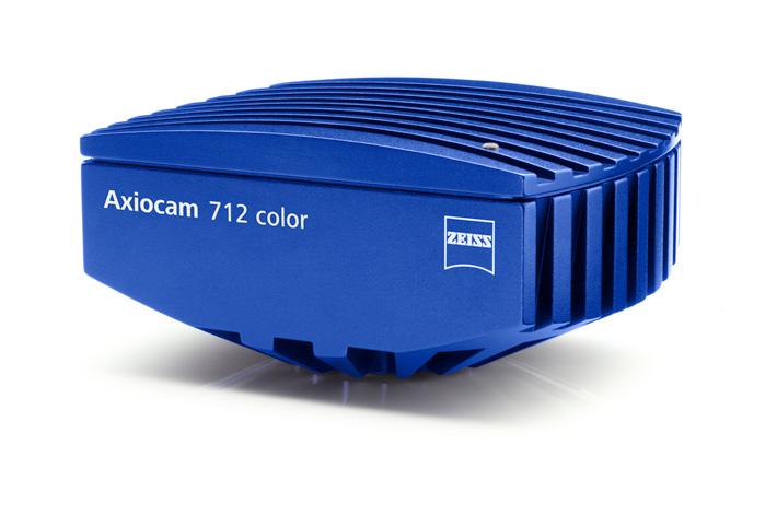
ZEISS Axiocam 712 color
Your All-round 12 Megapixel Microscope Camera for True Color Acquisition of Large Specimen Areas in High Resolution
This 12 megapixel scientific grade camera combines a large image sensor, small pixel size, precise color rendition and fast imaging speed. The CMOS sensor delivers more than 20 frames per second with a 17.5 mm diagonal large field of view. You can now acquire large specimen regions quickly and with uncompromised image quality. The large field of view reduces the number of tiles required to image largest samples, and so drastically accelerates tiling experiments.
Axiocam 712 color is a highly evolved digital color camera addressing the needs of scientific microscopy, including documentation, reporting and analysis. Fast and artifact-free imaging with optimized color reproduction makes your work comfortable and efficient. In addition, exploring your sample on the screen, instead through the oculars, becomes a true and very convenient alternative.
Recommended for
• High-resolution microscopy
• Large region imaging
• Medical imaging
• Material science research
• Macroscopic imaging
• Pathology
• 12 megapixel cooled color CMOS sensor
• Large sensor with 17.5 mm diagonal for extended field of view
• Best-in-class color rendition
• Color and monochrome imaging modes
• 20 frames per second in full 12 megapixel resolution
• 30 frames per second of the entire field of view in live image mode
• Exclusive noise inhibition technology for lowlight imaging
• Dynamic range of 1:25,000 in high-dynamic range (HDR) mode
Simpler. More Intelligent. More Integrated.
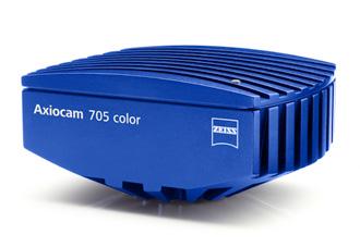
Your Fast, 7 Megapixel Microscope Camera for True Color Imaging of Large Fields of View
With its dual USB interface and state-of-the art CMOS sensor, this 7 megapixel color camera offers you amazingly fast live image and acquisition speeds. The large 17.6 mm diagonal sensor allows for efficient imaging of large fields of view.
This makes it the camera of choice when screening large sample areas as it efficiently reduces the required number of tile images. This is extremely beneficial when working with large, pathology tissue sections or large, colored materials samples.
Recommended for
• Color imaging applications in a broad range of fields from life sciences to materials research and geoscience
• Co-observation with fast high resolution live image in high quality color with a very large field of view
• Imaging of large pathology, cytology and materials samples
• Fast tile scanning applications
• Samples with a broad range of intensities and exposure times
More Intelligent. More Integrated.
• 7 megapixel CMOS sensor with 17.6 mm image diagonal
• 73 full-resolution color images per second recording (faster with sensor sub-region readout)
• High image contrast with 14-bit signal conversion
• 4.5 micron pixels for optimal resolution and sensitivity
• Fast read-out with global shutter architecture for distortion-free images
• Monochrome imaging mode
• Reproducible image quality due to active thermal stabilization of the sensor
• Easy to use high-speed dual USB 3.0 connection
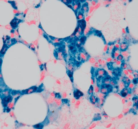
Prussian blue staining for hemosiderin (iron deposition) in brown adipose tissue of mice. Sample courtesy: A. Feuchtinger, Helmholtz Zentrum München, Germany
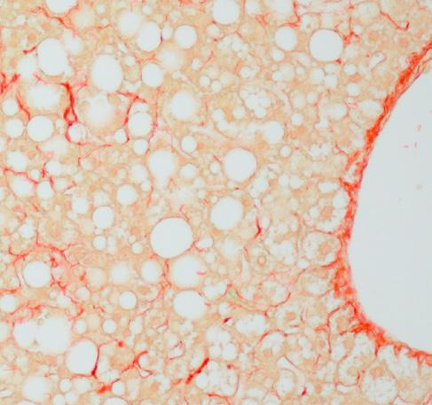
NASH mouse liver with connective collagenous tissue stained in red. Sample courtesy: A. Feuchtinger, Helmholtz Zentrum München, Germany
Simpler.
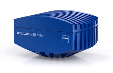
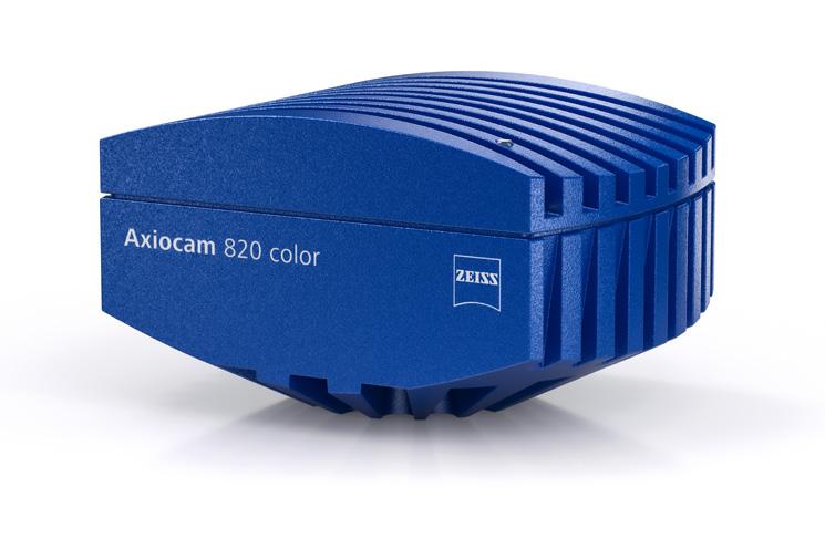
ZEISS Axiocam 820 color
Your Sensitive 20 Megapixel Microscope Camera for Demanding, True Color Imaging of Large Fields of View
Axiocam 820 color 20 megapixel camera combines high sensor resolution, color accuracy and a large sensor size for the most challenging samples. Its live image is 30 frames per second, providing navigation with the PC that is as smooth as looking through the eye pieces. The small pixels allow the use of low magnifications for high speeds and large fields of view without losing resolution.
Scan large areas faster than ever before due to its large 17.5 mm square sensor. This makes Axiocam 820 color the ideal choice for demanding color imaging in cytology, pathology, materials research, and life science applications.
Recommended for
• The most demanding color applications in life sciences, materials research and geoscience
• Co-observation with a fast, high-resolution live image in high quality color with a very large field of view
• Large sample area imaging in pathology, cytology and materials samples
• Fast tile scanning applications
• The broadest range of intensities by high dynamic range
Simpler. More Intelligent. More Integrated.
• 20 megapixel square CMOS sensor with 17.5 mm image diagonal
• 28 full-resolution color images per second (faster with sensor sub-region readout)
• Small 2.74 micron pixels for resolving the finest details at all magnifications
• High-quality noise inhibition technology
• Fast read-out with global shutter architecture for distortion-free images
• Best-in-class color rendition
• Color and monochrome imaging modes
• Reproducible image quality due to active thermal stabilization of the sensor
• Dynamic range of 1:25,000 in high-dynamic range (HDR) mode
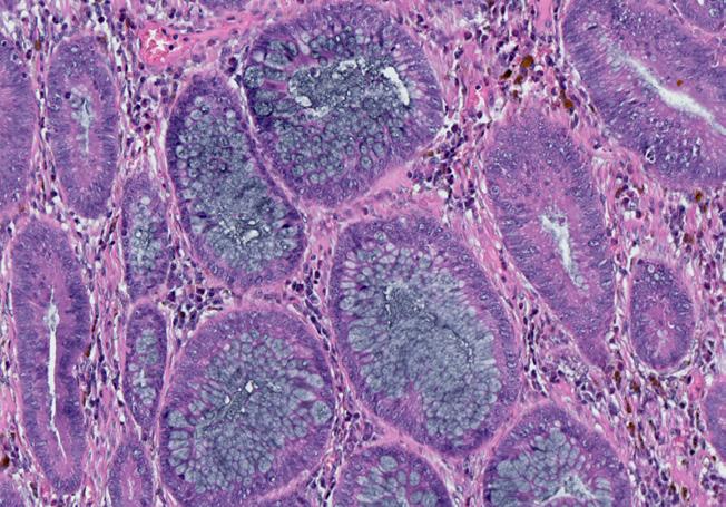
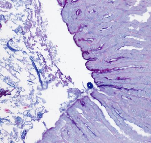
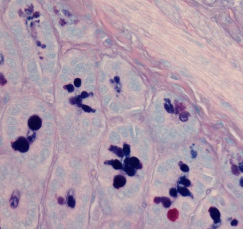
Mouse intestinal goblet cells with neutral (red) and acid (blue) mucine staining imaged with 20x/0.5 (left) and 63x/1.2W (right) objective. Sample courtesy: A. Feuchtinger, Helmholtz Zentrum München, Germany
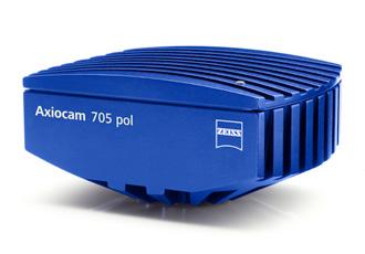
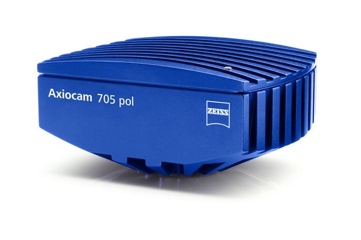
ZEISS Axiocam 705 pol
Your Scientific 5 Megapixel Microscope Camera for Single-shot Polarization Imaging
Axiocam 705 pol is your scientific 5 megapixel camera with a polarization filter mask enhanced sensor. A single image is sufficient to capture different polarization parameters such as angle of polarization, degree of polarization together with the image content. No special accessories such as an analyzer in your microscope are required. You simultaneously acquire polarization effects with one single exposure over the field of view which speeds up your imaging as no analyzer needs to be adjusted.
• Meaningful methods for visualization of multiple polarization parameters
• Low readout noise and analogue signal amplification
• Dynamic range of 1:25,000 in high-dynamic range (HDR) mode
• Small 6.9 µm per polarization pixel unit for high-resolution imaging
• Hardware triggering
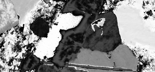
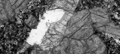
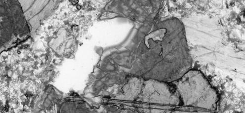
Representation of the acquired image information with Axiocam 705 pol
• Encoded Pseudo Color 4 Channel Image
• Channel 1 = Angle of Polarization
• Channel 2 = Degree of linear Polarization
• Channel 3 = Intensity
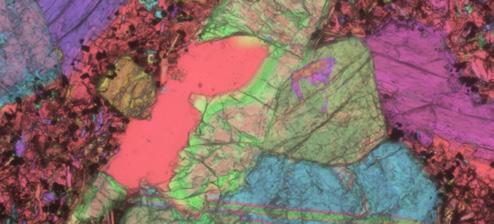
• Channel 4 = Color encoded information derived from above channels
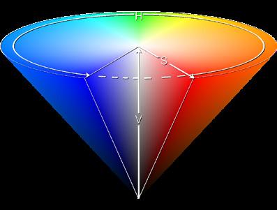
• Method of color encoding in HSV color space
• Hue = Angle of polarization
• Saturation = Degree of linear Polarization
• Value = Intensitiy
• = Intensity
https://de.wikipedia.org/wiki/HSV-Farbraum
Basalt thin section, Axiocam 705 pol, 4 channels
Simpler. More Intelligent. More Integrated.
High End Fluorescence Cameras
Capture Even Faint Fluorescent Signals
These sensitive monochrome cameras are dedicated to capture even faint signals from your living samples. Each Axiocam contributes a unique combination of resolution, sensitivity and speed to your most demanding live cell imaging experiments.
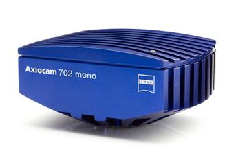
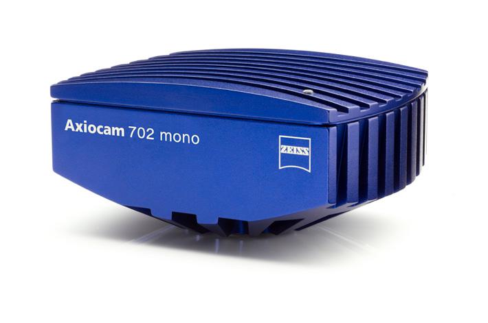
ZEISS Axiocam 702 mono
Your 2.3 Megapixel Microscope Camera for Fast Low Light and Live Cell Imaging
Fundamental to the investigation of weak and rapidly changing signals in biology is the recent advancement in camera techno logy. Axiocam 702 mono offers cell biologists and all other researchers a high-speed CMOS imaging solution that is very sensitive and affordable. If provides also distortion free high temporal resolution at a very budget friendly price.
This high-performance CMOS microscope camera has 2.3 megapixels and a 1/1.2″ sensor (diagonal 13.3 mm), making it the ideal choice for fast and sensitive fluorescence imaging.
Peltier cooling ensures low noise and reproducible image quality, particularly when you are dealing with long exposure times and dark areas in the sample.
Highest light sensitivity combined with low noise and high frame rates give you the temporal resolution that you always longed for in live cell imaging.
Recommended for
• Live cell imaging with high temporal resolution
• Low light applications
• Imaging at higher magnifications
• Monochrome CMOS microscope camera with 2.3 megapixels
• Sensor size of 1/1.2″ (diagonal 13.3 mm) and 1920 × 1216 pixels
• Pixel size of 5.86 µm
• Up to 128 fps at full resolution and up to 1000 fps @ 1024 × 128 pixels
• Dynamic range > 5000:1 (>74 dB) at typical < 6e read noise
• Thermo electrical cooled sensor
• Optional hardware trigger synchronization
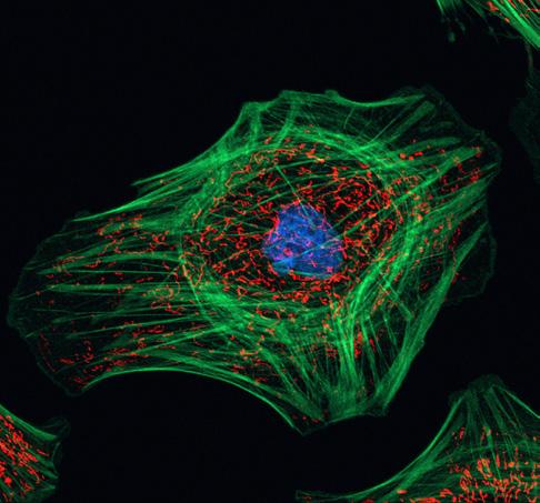
Indian Muntjac cultured cells. Sample courtesy of: M. Davidson, Florida State University, USA
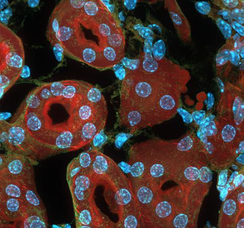
Mouse kidney section. Sample courtesy of: M. Davidson, Florida State University, USA
Simpler. More Intelligent. More Integrated.
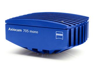
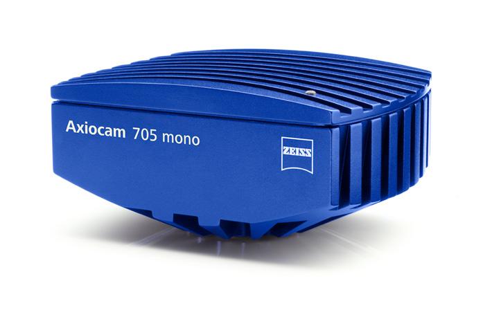
ZEISS Axiocam 705 mono
Your Fast 5 Megapixel Microscope Camera for High Resolution Imaging at High Speed
This 5 megapixel monochrome CMOS camera lets you capture time lapse sequences of the most dynamic processes in your sample. You can achieve more than 60 frames per second with full 5 megapixels. Or, you simply reduce the pixel count to accelerate your imaging even more – up to hundreds of frames per second. Hardware triggering delivers precise timing and enables extremely fast multidimensional imaging experiments. Active sensor cooling and low sensor readout noise make this microscope camera your ideal choice for fluorescence microscopy of dim and delicate specimens. Your Axiocam 705 mono employs analog pixel binning and amplification of signal to boost sensitivity. With high peak quantum efficiency of up to 72 % and a broad spectral sensitivity ranging from UV to near-IR light, you can tackle even the most challenging fluorescence imaging applications.
Recommended for
• High-resolution fluorescence microscopy
• High-framerate imaging
• Research
• Documentation
• Live cell imaging
• Low light microscopy
• 5 megapixel cooled global-shutter CMOS sensor
• 62 frames per second in full 5 megapixel resolution
• Wide sensitivity spectrum 350 nm – 1000 nm
• Exclusive noise inhibition technology for lowlight imaging
• Low readout noise and analogue signal amplification
• Dynamic range of 1:25,000 in high-dynamic range (HDR) mode
• Analogue pixel binning
• Small 3.45 µm pixels for high-resolution imaging
• Hardware triggering
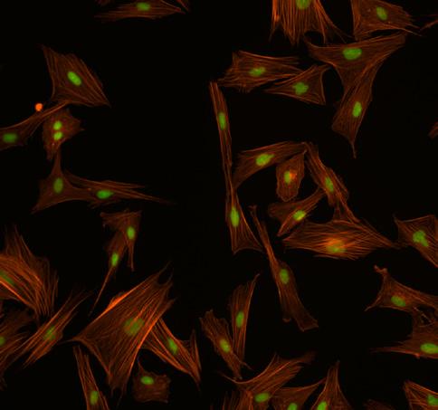
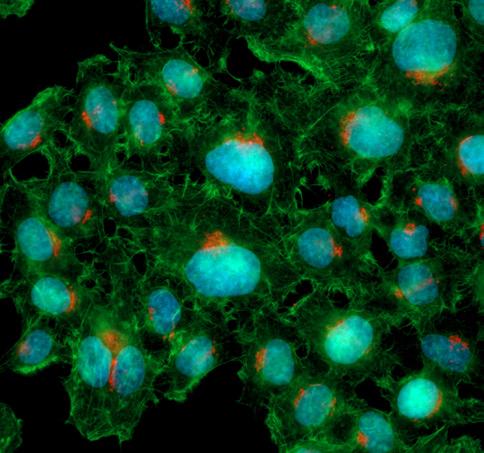
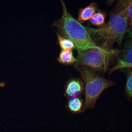
Fixed cultured HeLa cells
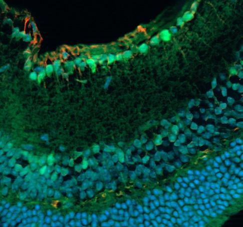
Fixed mouse retina section, acquired with ZEISS Apotome.2. Specimen courtesy of S. Nan and P. Heiduschka, Department of Ophthalmology, University Medical Center Münster, Germany.
Simpler. More Intelligent. More Integrated.
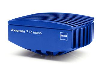
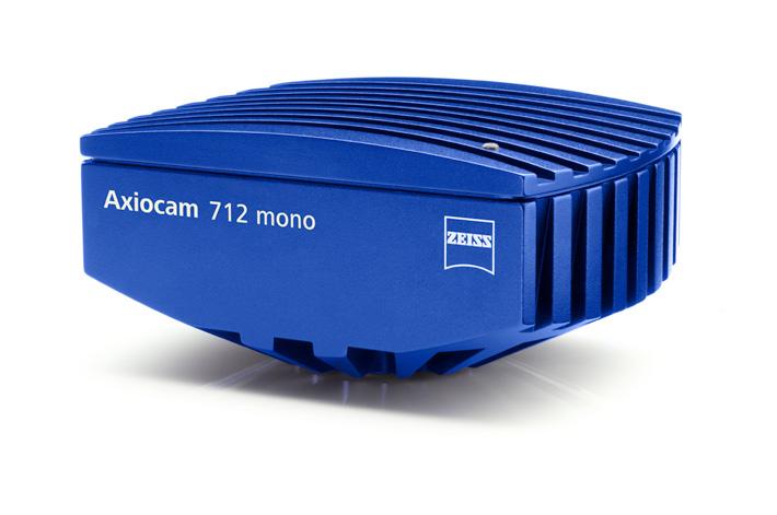
ZEISS Axiocam 712 mono
Your Flexible 12 Megapixel Microscope Camera for Fast High Resolution Imaging of Large Specimen Areas
This camera brings lowest noise and high quantum efficiency for those applications that require highest sensitivity.
Combining a large sensor with an abundance of small and sensitive pixels makes your Axiocam 712 mono a very flexible camera, suitable for countless different applications. The actively cooled CMOS sensor offers lowest readout noise and stable operation over long periods of time. Exposure times can range from 100 µs for the most dynamic specimens up to 60 s for detection of the dimmest signals. This camera delivers more than 20 frames per second at full pixel count and goes up to more than 100 frames per second with a reduced pixel count. Peak quantum efficiency of over 72 %, a broad detection spectrum and a high near-IR sensitivity complete the camera’s set of excellent features. That makes Axiocam 712 mono your all-in-one tool for monochrome imaging applications, ranging from imaging of large sample regions and dynamic specimens to high-sensitivity microscopy of fragile fluorescent specimens.
Recommended for
• High-resolution fluorescence microscopy
• Large region imaging
• Research
• Live cell imaging
• Macroscopic imaging
More Intelligent. More Integrated.
• 12 megapixel cooled global-shutter CMOS sensor
• Large sensor for extended field of view
• Wide sensitivity spectrum 350 nm – 1000 nm
• 20 frames per second in full 12 megapixel resolution
• 30 frames per second of the entire field of view in live image mode
• Low readout noise and analogue signal amplification
• Exclusive noise inhibition technology for lowlight imaging
• Dynamic range of 1:25,000 in high-dynamic range (HDR) mode
• Small 3.45 µm pixels for high-resolution imaging
• Hardware triggering
Spectral sensitivity
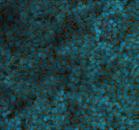
Polarized CACO-2 cells, filter-grown for two weeks. Specimen courtesy of C. Hartmann and K. Ebnet, Center for Molecular Biology of Inflammation, Institute of Medical Biochemistry, WWU Münster, Germany
Fixed mouse retina section, acquired with ZEISS Apotome.2. Specimen courtesy of S. Nan and P. Heiduschka, Department of Ophthalmology, University Medical Center Münster, Germany.
Simpler.
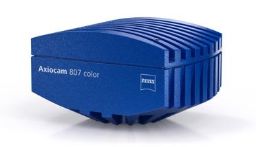
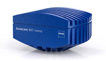
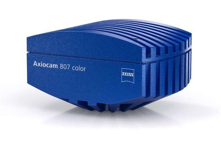
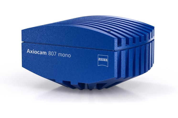
ZEISS Axiocam 807 mono
Your Fast, 7 Megapixel Microscope Camera for Live Cell Imaging of Large Fields of View
With its 17.6 mm diagonal, 7 megapixel CMOS sensor, Axiocam 807 mono allows acquisition of large fields of view with a single shot, resolving every detail of your specimen. The frame rate of 73 images per second at full sensor resolution captures even the fastest processes.
Distortion-free imaging is guaranteed by the global shutter technology. In combination with its high peak quantum efficiency of 78% and low readout-noise, high signal-to-noise ratios are ensured even at low light conditions.
Recommended for
• Fast imaging of dim fluorescent signals with a good signal-to-noise ratio
• Fast tile scanning applications
• Live cell imaging
• Flexible setups with varying applications
• 7 megapixel CMOS sensor with global shutter technology
• Large sensor with 17.6 mm diagonal for extended field of view
• Wide sensitivity spectrum from 350 nm to 1000 nm
• 73 full-resolution images per second
• High-quality noise inhibition technology and 78 % sensor quantum efficiency for lowlight imaging
• 4.5 micron pixels for optimal resolution
• Global shutter architecture for distortion-free images
• Reproducible image quality due to active thermal stabilization of the sensor
• Robust, very fast and easy-to-use dual USB 3.0 connection
• Hardware triggering
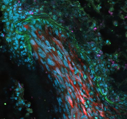
Murine lung tissue with tumor metastasis. Sample courtesy of H. Ishikawa-Ankerhold, Walter-Brendel-Zentrum für Experimentelle Medizin München, Germany
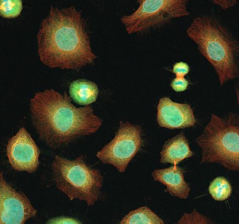
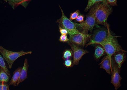
Optical section of mitotic cells created with ZEISS Apotome. Cells have DNA stained with Hoest33342, Aurora B with Alexa 488 und Tubulin with Alexa 568
Simpler. More Intelligent. More Integrated.
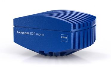
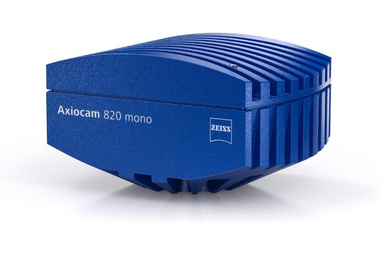
ZEISS Axiocam 820 mono
Your Sensitive 20 Megapixel Microscope Camera for Demanding, Large Field of View, Fluorescence Applications
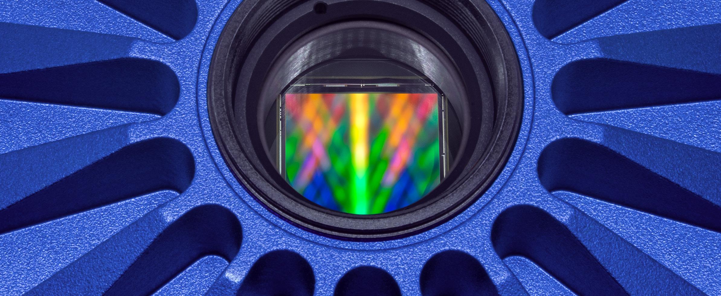
Axiocam 820 mono 20 megapixel camera is equipped with a large 17.5 mm diagonal back-illuminated CMOS sensor having a peak quantum efficiency of 86 %. In combination with its low readout noise, this camera is the perfect choice for imaging faint fluorescence signals in living or fixed samples. Its square sensor most efficiently utilizes the optical field of view of the microscope for capturing more details.
The dual USB 3.0 interface enables the highest acquisition speeds without sacrificing the robustness of a standardized interface. The high performance and unmatched flexibility of Axiocam 820 mono camera makes it the perfect choice for the most challenging samples in life science research.
Recommended for
• The most demanding fluorescence applications in life science research
• Imaging dim fluorescent signals with a good signal-to-noise ratio
• Fast tile scanning applications
• Low light imaging for the best sample protection with live cell samples
• Compatible with the most flexible setups for varying speed, resolution, and sensitivity settings
• 20 megapixel back-thinned square CMOS sensor with 17.5 mm diagonal
• 28 full-resolution images per second
• 30 frames per second of the entire field of view in live image mode
• Small 2.74 micron pixels for resolving the finest details at all magnifications
• High 86 % QE by back illuminated sensor architecture
• Low readout noise of 1.3 e- by high-quality noise inhibition technology for low light imaging
• Wide sensitivity spectrum from 350 nm to 1000 nm
• Fast read-out with global shutter architecture for distortionfree images
Spectral sensitivity
• Reproducible image quality due to active thermal stabilization of the sensor
• Robust, very fast and easy-to-use dual USB 3.0 connection
• Hardware triggering
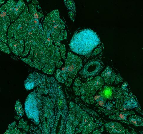
Mouse pancreas tissue stained for DNA (blue), insulin (green) and glucagon (red) on pancreas islet. Sample courtesy of A. Feuchtinger, Helmholtz Zentrum München, Germany
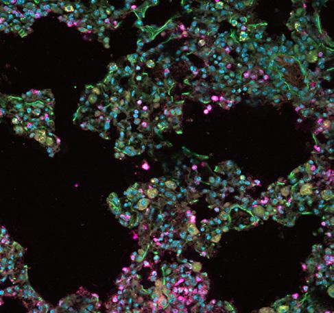
Murine lung tissue with tumor metastasis. Sample courtesy of H. Ishikawa-Ankerhold, Walter-Brendel-Zentrum für Experimentelle Medizin München, Germany
Simpler. More Intelligent. More Integrated.
Knowledge Base
Your Resource for Camera Terminology
Learn about fundamental terms of camera technology and their meaning. See how sensor type, resolution, frame rate and sensitivity are interconnected and influence your results.
Sensor Size vs Camera Adapter vs Field of View (FOV)
Use a c-mount camera adapter to mount your camera onto your microscope. Depending on the magnification factor of the adapter, the camera’s sensor may cover more (lower
magnification) or less (higher magnification) of the image coming out of your microscope (intermediate image). Typical intermediate image sizes are 25 mm for Axio Imager, 23 mm for Axio Observer or 23 mm for Axio Zoom.V16.
Typical image sensor diameters are 7.9 mm (1/2″ format), 11 mm (2/3″ format) or 16 mm (1″ format).
Different C-mount adapter magnifications are 1×, 0.63×, 0.5×.
Using a lower adapter magnification such as 0.63× causes into a:
• Demagnification of the intermediate image, resulting in a larger field of view for the final image
• Enlargement of pixel size, thus increasing light intensity detected by the sensor
• Enlargement of pixel size, which reduces the effective camera resolution
Resolution
The spatial resolution of a digital camera is related to the pixel density, which is defined by the pixel count per sensor area. The smaller the pixel aperture, the finer is the sampling of the presented structure. The reproduction of fine structures (lines) requires at least two pixels per structure sequence (line pair).
Depending on the spectral composition of the signal, the optical resolution of color cameras can be slightly lower compared to monochrome cameras because of the color filter array. However, elaborate interpolation algorithms allow color cameras to provide optimal image quality.
The unit cell size can be estimated by taking the geometrical length (height) of one sensor line (column) and dividing it by the number of all pixels in one line (column).
Effects of pixel size:
Smaller pixels are
• good for higher resolution
• lower in dynamic range
• less light sensitive
• noisier
Larger pixels are:
• good for better light sensitivity
• less noisy
• higher in dynamic range
• reducing the spatial resolution
The best pixel size is a balance between sensitivity (larger pixel) and resolution (smaller pixel) to get the best possible compromise for the imaging requirements at a given optical setup.
Name of Effect Related Limitation Counter Measure
Dark current
Spurious signal by thermally generated electrons inside the sensor silicon material.
Maximum exposure time, Low light sensitivity, dynamic range, single pixel defects (hot pixels)
Given for a specific sensor-technology, active thermo-electrical cooling
pixel is the smallest effective area on the sensor which is to become
This signal varies from pixel to pixel and causes an exposure time dependent signal offset for each individual pixel.
In addition it contributes to the signal noise.
Readout noise
Noise added to the signal during read-out
Photon shot noise
Physical property of light, proportional to square root of produced electrons
ADC effects
Differential and integral linearity effects, quantitation errors of Analog to Digital Converters
Static sensor artefacts
Defective pixels, non-uniformity effects of photo response, dark current, dark offset, electronic glow, hot pixels, column or row offsets, black offset non-uniformities
Dynamic sensor artefacts
Blinking pixels, hot pixels, pixel and line offset flicker effects, electro-magnetic crosstalk of high frequency interference effects, etc.
Low light sensitivity, directly limiting the potential low light detection threshold, dynamic range Sensor design and analog signal management dependent, signal amplification by EMCCD architecture
Detection precision at high intensity levels, noisy, low light images
Detection precision, intensity errors
Visible cosmetic defects, fixed patterns in image overlaying image information
Visible cosmetic defects, traveling overlaid patterns in image, subsequent artefacts in multi channels or Z-stack images causing errors in 3D renderings, errors in post processing algorithms like segmen tation, counting, etc.
Theoretical and practical limit of detection is absolute, therefore no direct countermeasures
Use of good ADCs, use more bits than needed, software calibration algorithms
On the fly processing of the image data with correction algorithms, black reference, pixel wise dark current maps, use of selected sensors, Correction by calibration of static effects, dead pixel storage memory in camera
High quality electronic design, electronic shielding, high quality cables and connectors, on the fly dynamic correction algorithms, selection of high quality components, high quality sensors and dark current calibration.
The pixel size defines the resolution.
One
Even when the pixel count is the same, the image taken with the larger-sized pixels is less noisy because the CCD sensor is larger.
Noise
Noise in a digital camera is a random fluctuation of the image signal which causes a detection error. Noise can come from various physical sources and it limits the detection capability of a given camera. Post-processing algorithms can be used to minimize noise, but this sacrifices other image factors such as resolution.
Sensor Cooling
Cooling is used to minimize the thermal generation of electrons (dark current) in the sensor silicon material and the resulting dark current noise. You can reduce the dark current by approximately a factor of two by lowering the sensor temperature by 7 °C. Active thermo-electrical cooling prevents the sensor from being heated by the power dissipation inside the camera electronics.
Description Explanation Advantage Disadvantage
Analog Gain Amplification of the analog voltage signal at the output of an image sensor before the Analog-Digital-Converter (ADC)
• Increases the brightness impression of the signal
• Needed to optimally adapt the analog signal output from the camera sensor to the input range of the Analog-Digital Converter (ADC) within the camera electronics
Special case: in the case of a bottleneck from the ADC input range analog gain can be used as sensitivity improvement
EM-Gain Electron Multiplication-Gain. Dedicated on-chip high-voltage acceleration stage
• Compensation for read noise limitation real detection improvement of low light image signals
• In combination with back thinning technology and large pixels providing best possible low light sensitivity
• Standard case: when the ADC can handle the full signal amplitude of the sensor no sensitivity improvement can be achieved by analog gain
• Images look very noisy
• Reduction of available intra-scene dynamic range
Heat sink for
Digital Gain Multiplication of the digital pixel value by a numerical factor
• Mathematical way to increase brightness
• Commonly used for adapting different intensities to display different fluorescence channels in a multichannel image
• Image affected by new noise source random bright pixel events minimization of EM-gain required
• Gain efficiency affected by ageing limited durability of EM gain
• Reduction of available intra-scene dynamic range
• No increase in detection sensitivity
• Histogram representation affected gaps in the histogram data
• Reduction of available intra-scene dynamic range
Cooling requires a heat sink to dissipate power from the thermo-electrical cooler itself. Additional measures are needed to prevent condensation from humidity on the cold sensor surface. Modern sensors show a vastly reduced amount of dark current compared to devices from the past.
Exposure time shorter = faster:
Extremely low temperatures – say, –20 °C – are not always required. Cooling is still unavoidable for EMCCD cameras, due to their specific working principle. All other camera technologies have a benefit by cooling only at long exposure times (after some 30s and more), when the low dark current sums up and gets disturbing again.
Binning
Camera sensitivity can be increased by combining photo generated signal charges from neighboring pixels during read-out. This also increases the camera frame rate. One side effect is the loss of image resolution. Binning factors can range from 1 × 1 (no binning) up to multiple pixels such as 5 × 5. Multiple charge binning is mainly available for CCD sensors. Binning in CMOS camera sensors is traditionally done in the digital domain by adding neighboring pixel values, which gives no extra sensitivity.
Frame Rate
The frame rate of a digital camera denotes the number of images which can be delivered per second (fps = frames per seconds). Unlike TV cameras, scientific cameras are not limited to standard video frame rates. Digital camera frame rates depend on various parameters:
Exposure time limits the absolute frame rate independent from all other technical factors. If the time to collect photons lasts for 100 ms, the maximum achievable frame rate is 1 / 100 ms = 10 fps.
Total time to readout: accumulate photon signal+ conversion into a digital signal + transmission to a PC.
Exposure and Readout correspond to a full cycle of an image acquisition.
Pixel count less = faster:
The more pixels, the longer the readout cycle, the slower the frame rate.
The interface bandwidth can become the bottleneck if the pixel count cannot be transferred within the sensor readout time.
Sensor sub frame region of interest smaller = faster:
Definition of sensor sub areas (ROI) help reduce the amount of transmitted image data
frame rates can be increased,
Prerequisite: exposure time is shorter then readout time of ROI
Bandwidth of digital interface higher = faster:
Data transfer capacity of the interface. Effective USB 3.0 bandwidth is approximately 320 Mbytes / s.
Parallel readout architecture of CMOS sensors more = faster:
CMOS sensors exceed the frame rates of comparably sized CCD sensors due to significantly more parallel output structures on the sensor.
The interface bandwidth is more likely to be the data transfer bottleneck.
Trigger signal synchronization
Synchronization of external trigger components with image acquisition
reduction of the maximum achievable frame rates with improvement of precision.
Overlapping readout and exposure Special optimization for fast time series acquisition (fast time-lapse) without switching external components
overlap of exposure event while readout of the previous image.
Only if exposure time is longer than readout frame rate limited by exposure time.
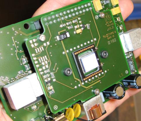
Thermo electrical cooling helps to minimize dark current effects of CCD and CMOS image sensors.
Peltier cooler (white plate), mounted on heat sink
Sensor Socket
cooler, connected with housing for dissipation
Hot Pixel
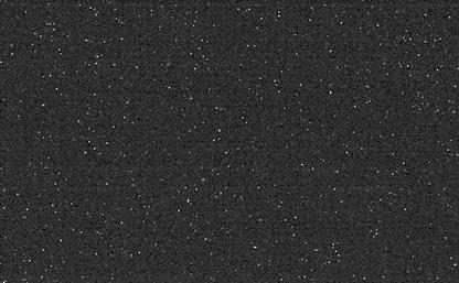
Cosmetic sensor defects are caused by a local emission of electrons in the sensor material. Hot pixels are visible as static single bright pixels against the black background. Their intensity varies widely and scales with exposure time and sensor temperature. The signal cannot be differentiated from photon generated electrons. If the sensor is temperature stabilized, the dark current can be compensated for by subtracting the spurious signal in correspondence with exposure time. Saturated pixels need to be interpolated because image information in these pixels is lost and cannot be reconstructed. Cosmic radiation can induce new hot pixel defects over time.
Spectral Sensitivity / Quantum efficiency
All kind of light detectors show a wavelength dependent light sensitivity. The conversion efficiency is the ratio of incoming photons to generated signal electrons stated as a percentage.
Detection range of silicon based sensors like CCD or CMOS can stretch from approximately 350 nm up to 1000 nm, with a peak between 500 nm – 600 nm. For detection of radiation outside of this spectral range, other materials need to be used.
Modern front illuminated devices offer a typical QE in the range of 70 %. Monochrome peak QE can be improved with back thinned technology by up to 95 % in peak.
The spectral sensitivity of color cameras is lower than monochrome cameras. The color filter dyes on the pixels reduce the peak spectral QE by approximately 15 %. Color cameras also need an IR filter as color is only defined in the visible spectrum.
Color Temperature
White Balance
The color of the illuminating light source influences the color of an object. The relative intensity of the color channels of a color camera needs to be adjusted to assure a neutral color reproduction. For this, you will need a manual or automatic selection of a neutral (grey) point in the image. Fine-tune the color reproduction by assigning slightly shifted target values for the neutral point. Adjust the color temperature of the monitor (i.e. 3,200 K) to reach the desired color reproduction.
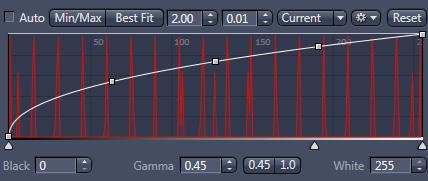
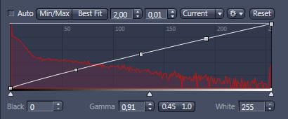
Display Curve
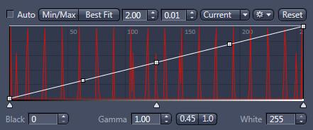
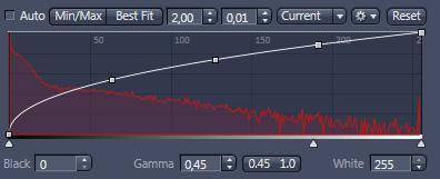
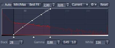
The image display curve is a powerful tool in ZEN imaging software, used to define how image data is displayed on a computer screen without changing the raw image data. Use this tool to adjust dark areas of your image visually by selectively changing the curvature or the steepness of the curve. Shift the minimum or maximum points to allow for the limitation of the visualized intensity range. The color rendition can be influenced by a Gamma curvature. Image characteristics are applied to the image data, if the image gets exported into non .CZI image formats.
Color temperature is a temperature value (in Kelvin) of a light source and is used to describe the spectral characteristic of the corresponding spectral emission. It indicates the color impression of a light source: lower temperatures are more red, higher temperatures are more blue.
The color temperature of the light influences how the human eye perceives color.
ZEISS Axiocam
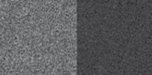
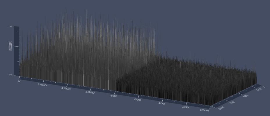
Left: Dark background, non uniformity from common CMOS sensor at 10s, Right: ZEISS Axiocam 702 mono with modern CMOS sensor at 10s with very dark background, low non uniformity
CMOS Sensor with high dark current, 10s, 4 °C ZEISS Axiocam 702 mono, 10s, 15 °C
CMOS sensor with long exposure times
ZEISS Axiocam 702 mono
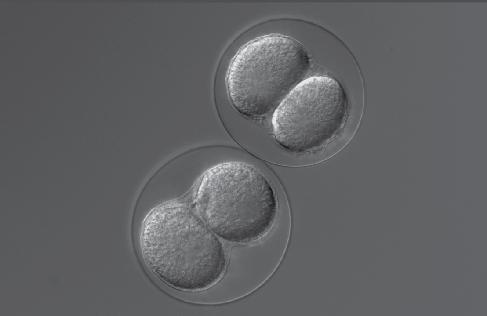
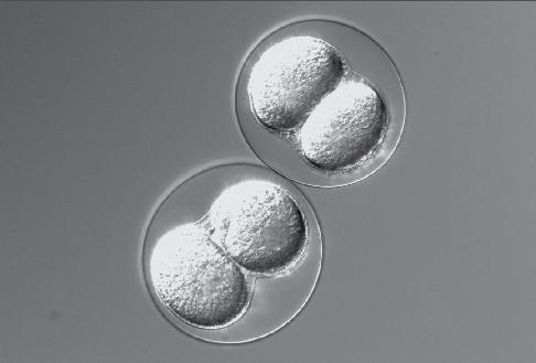
A nearly linear Gamma over the whole dynamic range delivers a rather dark display of this transmitted light image.
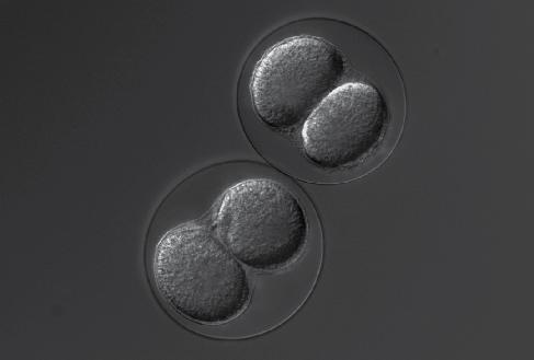
As seen here, a nonlinear Gamma curvature in the range of 0.45 over the whole dynamic range often delivers good results for transmitted light images.
The same image displayed with a steep display curve – cutting away some of the dark and bright information – shows too much contrast.
Dynamic Range
The available range of measurable intensities within one single image can be computed as the ratio between the brightest and the dimmest point in an image.
Maximum range is the difference between the saturation of the sensor (full well capacity) and the noise floor (read noise).
Sensor Technology Explanation Advantage
CCD “Charge Coupled Device”, Proven reliable technology with a long history of optimization. Stable technology and quality
For example, full well 15,000 e⁻/ read noise 6 e⁻ = 2,500 resolvable intensity values in one image. In this case, a 12 bit analog-digital converter (ADC) is necessary to properly display these values.
High sensitivity, good dynamic range, very homogenous image quality, low number of image artefacts,
Usable for long exposure times with cooling.
Minimum amount of post-processing needed,
Global shutter architecture for simultaneous acquisition,
Front illuminated and back illuminated solutions,
Wide selection of pixel counts and pixel sizes available, different architectures (Interline global shutter, frame transfer)
CMOS “Complementary Metal Oxide Semiconductor” Successor of CCD technology, recent breakthrough for mass production of quality products, currently high innovation rate
Advantages of Monochrome Cameras for Fluorescence Applications
Monochrome cameras are better suited for fluorescence imaging than color cameras due to multiple reasons:
Feature Explanation
Spectral
Sensitivity Range Full spectral range of the silicon, effective range from 350 nm up to 1000 nm due to for the lack of an IR blocking filter.
Absolute QE Higher quantum efficiency of +8 % up to +30 % depending of the wavelength, due to no color filters on the pixels.
Spatial Resolution Higher spatial / optical resolution, since there is no color filter pattern on the pixels. With a color camera, the pixels are 25 % red, 25 % blue and 50 % green. With the monochromatic signal from fluorescence, only a fraction of these pixels is then stimulated, and is thus less efficient.
Advantages of Monochrome Cameras for Fluorescence Applications
sCMOS “Scientific Complementary Metal Oxide Semiconductor” High end CMOS
Products with broad range of different quality and performance levels.
Fastest image readout due to massive parallel readout architecture, highest dynamic range, high light sensitivity, rolling and global shutter technology available, high quality mass production technology, sensor control and signal processing including on-chip ADC, wide selection of pixel counts and pixel sizes, Front illumination as standard, growing mass production of back thinned global shutter devices.
Very low average readout noise enables very good low light signal detection, high dynamic range, high frame rates possible, sensor control and signal processing including on-chip ADC, large field of view
sCMOS Back Thinning High end CMOS technology with back thinned technology for higher QE
Further improved sensitivity by higher QE up to 95 %
External driver electronics and ADC required, Relatively high heat production from external support circuitry, Limited readout speed due to charge transport mechanism, Speed limitation due to architecture
EMCCD Electron multiplication CCD, Back thinned Frame Transfer CCD with dedicated structure for amplification of photo generated electrons
Highest available detection sensitivity with semiconductor imagers, best choice for super low light imaging requirements, amplification architecture is built to skip the read noise limitation for detection of lowest signals
Limited range of usable exposure time, massive post-processing of image data due to high amount of non-uniformities and cosmetic defects, Widely used rolling shutter architecture can cause geometrical distortions from moving objects. Charge binning feature is not commonly available.
Only rolling shutter, mandatory postprocessing of image data due to nonuniformities and cosmetic defects, limited exposure range, cooling required due to high dark current, blinking pixel noise, extreme bandwidth requires dedicated interface technology, i.e. camera link currently no charge binning feature
Expensive, low volume manufacturing, Only rolling shutter architecture, mandatory post-processing of image data due to non-uniformities and cosmetic defects, limited in maximum usable exposure time, cooling required to suppress higher dark current, new type of noise, currently no charge binning feature
Low resolution, low pixel count, Possible artefacts due to frame transfer architecture, limited dynamic range, ageing effect of on-chip amplification structure, deep cooling mandatory for correct function, very high pricing
Technical Data
Color Cameras
30 fps at 4K/1080p (H.264) USB 3.0: 30 fps at 4K/1080p (MJPEG) HDMI: 30 fps at 4K/1080p Not recommended for timelapse imaging in ZEN
30 fps at 5 MP 67 fps at 1920 × 1080 (ROI in HD format) 136 fps at 512 × 512 (ROI)
up to 430 fps at 1020 × 120
Live 25 fps 5 MP (values only for monochrome or fast color modes),
Live 30 fps at 1920 × 1080 60 fps at 2464 × 2056 (5MP) 115 fps at 1920 × 1080 (HDTV format)
436 fps at
Technical Data
Monochrome Cameras
Analog Gain
Frame rate live image / Time Lapse Recording Live Ethernet: 30 fps at 1080p (H.264)
USB 3.0: 30 fps at 1080p (MJPEG)
HDMI: 30 fps at 1080p Not recommended for Timelapse imaging in ZEN Live 19 fps at 6 MP 19 fps at 2752 × 2208; 32 (ROI in HD format) 33 fps at 917 × 733; 51 fps at 550 × 440
30 fps at 2.4 MP time lapse: 128 fps at 1920 × 1216 210 fps at 1929 × 720 534 fps at
30 fps at 2048 × 1504, Live 20 fps at full frame 23 fps full frame
fps at 1920 × 1080 (HDTV) up to 430 fps at 1020 × 120
at 30 fps at 7 MP
fps at 3216 × 2208
fps at 1920 × 1080 (HDTV)
fps at 1608 × 1104 487 fps up to 1920 × 256
Applications
Cameras
Monochrome Cameras
Service and support for your ZEISS microscope system.
ZEISS moments are about passion. It is this passion with which we service and optimize your ZEISS microscope and keep it at the latest state of the art, so that your work can systematically lead to success.
Experience service that lives up to its name.
Your microscope system from ZEISS is one of your most important tools. For over 160 years, the ZEISS brand and our experience have stood for reliable equipment with a long life in the field of microscopy.
You can rely on us to ensure that you can always use your microscope’s full performance. With repair services and spare and replacement parts, our skilled ZEISS service team makes sure that your microscope is always ready for use. Our experts keep on working even after you have chosen ZEISS, with a wide range of additional services to ensure that you can experience those special moments –those special moments that inspire your work.
Maintenance and optimization
Your ZEISS Protect service agreement provides all-around security for your microscope system. There are no unexpected operating costs, and the availability of your system is increased. With preventative maintenance as a fundamental part of the service agreements, you benefit from optimized system performance. We’ll work with you to select the service package that best meets your needs, that corresponds to the equipment that you have, and that is tailored to the specific requirements of your applications.
Enhance your microscope system
Your ZEISS microscope is designed to be future-proof. Open interfaces allow you to extend your system. You can add your choice of accessories to keep up with the state of the art and thus extend your microscope’s useful life.
We would be happy to help you to find which accessories are available for your microscope that ideally match your application.