45 minute read
Foreword to Cryostasis Revival by Gregory M. Fahy, Ph.D


Foreword by Gregory M. Fahy, Ph.D.
Dr. Greg Fahy is a complex systems cryobiologist and the Executive Director and Chief Scientific Officer of the cryobiological biotechnology company, 21st Century Medicine, Inc. His scientific research has been recognized by the Society for Cryobiology, which made him a Fellow in 2014.1 His comments are his own and do not reflect the views of any affiliated organization.
Advertisement
A Glimpse of the Future
This is a book that is ultimately about the technical feasibility of cryonics. Cryonics is the practice of preserving individuals at cryogenic temperatures today for possible revival in the future. Human cryopreservation could in theory have several uses, ranging from the exploration of the cosmos in the distant future to “medical time travel” in the present. Regarding the latter, if medical conditions that are incurable today, including even the “medical condition” of being preserved by today’s imperfect methods under often imperfect circumstances, will become curable in the future, then putting the course of today’s fatal illnesses on hold by cryopreservation may provide hope for those who are deemed to be beyond hope by today’s medicine.2
But “if” is a large word in this context. Without a clear answer, cryonics has been inevitably controversial. In the meantime, in the absence of being able to foretell the future, cryonics has historically been defended on the basis of two fundamental empirical observations.3
The first observation is the fact that humans have been creating more and more advanced technology, and a deeper and deeper understanding of the world, since our inception as a species, and that this creative process of discovery and improvement is likely to continue. Carried to its logical conclusion, it seems inevitable that, if the species survives and progress is allowed to continue indefinitely, we will eventually acquire the ability to accomplish any goal that does not violate physical and economic law.
The second observation is that cryobiology, the study of the effects of low temperatures on living systems, teaches us that viable mammalian cells can be stored for very long times at cryogenic temperatures and then revived successfully in a distant future. For example, present estimates suggest that it would take 32,000 years for background radiation to kill 90% of cultured cells after freezing to low temperatures.4 Neurons in the brain are less sensitive to radiation damage than tissue culture cells and so might last longer, and storage times may also be extended by enhanced post-thawing DNA repair alone and by the presence of very high concentrations of cryoprotective agents, which are used in ice-free cryopreservation by vitrification5 and may be radioprotective.6 And even longer survival times have been claimed under natural conditions.7
Potentially tens of thousands of years is a long time for technological achievements to accumulate. Even today, it seems apparent that advances in all areas of science and technology are proceeding at exponential rates. Some have even projected the arrival of a “singularity,”8 which is a point in time beyond which progress becomes so transformational that it is impossible to imagine what it will mean for our experience of life, just decades from now.
So it does seem relevant to ask: are the tasks that would be required to revive individuals after previous cryogenic storage with present imperfect techniques forbidden by physical law? The answer depends on two factors: how much and what kind of damage must be repaired, and what repair capabilities are possible in principle?
The first question has been addressed experimentally, as reviewed in depth in this book. From the perspective of cryobiology, there are some positive findings, but also still many unknowns and limitations. When conventional ice crystal damage to whole organs was prevented by vitrification, a rabbit kidney was able to support life indefinitely after cooling to -130 ᵒC, rewarming, and transplantation.9 Regarding the brain in particular, which is of central importance for the proposition of cryonics, it has been found that, as Freitas reviews in this book, partially frozen animal brains have registered coherent electroencephalograms after thawing from -20 ᵒC,10 frozen hamsters have recovered despite conversion of more than half of their brain water into ice at -1 ᵒC,11 and both frozen and vitrified worms have retained memories after rewarming that were instilled before cryopreservation.12 Hippocampal slices, also, have been vitrified and rewarmed with good retention of viability,13 ultrastructure,14 electrical responsiveness,15 and the ability to generate long-term potentiation.16 The most direct observations that can be made, however, are those that can be made on the brains of actual human cryonics volunteers. Beyond the macroscopic studies reviewed in this book, there has been one study, whose results are presently being analyzed and prepared for publication, on brain fine structural integrity.17 Preliminary indications are that brain structure was well preserved on both the histological and electron microscopic level, and was actually preserved much better than in animal brains in past studies. Ice formation was successfully avoided in all brain biopsies examined by differential scanning calorimetry, indicating excellent perfusion of the brain after cardiac arrest and transportation, and whole
brain fracturing was not observed after cooling to -146 ᵒC. These results suggest that, when cryonics is carried out under favorable conditions, and when ice formation is prevented by vitrification, it has every appearance of preserving the structure and the molecular inventory of the brain.
However, cryonics is often attempted under poor conditions, and although the effects of disease and ischemia are reviewed in this book in detail, there is no published information on the effects of these pre-cryopreservation factors on the outcomes of preparation for cryopreservation (although some unpublished studies involving 1 hour of warm ischemia followed by 24 hours of cold storage before cryopreservation have been promising18). There are also few19 published studies of human or other mammalian brain viability after vitrification, and past successful studies involving partial brain freezing20 did not involve cooling to temperatures low enough for long term storage. Factors that may affect brain viability after vitrification (including brain shrinkage, background hypothermic injury, and possibly protein denaturation by cryoprotective agents21) have been individually addressed with success in separate model systems, but whole brain viability studies are lacking. The final reality is that, after all, it is still true that today’s methods, if applied to the cooling of either a brain or a whole mammal to cryogenic temperatures, even under good conditions, would not be expected to result in spontaneous recovery, and superimposing variable degrees of cardiac arrest prior to such experiments would make failure even more likely. In addition, preserving the brain is far from being the only problem that must be overcome to restore cryopreserved individuals to life.
Therefore, despite several favorable signs, injury induced by present cryonics procedures cannot be repaired without highly advanced future repair technologies that can be applied to cryopreserved systems. Therefore, the second question, pertaining to the ultimate feasibility of these technologies, remains essential for the evaluation of cryonics, and is fortunately the major issue addressed in this book.
Much has been written previously about the general technical feasibility of what K. Eric Drexler named “nanotechnology” (and later, “molecular nanotechnology”), which is the atomically precise engineering and fabrication of molecular machines and molecularly defined materials. Further, the offspring of nanotechnology, “nanomedicine,” has been explored in overwhelming detail in previous works by present author Robert Freitas. Ralph Merkle, too, has contributed much to an understanding of the global feasibility of molecular repair of cryopreserved brains, and other scenarios of repair have been suggested and are reviewed in depth in this volume. However, the present book is the only resource that comprehensively puts together the entire picture of nanomedical repair of cryonics patients as only Rob Freitas can do. “Conventional” nanomedicine must confront modifications to a body that is for the most part already maintaining its own viability. It is an entirely different problem to repair patients who are in a solid state at cryogenic temperatures. For that, new tools must be devised, and in this work, Freitas supplies the closest look we have had so far at what these future tools might include.
Freitas’ approach to repair proceeds in a logical fashion. First, survey the damage. Second, gain access to what needs to be repaired by tunneling through inconsequential material, particularly in the vascular system and similar conduits. Third, stabilize structures that must be repaired and install structures necessary for later repair and more detailed surveys. Fourth, figure out what needs to be done based on what has been learned. Fifth, start by stabilizing fractures and then warming sufficiently to enable fracture faces to be fused back together again. And sixth, proceed with the rest of the details (of which there are a great many). It is hard to argue with this staging of events.
Even while attempting to describe as many details of the repair process as possible, however, it must be understood that concepts of repair depend on concepts of the damage that requires repair, and, as noted above, the latter remain incomplete. To his credit, Freitas identifies a myriad of topics worthy of future research throughout these scenarios, an acknowledgement that the present work is not and cannot be the final word, but it does not need to be. Extracting and then replacing molecules like glucose and oxygen, attempting to artificially manipulate osmosis, and artificially resealing membranes with altered semipermeability may all be unnecessary. However, if they are technically possible, then it becomes plausible that lesser capabilities will be able to do lesser tasks that actually are necessary. If the real goal is to plumb the depths of the possible, the present volume accomplishes that goal extraordinarily well.
Will the overall possibilities outlined here someday be realized? Will lives be saved? Will new adventures emerge as the people of the present engage with the entities of the future? Only time will tell. The proposals in this book cannot by themselves prove that cryonics will succeed, or define precisely what conditions of preservation will be required for cryonics to succeed. The totality of the arguments presented does, however, elevate the discussion to an unprecedented level of specificity and detail, and must figure prominently in further scientific evaluation of the proposal of cryonics.
Cryonics, uniquely, is a present practice that is largely based on the possibilities of future technology, but the future has always been hard to see. Now, exactly 60 years after cryonics was first proposed in 1962,22 Freitas has provided a highly concrete glimpse into a future in which a set of definable and technically defensible future technologies could be equal to the task of repairing virtually any degree of biological injury associated with cryonics. His new comprehensive and falsifiable framework for debate and discussion may well lead to less dismissal and more serious consideration of the proposition of cryonics from this point forward. In that sense, just possibly, cryonics itself, if
not yet those who have undergone it, may be at the threshold of a new awakening. Gregory M. Fahy, Ph.D. Corona, California January, 2022
References
1. https://en.wikipedia.org/wiki/Greg_Fahy.
2. Ettinger RCW. The Prospect of Immortality. New York:
Doubleday; 1964.
3. Ibid.
4. Ashwood-Smith MJ, Grant E. Genetic stability in cellular systems in the frozen state. In: Elliott K, Whelan
J, editors. The Freezing of Mammalian Embryos.
Amsterdam: Elsevier / Excepta Medica /North-Holland 1977. p. 251-67.
5. Fahy GM, Wowk B. Principles of ice-free cryopreservation by vitrification. In: Wolkers WF,
Oldenhof H, editors. Cryopreservation and freeze-drying protocols (Methods Mol Biol 2180). New York: Humana
Press; 2021. p. 27-97.
6. Ashwood-Smith MJ. Current concepts concerning radioprotective and cryoprotective properties of dimethyl sulfoxide in cellular systems. Ann N Y Acad Sci. 1975;27:243-6.
7. https://www.livescience.com/63187-siberian-permafrostworms-revive.html?utm_source=notification.
8. Kurzweil R. The Singularity is Near: When Humans
Transcend Biology. New York: Viking; 2005.
9. Fahy GM, Wowk B, Pagotan R, Chang A, Phan J,
Thomson B, et al. Physical and biological aspects of renal vitrification. Organogenesis. 2009;5:167-75.
10. Suda I, Kito K, Adachi C. Viability of long term frozen cat brain in vitro. Nature. 1966;212:268-70. Suda I,
Kito K, Adachi C. Bioelectric discharges of isolated cat brain after revival from years of frozen storage. Brain
Research. 1974;70:527-31.
11. Lovelock JE, Smith AU. Studies on golden hamsters during cooling to and rewarming from body temperatures below 0oC. III. Biophysical aspects and general discussion. Proc Roy Soc, B. 1956;145:427-42. 12. Vita-More N, Barranco D. Persistence of long-term memory in vitrified and revived Caenorhabditis elegans.
Rejuvenation Res. 2015;18(5).
13. Pichugin Y, Fahy GM, Morin R. Cryopreservation of rat hippocampal slices by vitrification. Cryobiology. 2006;52:228-40.
14. Ibid.
15. Fahy GM, Guan N, De Graaf IAM, Tan Y, Griffin L,
Groothuis GMM. Cryopreservation of precision-cut tissue slices. Xenobiotica. 2013;43:113-32.
16. Ibid.
17. Fahy GM, Wowk B, Vargas V, Hixon H, Graber S,
Thomson B, et al. Ultrastructural cryopreservation of the mammalian brain. (in preparation).
18. Fahy GM, et al. Vitrification of whole rabbits under good and compromised conditions. (unpublished observations)
19. Pichugin Y, Marchenko VS, Shilo AV, editors. Bioelectric activity of cryopreserved brain pieces of a rabbit. The 14th Conference on Preservation of Genetic Resources; 1996; Pushchino: Academpress.
20. Suda I, Kito K, Adachi C. Viability of long term frozen cat brain in vitro. Nature. 1966;212:268-70. Suda I,
Kito K, Adachi C. Bioelectric discharges of isolated cat brain after revival from years of frozen storage.
Brain Research. 1974;70:527-31. Lovelock JE, Smith
AU. Studies on golden hamsters during cooling to and rewarming from body temperatures below 0oC. III.
Biophysical aspects and general discussion. Proc Roy
Soc, B. 1956;145:427-42.
21. Fahy GM, Wowk B. Principles of ice-free cryopreservation by vitrification. In: Wolkers WF,
Oldenhof H, editors. Cryopreservation and freeze-drying protocols (Methods Mol Biol 2180). New York: Humana
Press; 2021. p. 27-97.
22. Duhring N. Immortality: Physically, Scientifically, Now. Washington, D.C.: 20th Century Books Foundation; 1962.
Cryostasis Revival: The Recovery of Cryonics Patients through Nanomedicine
By Robert A. Freitas Jr.
The technology of cryopreservation has dramatically improved in the 50 years since Alcor’s founding in 1972. But in all that time the cryonics community has had only vague answers to the difficult question of revival. Yes, physical structures can be excellently preserved at low temperatures. But exactly how do we plan to breathe life back into our cryopreserved patients? The recently-published1 700-page technical book Cryostasis Revival was written to provide a detailed answer to this question. The processes proposed in the book make extensive use of a mature nanotechnology and represent “the first comprehensive conceptual protocol for revival from human cryopreservation, using medical nanorobots.”
The restorative methods presented in Cryostasis Revival generally involve three phases of work: (1) collecting information from preserved structure, (2) computing how to fix damaged structure, and (3) implementing the repair procedure. The first and last of these phases employ sophisticated nanorobots small enough to pass through blood vessels and other microscopic tissue corridors, as well as a nanorobotic support infrastructure called the “vasculoid” that temporarily coats the inner surface of these spaces with atomically precise machinery. The activity in the second phase is primarily computational and takes place outside of the body using an external high-performance computer and specialized software.
Ultimately, it all depends on nanotechnology.
Will Nanotechnology Work?
If a mature nanotechnology is the key to revival, how can we be sure it will exist when we need it, and will actually work when we use it? We know that molecular machines such as nanoscale bearings, ratchets, pumps, motors, conveyors, and the like exist in various forms in biological systems. And they work! Additionally, these basic molecular machines have been assembled into complex micron-scale biological devices called cells, which have many capabilities analogous to those envisioned for medical nanorobots. In turn, these molecular machines and micron-scale biological devices have been assembled into highly-differentiated macroscale systems including large organisms such as human beings. Human beings can manufacture more of themselves, thus increasing total biological productive capacity, much as is envisioned for nanofactories that will someday manufacture more nanofactories, along with medical nanorobots. Because these molecular machines already exist in biological systems, they clearly violate no fundamental physical laws.
It is also important to note that the emergence of biological systems required a continuous chain of incremental evolutionary steps that imposed very stringent design limitations on these systems (e.g., must forage for their own food, defend themselves from predators, not differ markedly from parental systems, carry their own instructions for replication, etc.). Medical nanorobots, on the other hand, can be designed de novo at any easier-tobuild point in the design space and will have far less stringent design limitations (e.g., can use optimal feedstock materials and energy conveniently supplied externally, can utilize a wider range of building materials, need no defense from predators during fabrication, has no need to self-replicate, etc.), hence can be much simpler systems than biological organisms.
As a result, we have high confidence that medical nanorobots can exist and can be simpler to design and operate than biological systems. How long might it take human technology to fabricate such complex nanosystems? Natural evolution required ~750 million years to evolve the first simple replicating cells via a ponderously slow incremental random walk through a very large design space. In contrast, human scientists can apply intelligence, creativity, selectivity, computer simulations, the physical tools of engineering, and the inspiration of a worked example (i.e., biology) to inform and vastly speed the development process. Human engineers built the first mechanical self-replicating systems in less than 750 years of effort.2 That’s a million times faster than nature required to blindly evolve the first self-replicating cells. So, how long until we have molecular manufacturing? Perhaps centuries? Possibly decades? Opinions on timing differ widely, and the development speed obviously depends on how well the effort is funded, but there are no fundamental scientific or technical reasons why it cannot be done.
Indeed, the earliest parts of this work have already been successfully concluded. Pure mechanosynthesis – the sitespecific making and breaking of covalent chemical bonds on specified individual atoms using only mechanical positioning and mechanical forces – was first demonstrated experimentally in 2003.3 It has been repeatedly confirmed over the years with various chemical elements in numerous experiments,4 debunking
early objections.5 The first detailed mechanosynthetic reaction sequences for building small diamondoid structures, validated by 100,000 CPU-hours of computational quantum chemistry simulations, were first published in 2007.6 This is the gateway to molecular manufacturing.
It’s just a question of when, and not if, we’ll have medical nanorobots. Cryopatients can afford to wait in their dewars as long as necessary for nanorobotic technology to sufficiently mature.
Recovery of Personal Identity
A crucial aspect of revival from cryostasis is the strong desire to recover the patient’s entire memory intact. Do we need to restore the patient perfectly down to the last atom, or will some lesser repair protocol suffice to preserve full personal identity? Each of us believes our mind to be a unique and enormously complex treasure house of knowledge. We might worry that even the tiniest error or omission in scanning or repairing a synaptic structure could result in some significant loss of memory, personality, or personal identity. Such concerns may be grounded in our modern experience with digital computers. In computers, it is often possible for one or a few flipped bits of data, if strategically located on a hard drive or in a software program, to produce disastrous consequences.
But there is a compelling argument that human long-term memory is vastly more robust than this. In 1986, Bell Labs scientist Thomas Landauer estimated that the average rate at which humans accumulate information into long-term memory during the normal activities of life, such as reading text or exposure to visual images, approximates 1-2 bits/sec, asymptotically approaching a stable lifetime total (integrating memory gains with losses) of ~2 x 109 bits for adults.7 This figure includes a generous allowance for motor memory – the information storage required when learning to play a piano, ride a bicycle, or perform gymnastics – and also incorporates an analysis of competing rates of both learning and forgetting. A 2 gigabit human mind is roughly equivalent to the content of a library of ~400 consciously-recallable books of text, each with 250 pages, 400 words per page, 6 characters per word, and 8 bits per character.
These 2 x 109 bits should be compared to the best current estimates of ~86 x 109 neurons in the average human brain,8 ~2 x 10¹⁴ synapses in the adult human neocortex,9 and ~106 protein molecules per synapse.10 While neurons and their synapses clearly perform many tasks unrelated to long-term memory storage, it would appear that up to ~43 neurons, ~100,000 synapses, and ~1011 protein molecules may be associated with each single bit of experienced, recallable, usable human memory. If longterm memory is truly this super-redundant, then it seems highly unlikely that the random loss of a single neuron, or the random corruption or misrepair of thousands of synapses or millions of proteins, could flip the associated single bit from “1” to “0” and destroy the tiniest piece of mind. This apparent robustness of the structures embodying long-term memory is consistent with the observation that human long-term memory persists over periods of decades despite a turnover rate of ~0.7%/hour for synaptic proteins – a half-life of only 2-5 days.11 Such turnover means that every few days, on average, roughly 1 out of every 20 proteins in every synapse is replaced with a new protein incorporating at least one random peptide sequencing error12 – yet memory and personal identity persist, in many cases over a lifetime.
As reviewed at length in Cryostasis Revival, the physical dimensions of almost all significant dendritic features and synaptic structures seem to be larger than ~100 nanometers (~0.1 micron) in size. Many of the smaller subcomponents composing these features and structures are generic or can be inferred (a) from the patient’s DNA; (b) from neuronal connectivity patterns; (c) from synapse type or size and shape,13 indirectly evidencing relative synaptic strengths; (d) from general knowledge of subcellular structures of specific types; and from other means. Future research will determine if there are any sub-100 nm-scale or molecular-scale structures that might need to be precisely scanned and precisely repaired in order to recover personal identity. This is a key point for revival, since it appears that nonmolecular and relatively non-invasive scanning methods can be used to map a cryopreserved body and brain down to ~100 nm resolution.
If research confirms that most or all surviving personal identityrelevant structures can be restored using scans at ~100 nm resolution to plan and execute the repairs, and if a patient receives a “good” (i.e., thoroughly vitrified) cryopreservation, then revival may be successfully accomplished using a relatively less expensive and less complex method called conventional cell repair (“Plan A”) which is entirely nondestructive and only moderately invasive. This method should recover all of the connectome and most of the synaptome as well.
On the other hand, if the identity, number, and location of structures smaller than ~100 nanometers is determined to be essential to recover personal identity, and if these structures are so damaged by cryopreservation that the repair process requires detailed sub-100-nm knowledge of them in order to infer and restore the correct original state, then conventional cell repair likely will not suffice and a fully invasive molecular scan and molecular reconstruction (“Plan B”) may be required for the revival of cryonics patients. This process might also be required in cases of extensive damage or extremely poor cryopreservation.
Plan A: Conventional Cell Repair
Conventional cell repair relies on nanorobotic systems that are deployed, first throughout the patient’s solid (cryogenic) body, and later throughout the “reliquidified” (~0 °C) body, without disturbing the molecular structure of tissues except to
make repairs. In this process, we scan and record all relevant physical structures to subcellular (~100 nm) resolution from within the vasculature while the patient is still in the solid state. After this information has been obtained and processed into a plan for repair, the patient is warmed sufficiently to allow rapid extraction of all metabolic and degradative molecules as cells reliquidify, quickly establishing complete biological stasis at the higher temperature. Conventional medical nanorobots14 can then be introduced to comprehensively restore at the subcellular level the cryopatient’s previously recorded (and now therapeutically editable) physical structure, over an extended period with reduced time urgency.
Specifically, this method for cryonics revival involves executing the following 15 operational steps, each of which is described in much greater detail in the book:
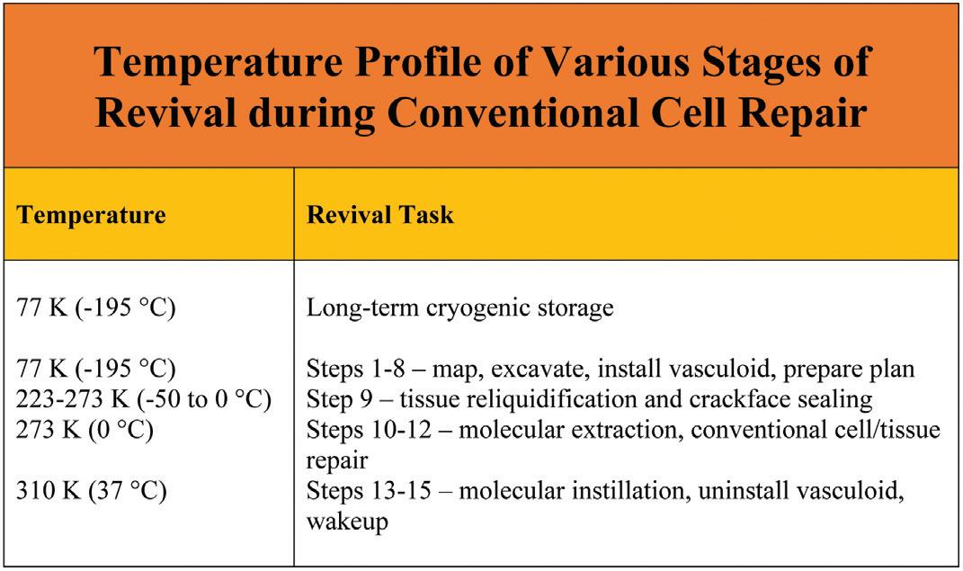
Step 1. Millimeter Vascular Scan. In a cryopreserved patient stored at ~77 K (196 °C), noninvasively scan and map all major blood and lymphatic vessels down to 0.1 mm (100 microns) in diameter.
Step 2. Large Vessel Excavation. Employ nanorobots or suitable macroscale technical means to mechanically excavate interior ice or vitrified material from all major blood and lymphatic vessels down to 0.1 mm in diameter.
Step 3. Microvascular Scans. Scan and map the blood and lymphatic microvasculatures, including all arterioles, venules, capillary beds, and lymphatic precollecting ducts, to micron resolution.
Step 4. Microvascular Excavations. Deploy nanorobots to mechanically excavate interior ice or vitrified material from all blood and lymphatic microvasculatures, all void spaces between crackfaces, all exposed perimeter surfaces of organs and other tissues, and some extracellular spaces.
Step 5. Recondition and Map Exposed Ice Surfaces.
Clear excavation debris from all exposed ice surfaces, then recondition those surfaces. Geometrically and biochemically map the reconditioned exposed ice surfaces to ~1 nm resolution, locating and identifying all vascular faults and fracture planes in crackfaces throughout the ice.
Step 6. Install Vasculoid. Install the vasculoid appliance, a mechanical ciliary transport system previously proposed15 as a means for replacing the vascular transport function in a living person. This provides rapid and reliable conveyance of nanorobots and materiel throughout the excavated vasculature of the cryopreserved human body. Vasculoid basic plates cover the lumenal walls of the entire vasculature, bridge any empty gaps across crack voids, and are installed across all major crackfaces using periodically-spaced anchors into the ice to temporarily stabilize the faces.
Step 7. Submicron Tissue Scans. Using sensor components mounted on the ubiquitous vasculoid, all tissues in which the vasculoid is embedded are scanned and mapped to ~100 nm feature resolution in three dimensions, clearly identifying most major organelles in all tissue cells and all other cytoplasmic and extracellular structures down to ~100 nm in size including neuronal synapses and boutons.
Step 8. Compute Whole-Body Repair Plan. Compile existing scan data into detailed whole-body maps covering all exposed cryogenic surfaces, vascular faults, fracture planes, tissue components to 100 nm resolution in 3D, the neural connectome, and cell plasma membrane faults. These maps are used to create data-driven computational models to plan, simulate, and direct repairs.
Step 9. Prethaw and Crackface Fusion. The cryopreserved patient is rapidly warmed to 223-273 K (-50 °C to 0 °C), producing whole-body tissue reliquidification. During the warming process, thermal stresses in the cryogenic tissue are relaxed, allowing separated crackfaces on either side of ice fractures to be drawn together by contraction of vasculoid components, closing all crackface voids.
Step 10. Molecular Extraction. Extraction microprobes equipped with pumps having molecularly specific binding sites at their distal termini (aka. “sorting rotors”)16 are inserted from the vasculoid into reliquidified tissue cells at a 2-5 micron spacing. Tens of thousands of key fuel, metabolic, intermediate, and other molecules are rapidly extracted from the cells, establishing complete biochemical stasis throughout the tissues within ~1 hour of reliquification.17 The extraction microprobes are then withdrawn from the tissues.
Step 11. Reseal Plasma Membrane Compartments and
Rehydrate. Nanorobots are released from the vasculoid to repair all cellular plasma membranes, reseal all compartments against fluid leakage, and rehydrate the cells in part via extracellular water transfers.
Step 12. Conventional Cellular and Tissue Repair. Nanorobots are employed to remove unwanted cells and microbodies,
inspect and repair (or replace) existing cells, and then perform various supplemental repair tasks on tissues and neurons.
Step 13. Patient Warmup and Molecular Instillation. The patient is warmed to normal human body temperature (310 K). Microprobes inserted into cells from the vasculoid instill thousands of essential molecules into intracellular cytoplasm and organelles, omitting only those molecules that could restart active metabolism. The microprobes are then withdrawn from the tissues. Molecules capable of initiating active metabolism are loaded into storage nanorobots that are parked intracellularly, awaiting a future signal to release their cargoes.
Step 14. Uninstall Vasculoid and Finish Repairs. The vasculoid is rapidly withdrawn from the body and replaced with a temporary blood substitute that includes nanorobots capable of supporting normal metabolic and material transport functions, e.g., respirocytes.18 The patient’s metabolism, heartbeat, circulation, and respiration are restarted as key metabolic chemicals are released from the parked storage nanorobots (which are then removed), and final neural repairs are completed. The temporary blood substitute is replaced with manufactured natural blood.
Step 15. Patient Wakeup. Anesthetic agents are removed and the patient awakens to full consciousness.
The serial revival protocol described above for whole-body patients is estimated to require 512 days (~1.4 years) of calendar repair time to complete, using reasonably conservative assumptions. If we can shift cell repair from organelle repair/ replacement to exclusively whole-cell replacement operations, and if tolerable whole-body waste heat generation can be increased from 100 watts to 300 watts, then it may be possible to reduce the total calendar time for revival from 512 days to 244 days (~8.1 months). The nominal serial revival protocol for neuro patients is similarly estimated as 66 days (~2.2 months) or 46 days (~1.5 months) under the same two scenarios, and total repair time for both types of patients might be further modestly reduced by parallelizing some or all of these serial operations. The neuro repair estimates exclude the time required to print or regrow an acephalic replacement body and then reattach it to the fully repaired formerly cryopreserved cephalon. These tasks might also be parallelized to some extent.
Note that only lethal damage will be corrected during the revival process. Nonlethal conditions ranging from medical flaws to purely cosmetic issues will not be initially corrected, largely due to lack of informed consent and prioritized limited resources for revivals. Once a patient has been restored to life, a variety of elective procedures including genomic editing, whole-body rejuvenation,19 or exotic anatomical modifications can be performed at leisure using conventional medical nanorobotics. Nanostasis. Molecular extraction as summarized above in Step 10 for cryostasis revival is a new concept that also enables true suspended animation for living patients in a process called “nanostasis” or “warm biostasis.” One of the nanostatic methods described in the book uses only medical nanorobots injected into the patient’s body. To enter nanorobotic suspended animation, the patient would be sedated, cannulated, and cooled to hypothermic temperatures, after which a fleet of ~50 trillion nanorobots would be slowly introduced into all tissues and cells. Intravascular infusion of ~2 liters of compacted empty nanorobots suspended in ~2 liters of carrier fluid would require ~7 hours at a flow rate of ~10 cm3/minute. Once in their assigned locations inside tissues or cells, and upon receiving the command to proceed, the individual storage nanorobots simultaneously pump all target molecules out of the extracellular or cytosolic spaces in which they are parked and into the robots’ internal tankage volume, executing the molecular extraction process in the ideal progressive sequence and placing the patient into a state of reversible suspended animation in ~1 hour or less. While dormant in suspended animation the unconscious nanostatic patient remains susceptible to attack by bacteria and other external parasites. Microbivore-class20 nanorobots can thwart this invasion both internally and externally to the body using devices that are powered without using metabolically active chemicals (e.g., via acoustic power). The nanostatic patient should be stored in an inert environment (e.g., pure nitrogen) to avoid exposure to oxygen or other metabolically relevant molecules that might enter the body through the skin or elsewhere. The patient should also be kept isothermal by external means since no endogenous heat will be generated other than nanorobot thermal emissions. Revival is accomplished in similar time frames by reversing the molecule intake in a carefully staged manner to redistribute all essential biochemicals to their original locations in the ideal progressive order, then extracting the nanorobots from the body in under an hour via nanorobot washout or by other means, with final revival accompanied by warmup and ACLS21 or related conventional methods of resuscitation.
Nanotechnology. Of course, the success of the proposed Conventional Cell Repair procedure critically depends on the feasibility of diamondoid nanorobotics. In the unlikely event this technology proves infeasible, then some other method of revival would be required that is beyond the scope of the present work. Finding such other methods appears challenging for reasons enumerated in the book, but it could be a valuable service to the field of cryonics if someone could identify and describe at least one viable non-nanotech path to revival in book-length technical detail.
Plan B: Molecular Reconstruction
If it is determined that individual-unique structures smaller than ~100 nanometers are essential to recover personal identity, then conventional cell repair likely will not suffice and a fully invasive molecular scan, followed by molecular reconstruction (“Plan B”), may be required for revival from cryostasis.
Cryopreserved tissue at liquid nitrogen temperatures is literally as hard as solid rock, making nanorobotic locomotion prohibitively energy-intensive. But cryogenic solid materials can be disassembled or reassembled atom by atom using the techniques of mechanosynthesis22 – the emerging technology of positionally-controlled site-specific mechanically-driven single-atom chemical reactions. In Plan B, subtractive mechanosynthesis can be used to abstract one atom (or one small chemical moiety such as a methyl (–CH3) or amino (–NH2) group) at a time from a specific site on the patient’s physical structure. Additive mechanosynthesis can be used to donate one atom (or one small chemical moiety) at a time to a specific site. Recording the identity and precise location of every atom as it is removed or added creates an atomicallyprecise map of the entire cryopreserved body. The cryopatient’s physical structure is then known to a resolution of ~0.1 nm, which is roughly 1000-fold more detailed than the ~100 nm resolution potentially available using Plan A. This is the best resolution that is physically obtainable and virtually guarantees that all available structural information is captured and retained. After the initial scan data has been processed and corrected to eliminate medical flaws, the patient’s body can be reconstructed using the corrected scan data.
The first phase of a molecular reconstruction is to extract from the body all non-tissue and other loose matter that can later be replaced with fresh material. These items are not components of the patient’s persistent physical structure and make no essential contribution to structural integrity at the molecular scale, or to memory and personal identity, hence there is no need to retain or to map them to atomic precision. Their extraction reduces the total number of molecules that must be precisely mapped and later precisely repaired or replaced. Additionally, the removal process produces a coarse mapping of all interior void spaces that can provide a guide for the more precise atomically-precise mapping yet to come.
As noted, the revival process begins with coarse mapping and bulk extraction, similar to Plan A:
Step 1. Millimeter Vascular Scan. In a cryopreserved patient stored at ~77 K (-196 °C), noninvasively scan and map all major blood and lymphatic vessels down to down to 0.1 mm in diameter.
Step 2. Large Vessel Excavation. Employ nanorobots or other suitable macroscale technical means to mechanically excavate interior ice or vitrified material from all major blood and lymphatic vessels down to 0.1 mm in diameter.
Step 3. Microvascular Scans. Scan and map the blood and lymphatic microvasculatures, including all arterioles, venules, capillary beds, and lymphatic precollecting ducts, to micron resolution. Step 4. Microvascular Excavations. Nanorobots mechanically excavate interior ice or vitrified material from all blood and lymphatic microvasculatures, and from void spaces between crackfaces.
Step 5. Organ System Excavations. Deploy nanorobots to mechanically excavate ice from the interior gas or fluid volumes of the lungs, gastrointestinal tract, urinary bladder, heart, kidney, spleen, the ventricular system of brain and spine, gallbladder, synovial fluid capsules in joints, and the aqueous humor of the eyes. These excavations are done primarily to avoid the need to process informationally redundant bulk fluids during molecular reconstruction, which would be wasteful of time, energy, and manufacturing resources. All bulk substance removed in this manner can be restored during the whole-body fluid check, either as original or freshly manufactured material according to preference.
Step 6. Clear Excavation Debris. Clear excavation debris from all exposed ice surfaces.
Step 7. Reconstruction. Once nonstructural bulk materials have been extracted from the cryopreserved patient’s body, there are two broad pathways to revival that can be followed (as detailed in a 75-page chapter in the book), depending on the philosophical preferences and financial means available to the patient:
(7.1) Destructive Scan and Molecular Reconstruction of a Replacement Body. In a destructive molecular scan, the patient’s cryopreserved body is disassembled atom by atom, the precise location and type of atom is recorded in a data file, and the atoms are discarded as the process unfolds. After the initial scan file is digitally corrected to incorporate all necessary medical repairs, a new replacement body is manufactured via 3D printing that is a near-exact copy of the original cryopreserved body, but incorporating the specified repairs. This pathway appears to be somewhat faster and less expensive than (7.2).
(7.2) Nondestructive Scan and Molecular Reconstruction of the Original Body. In a nondestructive molecular scan, the patient’s cryopreserved body is temporarily progressively separated into its constituent atoms or molecules, but only a small piece at a time, during which the precise location and type of each atom is recorded in a data file, after which the same atoms are carefully reassembled back into the original molecules, and the original molecules are reassembled back into their original positions, maintaining the original physical cryopreserved body, completely intact. At any time during the nondestructive scan, fully 99.99999% of the patient’s solid body is undisturbed while
only one thin tissue slice ~200 nm thick is being processed over a period of ~10 sec. Successive slices are then scanned in turn, resulting in an estimated 39-month total scan time. Faster processing times are available by adding additional scan slices that are simultaneously processed. The initial scan file that results from this process is then digitally corrected to incorporate all necessary medical repairs. The original cryopreserved body is then repaired by repeating the nondestructive molecular scan, this time inserting the digital corrections incorporating the medical repairs. This pathway appears to be somewhat slower and more expensive than the destructive pathway in (7.1).
A crude cost estimate for cryostasis revival using either conventional cell repair (Plan A) or molecular reconstruction (Plan B) suggests that the key driver of operating expenses is the price of the energy required to power the nanorobots and computers. The total revival cost is estimated as ~$2 million for whole-body patients using Plan A assuming contemporary electricity costs, and similarly using Plan B assuming future energy costs become 100fold cheaper than today due to widespread commercial atomically precise manufacturing. Revival costs are somewhat reduced for neuro patients compared to whole-body patients because there is much less tissue to process. However, these savings are probably offset by the cost of obtaining and attaching a substitute body to the repaired cephalon.
Validation of the Revival Process
Once we have devised an experimental cryostasis revival protocol that we think will work, how do we test it to be sure? The obvious answer: test it on animals. Cryonics revival protocols can be validated using a variety of animal models including primates. Positive results from these tests should provide sufficient technical validation to warrant approval of the same protocols for the revival of cryopreserved human patients.
The validation tests should seek to confirm the following mental functionalities:
Simple Memory. Vita-More and Barranco23 conclusively established in 2015 that C. elegans nematode worms can survive cooldown to liquid nitrogen temperature and then be warmed back to normal temperature, with their memories of a trained simple behavior intact.
Complex Memory. We can start with rodents such as rats or mice that have learned complex tasks such as how to run a maze,24 and verify that, like the worms, these small mammals remember whatever they’ve been taught, demonstrating retention of complex memories after experiencing cryopreservation followed by our revival procedure.
Personality. We then proceed to highly intelligent mammals such as dogs who have learned to recognize their owner or trainer and have been taught a large number of tricks and word associations. One border collie25 was taught to recognize the labels of over 200 different items. The dog could infer the names of novel things by exclusion learning and could correctly retrieve those new items both immediately and four weeks after the initial exposure to the items. Besides these tests of specific memories and abilities, long-time pet owners know that their canine companions can: (1) express empathy, deception, and imitation; (2) develop demonstrable personalities that reflect how they interact with owners, friends, strangers, and other animals; and (3) display characteristic unique behaviors when confronted with challenges or during play. A dog that replicated its usual idiosyncratic behaviors after experiencing cryopreservation and experimental revival would provide good evidence that the animal’s personality had survived intact.26
Personal Identity. Chimpanzees and bonobos have cognitive capacities superior to those of dogs in self-consciousness, although dogs do better than chimpanzees at using the behavior of other animals, especially humans, as a cue. The logical animal model for the final phase of cryopreservation revival testing is probably a primate, given their physiological similarities to humans and their clear demonstration of self-awareness. Ideal animal subjects may be trained primates who have been taught language skills using sign language. Kanzi, a bonobo, is believed to understand more human language (after perhaps ~8 years of training) than any other non-human animal in the world.27 When animals like these are revived from cryopreservation, they can be tested on their memory of words, their ability to perform trained tasks, and their characteristic behaviors to determine the persistence of memory and personality. More importantly, these primates could, in principle, be directly interrogated to obtain answers to questions about their internal mental state – such as “how do you feel?” and “who are you?” to test if their sense of self has survived the revival procedure.
Whole-Brain Emulation (WBE) on Animals. Merkle28 described a WBE validation procedure that would likely be available in the future era of nanorobotic revivals and could be applied to laboratory animals: “We could record every nerve impulse in the brain by embedding a sufficient number of neurobots….We could then record data from neurobots in the brain of an experimental animal before they were cryopreserved, cryopreserve them, revive them, and then record data from neurobots in the brain of the revived experimental animal, giving us two sets of neuronal data: ‘before’ and ‘after’. Comparing the ‘before’ and ‘after’ data would let us tell if we had done a good job in cryopreserving and reviving the experimental animal.”
The larger great apes – chimps, orangutans, bonobos, and gorillas – have 30%-40% as many neurons as a human brain.29 Human, chimp, and rhesus macaque neural tissues show similar adult synaptic number densities at 0.3-0.5 synapses/ micron3, varying slightly with age.30 There are a few minor neuronal differences between humans and great apes. For
example, prefrontal area 10 has greater spacing among cortical minicolumns in humans than in chimpanzees.31 The pyramidal neurons of humans have significantly longer and more branched dendritic arbors in all cortical regions than similar neurons in chimpanzees, and the human prefrontal cortex contains a greater proportion of dendrites, axons, synapses, glial cell processes, and microvasculature relative to the space occupied by neuronal and glial somata than in chimpanzees.32 Post-differentiation, human and primate cultured neurons show slightly different firing rates with time.33 But these are all relatively minor differences in size, number, spatial distribution, and metabolic rate, not fundamental differences in kind that cell repair nanorobots would likely be able to handle in primates but unable to handle in humans. If we ever discover some exclusively-human physical neurocellular feature that absolutely must be repaired, nanorobots could practice and perfect such rare repair procedures on these specific human-unique features using brain tissue samples taken from fresh human cadavers.
If comparison of a before-cryopreservation WBE with an afterrevival WBE of a large primate reveals no significant operating differences when placed in the same simulated environment, and if in vivo neurobot scans reveal no fundamental structural differences in the neurons, dendrites, synapses, and connectomes of test primates before and after the revival procedure, then it is difficult to imagine how a human brain subjected to the same recovery process would fare differently, given that the cytoarchitecture, cell type composition, and neurogenic gene expression programs of humans and chimpanzees are remarkably similar.34
These results lead to our tentative conclusion that a successful primate validation of cryonics revival protocols should be sufficient evidence to warrant application of the same protocol to human cryopreservation patients. This tentative conclusion should be vigorously explored by careful comparison of human and nonhuman primate neurological ultrastructure and brain cytoarchitecture, and should be validated, nuanced, or challenged in future research.
Of course, there are literally hundreds of future research tasks – as enumerated in the book – that must be completed before we can have a reasonable prospect of successfully bringing back the first cryonics patient. My hope is that Cryostasis Revival will inspire, focus, and motivate this important work.
References
1. https://www.alcor.org/cryostasis-revival/.
2. Freitas RA Jr., Merkle RC. Kinematic Self-Replicating
Machines. Landes Bioscience, Georgetown, TX, 2004; http://www.MolecularAssembler.com/KSRM.htm.
3. Oyabu N, Custance O, Yi I, Sugawara Y, Morita S.
Mechanical vertical manipulation of selected single atoms by soft nanoindentation using near contact atomic force microscopy. Phys Rev Lett. 2003 May 2;90(17):176102; https://link.aps.org/pdf/10.1103/
PhysRevLett.90.176102.
4. Oyabu N, Custance O, Abe M, Moritabe S. Mechanical vertical manipulation of single atoms on the Ge(111)c(2x8) surface by noncontact atomic force microscopy.
Abstracts of Seventh International Conference on
Non-Contact Atomic Force Microscopy, Seattle,
Washington, USA, 12-15 Sep 2004, p. 34; http://www. engr.washington.edu/epp/afm/abstracts/15Oyabu2. pdf. Sugimoto Y, Pou P, Custance O, Jelinek P, Abe
M, Perez R, Morita S. Complex patterning by vertical interchange atom manipulation using atomic force microscopy. Science. 2008;322(5900):413-417; http:// www.sciencemag.org/cgi/content/full/322/5900/413.
Xie Y, Ma L, Zhang P, Cai X, Zhang W, Gan F, Ning
XJ, Zhuang J. Reversible atomic modification of nanostructures on surfaces using direction-depended tip-surface interaction with a trimer-apex tip. Appl Phys
Lett. 2009 Aug 18;95:073105; https://aip.scitation.org/ doi/abs/10.1063/1.3180814. Chen C, Zhang J, Dong
G, Shao H, Ning BY, Zhao L, Ning XJ, Zhuang J. Siteselective substitutional doping with atomic precision on stepped Al(111) surface by single-atom manipulation.
Nanoscale Res Lett. 2014 May 13;9(1):235; https://core. ac.uk/download/pdf/81056816.pdf. Kawai S, Foster
AS, Canova FF, Onodera H, Kitamura S, Meyer E.
Atom manipulation on an insulating surface at room temperature. Nat Commun. 2014 Jul 15;5:4403; http:// viesti.physics.aalto.fi/~asf/publications/Nature%20
Comm%20manip.pdf. Bamidele J, Lee SH, Kinoshita
Y, Turanský R, Naitoh Y, Li YJ, Sugawara Y, Štich
I, Kantorovich L. Vertical atomic manipulation with dynamic atomic-force microscopy without tip change via a multi-step mechanism. Nat Commun. 2014 Jul 31;5:4476; https://www.nature.com/articles/ ncomms5476. Huff TR, Labidi H, Rashidi M, Koleini
M, Achal R, Salomons MH, Wolkow RA. Atomic White-
Out: Enabling Atomic Circuitry through Mechanically
Induced Bonding of Single Hydrogen Atoms to a Silicon
Surface. ACS Nano. 2017 Sep 26;11(9):8636-8642; https://arxiv.org/pdf/1706.05287.
5. Smalley RE. Of Chemistry, Love and Nanobots. Sci Am. 2001 Sep;285(3):76-77; http://bcs.solano.edu/ewylie/ nanotech/Smalley%20SciAm%20sep%202001.pdf.
6. Freitas RA Jr., Merkle RC. A minimal toolset for positional diamond mechanosynthesis. J Comput
Theor Nanosci. 2008;5:760-861; http://www. molecularassembler.com/Papers/MinToolset.pdf.
7. Landauer TK. How Much Do People Remember? Some
Estimates of the Quantity of Learned Information in
Long-term Memory. Cognitive Science 1986;10:477493; https://onlinelibrary.wiley.com/doi/pdf/10.1207/ s15516709cog1004_4.
8. Azevedo FA, Carvalho LR, Grinberg LT, Farfel JM,
Ferretti RE, Leite RE, Jacob Filho W, Lent R, Herculano-
Houzel S. Equal numbers of neuronal and nonneuronal cells make the human brain an isometrically scaled-up primate brain. J Comp Neurol. 2009 Apr 10;513(5):53241; http://www.sakkyndig.com/psykologi/artvit/ frederico2009.pdf.
9. Pakkenberg B, Pelvig D, Marner L, Bundgaard MJ,
Gundersen HJ, Nyengaard JR, Regeur L. Aging and the human neocortex. Exp Gerontol. 2003 Jan-
Feb;38(1-2):95-9; http://citeseerx.ist.psu.edu/viewdoc/do wnload?doi=10.1.1.332.5850&rep=rep1&type=pdf.
10. This assumes the average chemical synapse is ~1000 nm tall, ~400 nm in diameter, ~1 kg/L in density, and composed of ~75 kilodalton protein molecules.
11. Cohen LD, Zuchman R, Sorokina O, Müller A, Dieterich
DC, Armstrong JD, Ziv T, Ziv NE. Metabolic turnover of synaptic proteins: kinetics, interdependencies and implications for synaptic maintenance. PLoS One. 2013
May 2;8(5):e63191; https://www.ncbi.nlm.nih.gov/pmc/ articles/PMC3642143/.
12. The typical error rate in protein synthesis is ~10-4 (https:// en.wikipedia.org/wiki/Kinetic_proofreading); assuming ~1000 residues/protein implies ~10% of all new protein molecules will contain at least one misincorporated amino acid.
13. Sorra KE, Harris KM. Occurrence and threedimensional structure of multiple synapses between individual radiatum axons and their target pyramidal cells in hippocampal area CA1. J Neurosci. 1993
Sep;13(9):3736-48; https://www.jneurosci.org/content/ jneuro/13/9/3736.full.pdf. Jones DG, Harris RJ. An analysis of contemporary morphological concepts of synaptic remodelling in the CNS: perforated synapses revisited. Rev Neurosci. 1995 Jul-Sep;6(3):177-219; https://pubmed.ncbi.nlm.nih.gov/8717635/. 14. http://www.nanomedicine.com.
15. Freitas RA Jr., Phoenix CJ. Vasculoid: A personal nanomedical appliance to replace human blood. J Evol
Technol. 2002 Apr;11:1-139; http://www.jetpress.org/ volume11/vasculoid.pdf.
16. Drexler KE. Nanosystems: Molecular Machinery,
Manufacturing, and Computation, John Wiley &
Sons, New York, 1992, Section 13.2.1(a) “Modulated receptors for selective transport: Basic concepts”; https://www.amazon.com/dp/0471575186/. Freitas
RA Jr. Nanomedicine, Volume I: Basic Capabilities,
Landes Bioscience, Georgetown, TX, 1999; Section 3.4.2, “Sorting Rotors”; http://www.nanomedicine.com/
NMI/3.4.2.htm.
17. Biochemical degradative processes that would proceed in ~6 minutes (the traditional nondamaging ischemic limit) at the normal human body temperature of 310 K require ~1.8 hours to occur at the pure water-ice melt temperature of 273 K, and even longer at slightly lower temperatures.
18. Freitas RA Jr. Exploratory design in medical nanotechnology: a mechanical artificial red cell.
Artif Cells Blood Substit Immobil Biotechnol. 1998
Jul;26(4):411-30; https://www.tandfonline.com/doi/ pdf/10.3109/10731199809117682.
19. Freitas RA Jr. Chapter 23. Comprehensive Nanorobotic
Control of Human Morbidity and Aging. In: Fahy GM,
West MD, Coles LS, Harris SB, eds, The Future of Aging:
Pathways to Human Life Extension, Springer, New York, 2010, pp. 685-805; http://www.nanomedicine.com/
Papers/Aging.pdf.
20. Freitas RA Jr. Microbivores: Artificial Mechanical
Phagocytes using Digest and Discharge Protocol. J. Evol.
Technol. 2005 Apr;14:55-106; http://www.jetpress.org/ volume14/freitas.pdf.
21. https://en.wikipedia.org/wiki/Advanced_cardiac_life_ support.
22. Freitas RA Jr., Merkle RC. A minimal toolset for positional diamond mechanosynthesis. J Comput
Theor Nanosci. 2008;5:760-861; http://www. molecularassembler.com/Papers/MinToolset.pdf.
23. Vita-More N, Barranco D. Persistence of Long-Term
Memory in Vitrified and Revived Caenorhabditis elegans.
Rejuvenation Res. 2015 Oct;18(5):458‐463; https://www. ncbi.nlm.nih.gov/pmc/articles/PMC4620520/.
24. https://en.wikipedia.org/wiki/Maze#Mazes_in_ psychology_experiments.
25. https://en.wikipedia.org/wiki/Rico_(Border_Collie).
26. In the mid-1980s, bloodless deep hypothermia experiments were performed on 6 dogs, who, once revived, showed “no neurological or other deficits” and demonstrated full “preservation of memory and personality” (https://www.alcor.org/library/alcorspioneering-total-body-washout-experiments/). In one example, a dog was anesthetized, her blood removed and substituted with a pumped and continuously oxygenated solution at 4.5 °C for 4.7 hours with no signs of life; once resanguinated and rewarmed, after some complications the dog recovered and “remembered her name and her tricks.” (Darwin M. Bringing Dixie Back –
A Research Diary. Cryonics 1985 Feb;6(2):6-15; https:// www.alcor.org/docs/cryonics-magazine-1985-02.txt; see also http://www.cryonet.org/cgi-bin/dsp.cgi?msg=26438.)
27. https://en.wikipedia.org/wiki/Kanzi; https://en.wikipedia. org/wiki/Great_ape_language.
28. Merkle RC. Revival of Alcor Patients. Cryonics 2018 May-Jun;39(3):10-19 and Cryonics 2018 Jul-
Aug;39(4):10-15; https://www.alcor.org/library/revivalof-alcor-patients/.
29. https://en.wikipedia.org/wiki/List_of_animals_by_ number_of_neurons.
30. Liu X, Somel M, Tang L, Yan Z, Jiang X, Guo S, Yuan
Y, He L, Oleksiak A, Zhang Y, Li N, Hu Y, Chen W, Qiu
Z, Pääbo S, Khaitovich P. Extension of cortical synaptic development distinguishes humans from chimpanzees and macaques. Genome Res. 2012 Apr;22(4):611-22; https:// www.ncbi.nlm.nih.gov/pmc/articles/PMC3317144/.
Bianchi S, Stimpson CD, Duka T, Larsen MD, Janssen
WG, Collins Z, Bauernfeind AL, Schapiro SJ, Baze WB,
McArthur MJ, Hopkins WD, Wildman DE, Lipovich
L, Kuzawa CW, Jacobs B, Hof PR, Sherwood CC.
Synaptogenesis and development of pyramidal neuron dendritic morphology in the chimpanzee neocortex resembles humans. Proc Natl Acad Sci U S A. 2013 Jun 18;110 Suppl 2(Suppl 2):10395-401; https://www.ncbi. nlm.nih.gov/pmc/articles/PMC3690614/. 31. Semendeferi K, Teffer K, Buxhoeveden DP, Park
MS, Bludau S, Amunts K, Travis K, Buckwalter J.
Spatial organization of neurons in the frontal pole sets humans apart from great apes. Cereb Cortex. 2011
Jul;21(7):1485-97; https://academic.oup.com/cercor/ article/21/7/1485/332179.
32. Bianchi S, Stimpson CD, Bauernfeind AL, Schapiro
SJ, Baze WB, McArthur MJ, Bronson E, Hopkins
WD, Semendeferi K, Jacobs B, Hof PR, Sherwood
CC. Dendritic morphology of pyramidal neurons in the chimpanzee neocortex: regional specializations and comparison to humans. Cereb Cortex. 2013
Oct;23(10):2429-36; https://www.ncbi.nlm.nih.gov/pmc/ articles/PMC3767963/.
33. Marchetto MC, Hrvoj-Mihic B, Kerman BE, Yu DX,
Vadodaria KC, Linker SB, Narvaiza I, Santos R, Denli
AM, Mendes AP, Oefner R, Cook J, McHenry L,
Grasmick JM, Heard K, Fredlender C, Randolph-Moore
L, Kshirsagar R, Xenitopoulos R, Chou G, Hah N, Muotri
AR, Padmanabhan K, Semendeferi K, Gage FH. Speciesspecific maturation profiles of human, chimpanzee and bonobo neural cells. Elife. 2019 Feb 7;8:e37527; https:// www.ncbi.nlm.nih.gov/pmc/articles/PMC6366899/.
34. Mora-Bermúdez F, Badsha F, Kanton S, Camp JG,
Vernot B, Köhler K, Voigt B, Okita K, Maricic T,
He Z, Lachmann R, Pääbo S, Treutlein B, Huttner
WB. Differences and similarities between human and chimpanzee neural progenitors during cerebral cortex development. Elife. 2016 Sep 26;5:e18683; https://www. ncbi.nlm.nih.gov/pmc/articles/PMC5110243/.
ORDER NOW! Preserving Minds, Saving Lives
The Best Cryonics Writings of The Alcor Life Extension Foundation
“Cryonics magazine introduced me to Alcor and cryonics at its best back in 1983. The visions and technological breakthroughs that you will read about in this book continue to shape Alcor’s mission to preserve life through science.” – Max More, Ph.D. Ambassador and President Emeritus of Alcor
Cryonics is an experimental medical procedure that uses ultra-low temperatures to put critically ill people into a state of metabolic arrest to give them access to medical advances of the future. Since its inception in the early 1960s, the practice of cryonics has moved from a theoretical concept to an evidence-based practice that uses emergency medical procedures and modern vitrification technologies to eliminate ice formation.
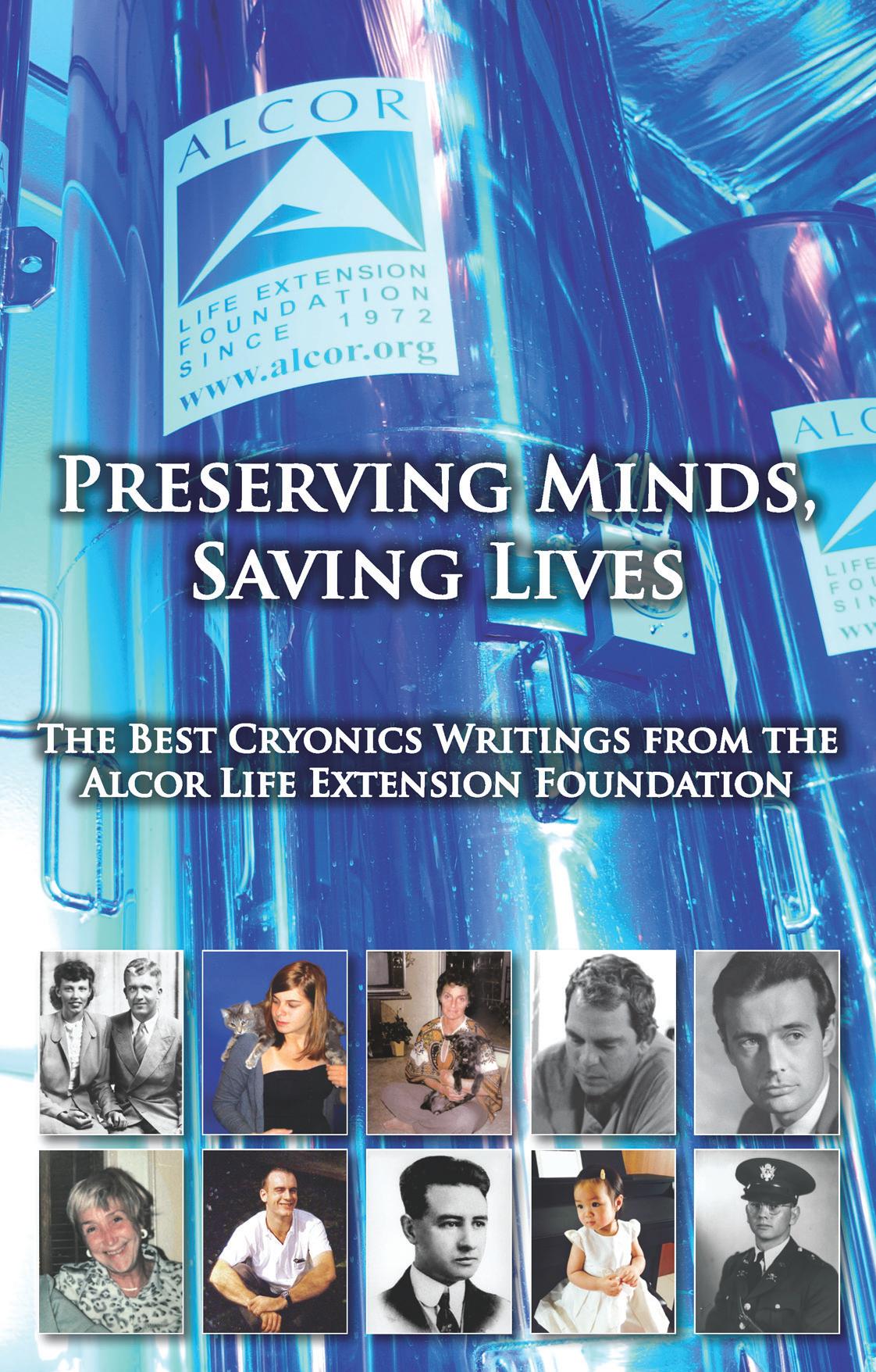
Preserving Minds, Saving Lives offers an ambitious collection of articles about cryonics and the Alcor Life Extension Foundation. From its humble beginnings in 1972, and its first human cryonics patient in 1976, Alcor has grown to a professional organization with more than 1,000 members, more than 150 human patients, and more than 60 pets, all awaiting a chance to be restored to good health and continue their lives.
This book presents some of the best cryonics writings from Cryonics magazine from 1981 to 2012. There are clear expositions of the rationale behind cryonics, its scientific validation, and the evolution of Alcor procedures. Also covered are repair and resuscitation scenarios, philosophical issues associated with cryonics, and debates within the cryonics community itself.


