4 minute read
Large Piercing Hole Causes Imaging Artifact, Case Study Finds

The following is a synopsis of the article “Severe radiographic artifact created by a large fenestration of the skin and labial mucosa following placement of a plate piercing: a case report,” which appears in the September/October issue of General Dentistry. Read the full article here.
It is well recognized that jewelry or medical devices worn during a radiographic examination may produce radiopaque artifacts that could result in interpretive challenges or misdiagnosis of a pathologic process.(1,2) However, there is little published information regarding radiographic anomalies associated with extensive soft tissue volume loss in an orofacial piercing site. In a new case report, Brooks et al describe a previously unreported type of artifact produced by an oral piercing site below the lower lip, despite removal of the jewelry prior to radiography.
Case Report
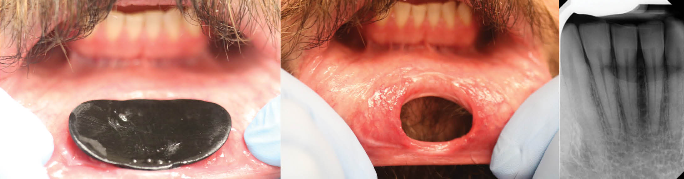
A man who sought comprehensive dental care had multiple facial piercings, including a large plate piercing below the lower lip. The patient had undergone a 1-cm scalpel piercing of the skin under the lip and used progressively larger spacers to stretch the site over time. When the facial jewelry was removed prior to radiography, the aperture measured 28 mm and allowed a direct view of two mandibular incisors. The radiographic survey revealed a conspicuous horizontal, saucer-shaped radiolucency along the middle third of the mandibular incisors. The radiolucency, which mimicked the appearance of root resorption, severe dental caries, or cervical abfraction, was ultimately attributed to the extensive aperture below the lip.
Discussion
An upper or lower lip piercing, or labret, is inserted through a hole in the facial skin that opens into the labial mucosa.(3) Some individuals gradually dilate the aperture with spacers, and continued wear of the labret usually induces epithelialization of the tissue margins, hindering wound contracture and perpetuating the cleft.(4)
During radiography, inherent variations in tissue thickness and radiodensity may affect the degree of radiation absorption and scatter, potentially modulating object contrast.(5,6) Significant facial soft tissue loss may result in the creation of a distinct artifact within the radiographic field of view. In this case, an unusual artifact occurred following removal of the large labret and masqueraded as a severe resorptive defect, dental caries, or cervical abfraction. The authors propose that the anomaly resulted from the through-and-through soft tissue fenestration and absence of the otherwise superimposed, minimally radiodense cutaneous tissue and underlying labial mucosa. Analogously, loss of soft tissue following ballistic wounds may create a bullet track or cavitation that is discernible on computed tomographic imaging.(7)
Summary
The authors believe that this is the first published case of a simulated massive tooth or osseous defect found on a radiograph taken after removal of facial jewelry. The unusual artifact, associated with a through-and-through piercing of the facial skin and labial mucosa, highlights the importance of correlating atypical radiographic presentations with soft tissue defects to avoid unnecessary patient care.
Read the full article here.
References
Venkatraman S, Gowda JS, Kamarthi N. Unusual ghost image in a panoramic radiograph. Dentomaxillofac Radiol. 2011;40(6):397-399. doi:10.1259/dmfr/63151190
Liang H, Flint DJ, Benson BW. Why should we insist patients remove all jewellery? Dentomaxillofac Radiol. 2011;40(5):328-330. doi:10.1259/dmfr/77333052
Keddie G. Symbolism and context: the world history of the labret and cultural diffusion on the Pacific Rim. Paper presented at: Circum-Pacific Prehistory Conference; August 1-6, 1989; Seattle. https://staff.royalbcmuseum.bc.ca/wp-content/uploads/2015/11/LABRET-PAPER-1989-Grant-Keddie.pdf
Grohmann M, Weiland T, Tuca AC, Wimbauer JM. Earlobe correction of the pierced ear: a systematic review of the literature and principles for surgical reconstruction. Facial Plast Surg Aesthet Med. 2023;25(2):83-89. doi:10.1089/fpsam.2021.0269
Schropp L, Alyass NS, Wenzel A, Stavropoulos A. Validity of wax and acrylic as soft-tissue simulation materials used in in vitro radiographic studies. Dentomaxillofac Radiol. 2012;41(8):686-690. doi:10.1259/dmfr/33467269
Mallya SM. Film imaging. In: Mallya SM, Lam EWN, eds. White and Pharoah’s Oral Radiology. Principles and Interpretation. 8th ed. Elsevier; 2019:61-80.
Mazuchowski EL, Harcke HT. Incorporating radiologic imaging in the study of wound ballistics. Acad Forensic Pathol. 2013;3(2):154-163. doi:10.23907/2013.020
