
19 minute read
CPD: Post-inflammatory Hyperpigmentation in Darker Skin


PIH in Dark Skin
Dr Abirami Pararajasingam and Dr Sandeep Cliff explore treatment for post-inflammatory hyperpigmentation in individuals with Fitzpatrick skin types III-VI
Advertisement
What is post-inflammatory hyperpigmentation? Post-inflammatory hyperpigmentation (PIH) is an acquired hypermelanosis which occurs in all skin types, but it is more prevalent among individuals of darker skin, defined as Fitzpatrick type III-VI.1 Dyschromias, of which PIH comprise a significant proportion, are among the most common reasons for African and Asian patients to seek the care of a dermatologist.2-4 PIH can have a significant psychosocial impact on this cohort of patients as lesions often occur more frequently and persist for a longer duration, with greater severity.5 The condition typically arises following inflammation to the skin (for example with acne, psoriasis or atopic dermatitis), but can also occur after cutaneous injury including aesthetic interventions such as dermabrasion, laser and epilation, to name a few.6-9 When there is inflammation or injury to the epidermis, melanocytes are triggered to produce excessive melanin, which is then distributed to surrounding keratinocytes. Although the exact mechanism is unknown, the increase in melanocytic activity has been shown to be stimulated by prostanoids, cytokines, chemokines and other inflammatory mediators as well as reactive oxygen species.10-13 The excessive melanin manifests as dyschromic macules or patches, which in darker individuals can range in colour from red/purple to brown/black depending on baseline skin tone and depth of discolouration.14 If the basal layer is involved, melanin pigment is released and subsequently engulfed by macrophages in the papillary dermis. This can give the skin a blue-grey appearance and lesions may be exacerbated by exposure to sunlight. PIH may remain at the site of inflammation/injury long after the initial wound has recovered, typically resolving after many months to years.15 Management of the condition is challenging and therapeutic strategies often produce variable outcomes. Note that many of the studies mentioned below do not specify the exact Fitzpatrick type of the participants, so patients’ skin colour has been referred to using the same terminology as the study.
Management options for PIH in darker skin As with other medical conditions, prevention of PIH is better than cure. The importance of controlling the underlying source of cutaneous inflammation or injury must be emphasised. Shah et al. showed that early and efficacious treatment of acne in patients with darker skin helps minimise hyperpigmentation.16 This, of course, can be challenging in severe cases. For severe or persistent lesions, which often occur in darker-skinned individuals, there is a wide range of safe and effective treatment strategies which include topical agents, chemical peels, laser therapy and intense pulse light (IPL). There are also treatments that aim to conceal hyperpigmented lesions, remove the excessive pigment or regulate melanin production, or a combination.
Topical formulations There are several topical treatments which can help regulate melanogenesis by targeting the enzyme tyrosinase, which converts dihydroxyphenylalanine (DOPA) to melanin (Figure 1). The amino acid tyrosine undergoes hydroxylation to produce DOPA and subsequently oxidation to produce DOPAquinone; both reactions are catalysed by the enzyme tyrosinase.17 The addition of cysteine to DOPAquinone leads to the formation of eumelanin. In the absence of cysteine, DOPAquinone undergoes conversion to DOPAchrome, which leads to the formation of eumelanin.18
Hydroquinone Hydroquinone is an agent which acts on this pathway and continues to play an important role in the treatment of hyperpigmentation disorders. A concentration of 2-5% hydroquinone is commonly used for this indication for all skin types.19,20 It can be used as a monotherapy, or in formulation with other agents including corticosteroids and retinoids. Hydroquinone formulations including cortiscosteroid allows treatment of co-existent inflammation. In one study, hydroquinone combined with retinol 4% and 0.15% micro-sponge in patients with Fitzpatrick II-VI skin showed mean improvement in PIH of 39%, 77%, and 77% from baseline to weeks four, eight, and 12, respectively.21 Similarly in another study microencapsulated hydroquinone 4% and retinol 0.15% with antioxidants demonstrated 75% overall improvement in pigmentation at week 12 in 63% of subjects.22 Hydroquinone is currently only available on prescription and commercial use is highly regulated because of the recognised complications, which include contact dermatitis, permanent leukoderma and exogenous onchronosis; these are true for all skin types. Carcinogenicity of hydroquinone has been reported in animals, but there is no established increased risk of cancer in humans.23
Retinoids Another group of topical agents are the retinoids, which are structural and functional analogues of vitamin A. In a randomised controlled trial with 54 black patients, topical tretinoin cream 0.1% applied once daily for 40 weeks was more effective than placebo at lightening PIH lesions.24 However, 50% of patients developed dermatitis, which is an important consideration and advocates titrating retinoid concentrations
Tyrosine
DOPA
-cysteine
DOPAchrome DOPAquinone Tyrosinase
Tyrosinase
+cysteine
Cysteinyl-DOPA
Eumelanin
(Brown to black) Pheomelanin
(Yellow to Red)

and usage with time.24,25 Newer generation retinoids, which include adapalene and tazarotene, have also been shown to be efficacious in the treatment of dyschromias in people with a dark skin tone. 26,27 In one open label study of 65 African patients with acne associated with PIH, the severity of PIH significantly improved with 0.1% adapalene gel at four, eight and 12 weeks.38 Similarly, in a randomised controlled trial involving 74 dark-skinned patients with acne, 0.1% tazarotene cream was found to be effective against PIH in terms of overall disease severity.39 The oral form of retinoid, isotretinoin, is invaluable in the treatment of severe acne and in one report its use was successful in the treatment of PIH in an Asian patient.28
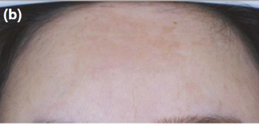
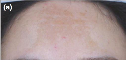
Other topicals Other topical treatments have also been implemented in the management of PIH, although evidence is limited. Azelaic acid is produced by malassezia furfur, an anthropophilic fungus which forms part of our natural skin flora. Its mechanism of action is complex and poorly understood, but its efficacy and safety in the treatment of PIH in individuals with a dark complexion has been demonstrated.29-32 One randomised vehicle-controlled double-blind trial of 52 patients (skin types IV-VI) with facial hyperpigmentation treated PIH with topical azelaic acid 20%. It demonstrated greater improvement in hyperpigmentation after 24 weeks of treatment versus vehicle, as measured by the investigator’s subjective scale and chromometric analysis. Side effects were mild and transient.33 Similar results were reported in a 16-week study looking at 15% azelaic acid in the treatment of acne-related PIH in 20 individuals with Fitzpatrick skin type IV-VI.34 Soybean is a legume that is native to East Asia and is a substance which has emerged as an effective skin-bleaching agent with promising results in the treatment of PIH. In a double-blind, placebo-controlled clinical study of African-American, Hispanic, and Asian patients with Fitzpatrick skin types III-V who had acneinduced PIH there was a significant improvement in PIH in patients using over the counter anti-acne medications containing soy compared to those without soy.35 Cysteamine (2-mercaptoethylamine) hydrochloride has been known to have depigmenting effects for decades, although exact mechanism remains unclear. Two randomised controlled trials showed positive outcomes in patients (skin types III-V) with melasma using a cream containing cysteamine compared to controls (Figure 2,3,4).36,37 Patients showed positive results after they were instructed to wash their face in the evening using a prescribed syndet bar and then apply a thin layer of the cream (placebo or cysteamine 5% w/w) to their lesions 30 min later.22 Although the exact pathophysiology underlying melasma and PIH are distinct, they are both considered disorders of hypermelanosis, thus further studies may reveal benefit of topical cysteamine in PIH patients. Other examples of depigmenting agents which have shown promising results in the treatment of dyschromias include vitamin C cream (or ascorbic acid), kojic acid (a potent inhibitor of tyrosinase by chelating copper at the active site of the enzyme), liquorice extract, niacinamide (physiologically active derivative of vitamin B3 or niacin), N-acetyl glucosamine (an amino sugar that is a precursor to hyaluronic acid), and arbutin.38-40 Evidence surrounding the efficacy of these products in PIH of all skin types is limited and further studies are required.
Concealment Skin camouflage is described as the application of pigmented creams that are designed specifically to mask skin discolouration or scarring.41 The pigment-rich products are matched to the patient’s normal skin colour for a natural appearance. Products are usually waterproof, non-comedogenic and long-lasting. As camouflage is non-invasive and avoids trauma to the skin, there is a favourable safety profile, particularly for individuals with a darker skin tone who are inherently more prone to developing PIH.42
Chemical peels Chemical peels, which involve the application of acid to remove dead skin cells, are one of the most common non-surgical cosmetic procedures performed in the UK.43 Most chemical peels are brief procedures that require little recovery time, although multiple treatments are usually necessary to achieve best results. There is a long-standing myth that chemical peels are not safe to use in

Before After
Figure 2: A 37-year-old woman with a five-year history of melasma, before and after four months of treatment with topical cysteamine cream. Fitzpatrick type not specified, however only those with Fitzpatrick skin types III, IV and V were recruited to the study.36 Images courtesy of Scientis Pharma SA, Geneva, Switzerland.
Before After
Figure 3: Another 37-year-old woman with a five-year history of melasma, before and after four months of application of cysteamine cream.36 Images courtesy of Scientis Pharma SA, Geneva, Switzerland.
Before After
Figure 4: A 36-year-old woman with a three-year history of melasma, before and after four months of treatment with topical cysteamine.36 Images courtesy of Scientis Pharma SA, Geneva, Switzerland.
darker-skinned individuals due to risks of pigmentation disorders;44 however, careful selection of the type and strength of peel can help avoid this potential complication and achieve positive results. Options include glycolic acid, salicylic acid, mandelic acid and trichloroacetic acid peels. Glycolic acid peels, in combination with tretinoin 0.05%, hydroquinone 2%, and hydrocortisone 1% have been shown to produce more rapid reduction in pigmentation compared to topical treatment alone for PIH in dark-complexioned individuals.45,46
Light therapies Lasers have been described to treat skin conditions for more than 40 years. Although topical agents remain first line for treatment of PIH, laser and light therapies may serve as useful adjuncts. Melaninspecific high energy Q-switch laser systems can successfully lighten or eradicate pigmented lesions in all skin types.47 The short pulse laser systems effectively treat lesions by confining their energy to the melanosome (tiny granules within pigment cells which contain melanin). The results of the laser treatment depend on the depth of the melanin and the colour of the lesion and is, to some degree, unpredictable. Typically, energy from short wavelength lasers is more efficiently absorbed by superficial or epidermal melanin, while longer wavelengths penetrate deeper with more selective absorption by dermal targets. A greater safety margin can be achieved using longer pulse durations and cooling devices, while still maintaining efficacy in darker-skinned individuals.48 In one study, the 1064 nm Q-switched Nd:YAG laser with low-fluence was shown to be effective in the treatment of PIH caused by procedures such as laser surgery and chemical peeling in five Asian patients.49,50 A pulse-in-pulse mode IPL worked well for facial PIH in 25 Korean patients, with 23 patients showing more than 50% improvement.51 Use of the picosecond 755 nm Alexandrite laser produced similar results in three Korean patients (skin type IV) with melasma or PIH.52 Combined therapy using Q-switched ruby laser and cutaneous bleaching with tretinoin and hydroquinone was used in 18 Japanese patients with 38.9% and 44.4% showing excellent or good clearing of periorbital hyper pigmentation, highlighting a potential role for combined strategies in selected patients.53
Discussion With the treatment options discussed, particularly chemical peels and laser therapy, it is important to recognise that treatment itself may exacerbate or cause PIH particularly in darker-skinned individuals.54 Patients should be counselled appropriately prior to commencing interventions. Furthermore, although not considered as a treatment of PIH, sun protection also plays an integral role in management. In one study, regular use of sunscreen in 89 African-American and Hispanic patients (types IV-VI) for eight weeks showed an improvement in dyschromia and skin darkening as measured by colourimetry, which is a device used to test the concentration of a solution by measuring its absorbance of a specific wavelength of light.55 Daily use of broad spectrum sunscreen (SPF 30 or more) and sun-protective measures should be advised to patients in order to minimise the darkening of lesions of PIH.56,57 This must be emphasised to darker-skinned individuals with PIH as sun protection measures are often used less frequently by this cohort of patients.58
Conclusion PIH is common among those with darker skin and can cause a significant psychological burden. Understanding available treatment options are important to allow physicians to tailor an approach that is most acceptable and efficacious for the patient. There are currently a wide range of safe and effective therapies for this condition which we have discussed here. Although some options like hydroquinone and laser therapy are well-established treatments, others have less evidence underpinning their usage, highlighting a need for more research in this area. Regardless of the chosen approach, it is important to initiate treatment early, including the use of regular sun protection and controlling the underlying inflammatory dermatosis.
Dr Abirami Pararajasingam has completed a Bachelor of Medicine and a Bachelor of Surgery with Honours at the University of Liverpool, as well as a Bachelor of Science in Neuroscience at University College London. She is a core medical trainee at East Surrey Hospital and has a keen interest in dermatology as well as teaching and research. Her special interests include inflammatory dermatoses and skin cancer.
Dr Sandeep Cliff is a consultant dermatologist at a university hospital and has a particular interest in skin cancer and inflammatory dermatosis. He has lectured and demonstrated extensively throughout the world on various non-invasive techniques for facial rejuvenation, including lasers, dermal fillers and toxins. He has been principal investigator for over six clinical research trials and is a clinical subdean at Brighton and Sussex Medical School.
REFERENCES
1. Alexis AF, Sergay AB, Taylor SC. Common dermatologic disorders in skin of colour: a comparative practice survey. Cutis. 2007;80:387–394 2. Taylor S, Grimes P, Lim J, Im S, Lui H. Postinflammatory hyperpigmentation. J Cutan Med Surg. 2009;13(4):183–91 3. Halder RM, Nootheti PK. Ethnic skin disorders overview. J Am Acad Dermatol. 2003;48(6
Suppl):S143–8. 4. Sofen B, Prado G, Emer J. Melasma and Post Inflammatory Hyperpigmentation: Management
Update and Expert Opinion. Skin Therapy Lett. 2016 Jan;21(1):1-7. 5. Park JH, Kim JI, Kim WS. Treatment of persistent facial postinflammatory hyperpigmentation with novel pulse-in-pulse mode intense pulsed light. Dermatol Surg. 2016;42(2):218–24. 6. Epstein JH. Postinflammatory hyperpigmentation. Clin Derma- tol. 1989;7(2):55–65. 7. El-Essawi D, Musial JL, Hammad A, Lim HW. A survey of skin disease and skin-related issues in
Arab Americans. J Am Acad Dermatol. 2007;56(6):933–8. 8. Sanchez MR. Cutaneous diseases in Latinos. Dermatol Clin. 2003;21(4):689–97. 9. Dunwell P, Rose A. Study of the skin disease spectrum occur- ring in an Afro-Caribbean population.
Int J Dermatol. 2003;42(4):287–9. 10. Chang MW. Disorders of hyperpigmentation. In: Bolognia JL, Jorizzo JL, Rapini RP, editors.
Dermatology. 2nd ed. Elsevier Mosby; 2009. pp. 333–389. 11. Taylor SC, Grimes PE, Lim J, et al. Postinflammatory Hyperpigmentation. J Cutan Med Surg. 2009;13:183–191. 12. Ortonne J. Retinoic acid and pigment cells: a review of in-vitro and in-vivo studies. Br J Dermatol. 1992;127(Suppl 41):43–47 13. Nordlund JJ, Abdel-Malek ZA. Mechanisms for post-inflammatory hyperpigmentation and hypopigmentation. In: Bagnara JT, editor. Advances in Pigment Cell Research: Proceedings of
Symposia and Lectures from the Thirteenth International Pigment Cell Conference. New York, NY:
Liss; 1988. pp. 219–239. Tucson, AZ; October 5–9 1986 14. Pandya AG, Guevara IL. Disorders of hyperpigmentation. Dermatol Clin. 2000;18(1):91–98. 15. Park JH, Kim JI, Kim WS. Treatment of persistent facial postinflammatory hyperpigmentation with novel pulse-in-pulse mode intense pulsed light. Dermatol Surg. 2016;42(2):218–24. 16. Shah SK, Alexis AF. Acne in skin of color: practical approaches to treatment. J Dermatolog Treat. 2010 May. 21(3):206-11 17. Chaowattanapanit et al. Postinflammatory hyper pigmentation a comprehensive overview.
American Academy of Dermatology 2017. 18. S A. N. D’Mello, G J. Finlay, Bruce C. Baguley, M E. Askarian-Amiri Signaling Pathways in
Melanogenesis. Int J Mol Sci. 2016 Jul; 17(7): 1144. 19. Grimes PE. Management of hyperpigmentation in darker racial ethnic groups. Semin Cutan Med
Surg. 2009;28:77-85. 20. Spencer MC. Topical use of hydroquinone for depigmentation. JAMA. 1965;194:962-964 21. Grimes PE. A microsponge formulation of hydroquinone 4% and retinol 0.15% in the treatment of melasma and postinflam- matory hyperpigmentation. Cutis. 2004;74(6):362–8. 22. Cook-Bolden FE, Hamilton SF. An open-label study of the efficacy and tolerability of microencapsulated hydroquinone 4% and retinol 0.15% with antioxidants for the treatment of hyperpigmentation. Cutis. 2008;81(4):365–71. 23. Nordlund JJ, Grimes PE, Ortonne JP. The safety of hydroquinone. J Eur Acad Dermatol Venereol. 2006;20:781-787. 24. Bulengo-Ransby SM, Griffiths CE, Kimbrough-Green CK, et al. Topical tretinoin (retinoic acid) therapy for hyperpigmented lesions caused by inflammation of the skin in black patients. N Engl J
Med. 1993;328:1438-1443. 25. Callender VD. Acne in ethnic skin: special considerations for therapy. Dermatol Ther. 2004;17:184–195. 26. Jacyk WK, Mpofu P. Adapalene gel 0.1% for topical treatment of acne vulgaris in African patients.
Cutis. 2001;68(4 suppl): 48-54. 16. Kligman AM, Willis I. 27. Grimes P, Callender V. Tazarotene cream for postinflammatory hyperpigmentation and acne vulgaris in darker skin: a double-blind, randomized, vehicle-controlled study. Cutis. 2006 Jan. 77(1):45-50.
28. Smit N, Vicanova J, Pavel S. The hunt for natural skin whitening agents. Int J Mol Sci. 2009 Dec 10. 10(12):5326-49. 29. Woolery-Lloyd HC, Keri J, Doig S. Retinoids and azelaic acid to treat acne and hyperpigmentation in skin of color. J Drugs Dermatol. 2013 Apr. 12(4):434-7. 30. Balina LM, Graupe K. The treatment of melasma: 20% azelaic acid versus 4% hydroquinone cream.
Int J Dermatol. 1991;30:893–895. 31. Sarkar R, Bhalla M, Kanwar KJ. A comparative study of 20% azelaic acid cream monotherapy versus a sequential therapy in the treatment of melasma in dark-skinned patients. Dermatology. 2002;205:249–254. 32. Verallo-Rowell VM, Verallo V, Graupe K, et al. Double-blind comparison of azelaic acid and hydroquinone in the treatment of melasma. Acta Derm Venereol Suppl (Stockh) 1989;143:58–61. 33. Lowe NJ, Rizk D, Grimes P, et al. Azelaic acid 20% cream in the treatment of facial hyperpigmentation in darker-skinned patients. Clin Ther. 1998;20:945–959. 34. Kircik LH. Efficacy and safety of azelaic acid (AzA) gel 15% in the treatment of post-inflammatory hyperpigmentation and acne: a 16-week, baseline-controlled study. J Drugs Dermatol. 2011;10:586590. 35. Sah A, Stephens TJ, Kurtz ES. Topical acne treatment improves postacne postinflammatory hyperpigmentation (PIH) in skin of color [Poster] J Am Acad Dermatol. 36. Mansouri P, Farshi S, Hashemi Z, Kasraee B. Evaluation of the efficacy of cysteamine 5% cream in the treatment of epidermal melasma: a randomized double-blind placebo-controlled trial. Br J
Dermatol. 2015 Jul;173(1):209-17. 37. Farshi S, Mansouri P, Kasraee B. Efficacy of cysteamine cream in the treatment of epidermal melasma, evaluating by Dermacatch as a new measurement method: a randomized double blind placebo controlled studym J Dermatolog Treat. 2018 Mar;29(2):182-189. 38. Callender VD, St Surin-Lord S, Davis EC, Maclin M. Postin- flammatory hyperpigmentation: etiologic and therapeutic con- siderations. Am J Clin Dermatol. 2011;12(2):87–99. 39. Alexis AF, Blackcloud P. Natural ingredients for darker skin types: growing options for hyperpigmentation. J Drugs Derma- tol. 2013;12(9 Suppl):s123–7. 40. Nestor M, Bucay V, Callender V, Cohen JL, Sadick N, Waldorf H. Polypodium leucotomos as an adjunct treatment of pigmen- tary disorders. J Clin Aesthet Dermatol. 2014;7(3):13–7. 41. Antoniou C, Stefanaki C. Cosmetic camouflage. J Cosmet Dermatol. 2006;5:297–301. 42. Kayama et al. Camouflage Therapy for Post-Inflammatory Hyperpigmentation on the Face Caused by Fixed Drug Eruption. Journal of Cosmetics, Dermatological Sciences and Applications, 2013, 3, 8-10 Leyden J, Wallo W. The mechanism of action and clinical benefits of soy for the treatment of hyperpigmentation. Int J Dermatol. 2011 Apr. 50(4):470-7. 43. Cosmetics UK, Non-Surgical Treatments Statistics, 2015. <https://www.cosmedics.co.uk/nonsurgical-treatments-statistics/> 44. Castillo DE1, Keri JE Chemical peels in the treatment of acne: patient selection and perspectives.
Clin Cosmet Investig Dermatol. 2018 Jul 16;11:365-372. 45. Burns RL, Prevost-Blank PL, Lawry MA, Lawry TB, Faria DT, Fivenson DP. Glycolic acid peels for postinflammatory hyperpigmentation in black patients. A comparative study. Dermatol Surg. 1997
Mar. 23(3):171-4; discussion 175. 46. Sarkar R, Parmar NV, Kapoor S. Treatment of Postinflammatory Hyperpigmentation With a Combination of Glycolic Acid Peels and a Topical Regimen in Dark-Skinned Patients: A
Comparative Study. Dermatol Surg. 2017 Apr. 43 (4):566-573. 47. DermNet NZ, Lasers in dermatology, 2014. <https://www.dermnetnz.org/topics/lasers-indermatology/> 48. AF Alexis. Lasers and light-based therapies in ethnic skin: treatment options and recommendations for Fitzpatrick skin types V and VI. Br J Dermatol. 2013 Oct;169 Suppl 3:91-7. 49. Kim S, Cho KH. Treatment of procedure-related postinflam- matory hyperpigmentation using 1064nm Q-switched Nd: YAG laser with low fluence in Asian patients: report of five cases. J Cosmet
Dermatol. 2010;9:302-306 . 50. Rokhsar CK, Ciocon DH. Fractional photothermolysis for the treatment of postinflammatory hyperpigmentation after carbon dioxide laser resurfacing. Dermatol Surg. 2009 Mar. 35(3):535-7. 51. Park JH, Kim JI, Kim WS. Treatment of Persistent Facial Postinflammatory Hyperpigmentation With
Novel Pulse-in-Pulse Mode Intense Pulsed Light. Dermatol Surg. 2016 Feb. 42 (2):218-24. 52. Lee YJ, Shin HJ, Noh TK, Choi KH, Chang SE. Treatment of Melasma and Post-Inflammatory
Hyperpigmentation by a Picosecond 755-nm Alexandrite Laser in Asian Patients. Ann Dermatol. 2017 Dec. 29 (6):779-781. 53. Momosawa A1, Kurita M, Ozaki M, Miyamoto S, Kobayashi Y, Ban I, Harii K.Combined therapy using
Q-switched ruby laser and bleaching treatment with tretinoin and hydroquinone for periorbital skin hyperpigmentation in Asians. Plast Reconstr Surg. 2008 Jan;121(1):282-8. 54. Halder R, Munhutu M, Foltis P, Battie C, Verschoore M, Ore- sajo C. Evaluation and effectiveness of photoprotection com- position (sunscreen) on subjects of skin of color (abstract). J Am Acad
Dermatol. 2015;72(5 Suppl):AB215. 55. Halder R, Rodney I, Munhutu M, et al. Evaluation and effectiveness of a photoprotection composition (sunscreen) on subjects of skin of color [abstract]. J Am Acad Dermatol. 2015;72(5 suppl):AB215. 56. Ruiz-Maldonado R, Orozco-Covarrubias ML. Postinflammatory hypopigmentation and hyperpigmentation. Semin Cutan Med Surg. 1997;16(1):36–43. 57. Imokawa G, Yada Y, Miyagishi M. Endothelins secreted from human keratinocytes are intrinsic mitogens for human melano- cytes. J Biol Chem. 1992;267(34):24675–80. 58. Maymone MBC, Neamah HH, Wirya SA, Patzelt NM, Zanca- naro PQ, Vashi NA. Sun-protective behaviors in patients with cutaneous hyperpigmentation: a cross-sectional study. J Am Acad
Dermatol. 2017;76(5):841–846.e2.
Hyper Pigmentation
Whether you are treating PIH, melasma or photoageing, our portfolio of skin brightening products help protect, prevent and target hyper-pigmentation. Whatever pigment issue you are addressing, the sun, medication, hormones or inflammation, our comprehensive portfolio gives you the flexibility to tailor your patient’s treatment plan to their needs and budget. Find out more today by contacting AestheticSource.

TOPICAL CHEMICAL PEELS
• Cysteamine® • NeoStrata® Enlighten Range • Skinbetter® Even Tone and Alpharet® • Skin Tech Blending and Bleach & AD Aclarance® • NeoStrata® Pro System Peels • Skin Tech Peels • Skin Tech Bene Bellum® coming soon • RRS® Whitening
INJECTABLE
Meeting the needs of your business, delivering high satisfaction to your patients


