Produced exclusively for SCMG Enterprises Ltd by Scala Arts & Heritage Publishers First published in 2019 by Scala Arts & Heritage Publishers Ltd 10 Lion Yard Tremadoc Road London sw4 7nq, uk www.scalapublishers.com In association with Science Museum Exhibition Road London sw7 2dd www.sciencemuseum.org.uk
CONTENTS
DIRECTOR’S FOREWORD SIR IAN BLATCHFORD
6
FUNDERS’ FOREWORDS BARONESS ELIZA MANNINGHAM-BULLER, SIR PETER LUFF, DR PAULINE WILLIAMS
8
PREFACE
Every purchase supports the museum.
NATASHA McENROE 10
Texts © SCMG Enterprises Ltd, 2019 Science Museum ® SCMG Enterprises Ltd and designs © SCMG Enterprises Ltd This edition © Scala Arts & Heritage Publishers Ltd, 2019
1 COLLECTING MEDICINE
6 PREVENTING CERVICAL CANCER
ROUTES AND ROOTS OF MEDICINE AT THE SCIENCE MUSEUM
EMMA STIRLING-MIDDLETON
Editor: Charlotte Grievson Design: Peter Dawson, Alice Kennedy-Owen at gradedesign.com Printed and bound in China
SELINA HURLEY
isbn 978-1-78551-210-0 10 9 8 7 6 5 4 3 2 1
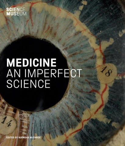
Every effort has been made to acknowledge correct copyright of images where applicable. Any errors or omissions are unintentional and should be notified to the Publisher, who will arrange for corrections to appear in any reprints. British Library Cataloguing in Publication Data. A catalogue record for this book is available from the British Library. Cover: Papier-mâché model of a human eye. Science Museum Group. Object number 1996-277/11 Frontispiece: Pharmaceutical glassware in the Science Museum stores. p. 7: Lancets and lancet cases from the Science Museum’s medicine collection. p. 11: Bottle containing wormwood (Artemisia absinthium).
7 HEARING DISTANT VOICES PSYCHIATRY, ASYLUMS AND THE LIMITS OF HISTORY
PEG-LEGS, PYLONS AND THE PIANIST’S ARM
OISÍN WALL
144
38
8 THE PHARMACY SHOP
3 THE PROBLEMATIC BODY
‘A GREAT RAGE FOR MAHOGANY, VARNISH AND EXPENSIVE FLOOR-CLOTH’
DISSECTION AND THE RISE OF THE ANATOMY SCHOOL ANNA MAERKER
126
12
2 PROSTHESES AT THE SCIENCE MUSEUM STEWART EMMENS
All rights reserved. No part of this book may be reproduced, stored in a retrieval system or transmitted in any form or by any means electronic, mechanical, photocopying, recording or otherwise, without the written permission of Scala Arts & Heritage Publishers Ltd and SCMG Enterprises Ltd.
VAGINAS, VACCINATION AND VENEREAL DISEASE
BRIONY HUDSON
154
66
9 MODELLING LIFE
4 THE CHILD IN THE IRON LUNG
EXPLORING THE HUMAN BODY
POLIO, PUBLIC HEALTH AND PROTECTION
MURIEL BAILLY
176
NATASHA McENROE 82
10 EMOTIONAL OBJECTS
5 CREATED THROUGH CONFLICT
FAITH AND FEELING IN THE MEDICINE COLLECTION
THE DEVELOPMENT OF MILITARY MEDICINE JACK DAVIES
98
SARAH BOND
202
NOTES 228 FURTHER READING 232 AUTHOR BIOGRAPHIES 233 INDEX 234 ACKNOWLEDGEMENTS AND PICTURE CREDITS 240
DIRECTOR’S FOREWORD
Medicine: The Wellcome Galleries form the biggest and most ambitious project that the Science Museum Group has undertaken in many decades. This permanent exhibition creates a centre for medicine that is of global importance, examining medical challenges of the past and the future through our world-class collections. In a series of five connecting galleries, our visitors can discover how we learn from the human body and how we influence the health of both individuals and communities. Intellectually stimulating and visually stunning, these galleries are relevant to us all. The Medicine collections at the Science Museum are composed of two elements. The historic collection of Henry Wellcome, whose personal treasure trove has been on long-term loan to us for over 40 years, is complemented wonderfully by the Science Museum’s own medical holdings. Often including cutting-edge medical technology, our active accumulation of material linked to scientific breakthroughs creates an ever-growing and everchanging historic resource. We are delighted to work closely with the Wellcome Trust in our shared commitment to engaging a wide audience with bio-medicine issues both past and present. We are grateful to our many funders of Medicine: The Wellcome Galleries, both organisations and individuals who have long supported the Science Museum and those who are newly involved. Their enthusiasm and support for this project has been truly inspiring. We would like to thank Wellcome (Title Funder), National Lottery Heritage Fund – and further support from GSK (Principal Sponsor), the Dr Mortimer and Theresa Sackler Foundation (Principal Funder), the Wolfson Foundation (Major Funder), Vitabiotics and the Lalvani family (Major Sponsor), with additional support from the Stavros Niarchos Foundation. A project as vast as Medicine: The Wellcome Galleries has involved an enormous number of people, to whom we are extremely grateful for their time and expertise. Current healthcare practitioners, experts in the academic field and, perhaps most importantly, patients themselves have all shared their stories with us. The project has been, in a very real sense, a team effort. At the Science Museum, one of our aims is to ‘Ignite Curiosity’ within all our visitors – something I am confident that we will achieve in this exceptional new space and through this book that acts as its companion.
SIR IAN BLATCHFORD DIRECTOR OF THE SCIENCE MUSEUM GROUP 6
text running over by 1 line now
FUNDERS’ FOREWORDS
WELLCOME TRUST
NATIONAL LOTTERY HERITAGE FUND
GSK
The pharmaceutical entrepreneur and philanthropist Sir Henry Wellcome – whose Will established the Wellcome Trust in 1936 – was also an avid collector. He sent his agents around the world in search of objects related to the ways in which people in different cultures have tried to preserve and restore their health over the centuries. His aim was to recreate humanity’s medical past in order to study the roots of contemporary practices and inform future discoveries and inventions. In his lifetime, Sir Henry accumulated more than a million items. Rich material for scholars, his collection was intended to inform, educate and entertain through public displays and exhibitions as well as research. It is gratifying, therefore, to see his collection at the heart of the new Medicine Galleries in the Science Museum. Around 115,000 objects from Sir Henry’s personal collection were placed on long-term loan to the Science Museum in the 1970s, and the links between our two organisations remain strong. With support from Wellcome, the Science Museum has been able to show many items from Sir Henry’s collection over the past four decades, giving visitors different perspectives on the human experience of sickness and healing. Wellcome’s mission is to improve health. We fund not only researchers investigating the biology of health and disease, but also those exploring the cultural and social contexts of science and medicine. Understanding how ideas develop is essential to driving change and tackling health challenges. Equally important is that we engage and involve people with science and research. The Medicine Galleries will help to ensure that Sir Henry’s collection continues to inspire people. They offer new opportunities to find significance and meaning in the diverse stories of health and medicine that these objects represent.
These fine galleries bring together three things in which the United Kingdom excels – medical innovation, curatorial skills and support for our heritage from the National Lottery. What the Science Museum has achieved, though, is far more than an engaging display of an unrivalled collection. The new Medicine Galleries will be a centre for international consultation and research. Informed and inspired by a wealth of extraordinary artefacts and documents, those using the new galleries to these ends will be powerfully reminded of the importance of the improved health and wellbeing in modern society. And it is entirely thanks to National Lottery players that the Fund I have the good fortune to chair has been able to provide almost £8 million towards these galleries, more than a third of the total project cost. Indeed, since the Fund’s inception 25 years ago we have invested more than £63 million in the Science Museum Group’s sites around the UK. Because our heritage is shared by all of us, and because our funds come from a wide cross-section of people, we are determined to support projects that reflect all aspects of that heritage and will have a wide appeal. This is exactly what the Medicine Galleries will do, with hands-on interactive technology that will appeal to people of all ages and will be fully accessible to individuals with disabilities. The new galleries will provide a thought-provoking and fascinating experience for the visitors who will flock to them. My hope and expectation is that, as they explore the story of medicine from Roman times to the present day, many of those visitors will be inspired to take up careers in medicine and related sciences, or perhaps to become curators. But I hope all of those old enough to do so will be inspired to play the National Lottery and so help to fund more great projects like this one for many years to come.
At GSK, we are extremely proud of our long-term partnership with the Science Museum. For over 25 years we have worked alongside the Museum – from supporting the original Health Matters gallery in the early 1990s to becoming a principal supporter of the new Medicine Galleries. We have watched the Museum develop and expand, constantly looking forward whilst retaining its standing as a global centre of excellence, a place where generations of children and young people have first had their eyes opened to the wonder of science and its possibilities. GSK, as a science-led global healthcare company, is fully committed to furthering science education, and looks to support programmes and projects that inspire every student to embrace the wonder and power of science, encouraging them to explore the amazing opportunities on offer. By bringing science to life, we aim to encourage the next generation of scientists and engineers because, without them, who will cure diseases and help solve the health challenges of the future? It has been a privilege to be part of the Museum’s exciting initiative to create a magnificent new home for its medicine collections based on Henry Wellcome’s original collection. The Medicine Galleries are now literally at the heart of the Museum, giving a greater focus and understanding to the importance and relevance that medical development and advancement plays in all our lives. We hope that you will enjoy your visit and this accompanying book will help to further your understanding and enjoyment of the galleries.
BARONESS ELIZA MANNINGHAM-BULLER CHAIR, WELLCOME TRUST
DR PAULINE WILLIAMS SENIOR VICE PRESIDENT, GLOBAL HEALTH RESEARCH & DEVELOPMENT, GSK
SIR PETER LUFF CHAIR OF THE NATIONAL LOTTERY HERITAGE FUND
8
GSK FOREWORD
9
PREFACE
The Medicine collection at the Science Museum in London is one of the finest in the world. It is made up largely of Sir Henry Wellcome’s personal collection, on long-term loan to the Science Museum since the 1970s. Wellcome, a pharmaceutical magnate, collected in the grand style of the Victorian age, believing that if he were to collect enough objects, it would be possible to calculate laws of humanity and learn the truth about our instinct for self-preservation. Since coming to the Science Museum, the collection has grown as curators have acquired objects relating to recent developments in health and medicine, adding to the historic material. Items range from a tiny Japanese netsuke carved from ivory to a large Victorian pharmacy shop. Endless rows of shelving in the Museum stores hold a multitude of blue and yellow ceramic jars once used to store drugs. There are countless drawers of surgical tools and whole rooms dedicated to X-rays and their associated ephemera, or to dentistry or fragile laboratory glassware. The collection includes groups of items brought together either for medical practice, or due to their association with a famous individual. There are teaching models and a whole range of curiosities linked to health. Caring for this vast and diverse collection takes a dedicated curatorial team who specialise in medicine and the role it plays in all our lives. The role of the curator is not only to understand their collection but to interpret it for the public, to add to it, to publicise and protect it. A curator who works specifically with medicine will surely never struggle to present their collection as relevant – we each have our own history when it comes to medicine and are profoundly and enduringly invested in the health of ourselves and our loved ones. The personal connection that we have with the subjects of health and medicine adds an immediacy to the subject matter – but it also carries a potential risk unless it is communicated with sensitivity. Medical collections contain some of the most problematic objects in any museum collection – real human remains, for example, and images of diseased body parts, nudity or children’s suffering. Historic collections such as that of Henry Wellcome also contain items collected during the colonial era that would have been acquired with none of the parameters set by current collection practices. Interpretation of all of these must be undertaken with great care and understanding. This book is published on the occasion of the launch of a series of new galleries dedicated to showcasing the Medical collection at the Science Museum in South Kensington. Medicine: The Wellcome Galleries, which opened in 2019, form the most extensive display of the history of medicine in the
world, telling the story of how we have managed our health at various points in time, and offering a broad perspective on medicine today. This book is not, however, a traditional catalogue of the galleries, nor does it seek to be an encyclopaedia of the history of medicine. Rather, it aims to express some of the interests and enthusiasms of the curators and historians who have created the new galleries, and whose professional research has been shaped by the collection itself. The authors of the book have acquired well over 100 years of curatorial experience between them and are united in their aim to share their passion for and knowledge of the history of medicine. Medicine: An Imperfect Science is formed of a series of standalone, but connected, chapters through which we will learn of the history of the Medicine collection and how it came together, and what the objects themselves can tell us about this area of science. Some objects show us how scientists have gone about their research and practice, often through the way they are made or the materials they are formed of. Others can communicate something of the experience of patients of the past in a manner quite different from a written account of mental health, or other kinds of evidence. Analysing information from our bodies, in many ways the object at the centre of this particular history – whether physical measurements, imaging or dissecting the dead – has long been a tool for increasing our knowledge of health. The models and images that were used to support teaching and record medical information in the past are often visually striking and can straddle a boundary between record and portraiture. The objects within our collection at the Science Museum offer us a unique opportunity to engage with the history of medicine in previously unexplored ways. As well as allowing readers to learn more about the ever-growing collection of objects in the care of the Science Museum, this book provides behind-the-scenes glimpses of life in a large museum and the huge task of creating a suite of new galleries. Each chapter of this book discusses, directly or indirectly, the problems inherent to medicine – whose status as a science can sometimes be called into question – and the telling of its stories. Medicine is an imperfect science and encompasses areas little understood today. The relationship of faith and trust between a patient and their practitioner and the support of loved ones have direct health outcomes, and even today, the immense power of the mind over the body is poorly understood. Through the historic Medicine collection at the Science Museum, we can reflect anew upon healthcare and our relationships to our own bodies. NATASHA McENROE KEEPER OF MEDICINE AT THE SCIENCE MUSEUM
10
EXTEND B/G
1 COLLECTING MEDICINE ROUTES AND ROOTS OF MEDICINE AT THE SCIENCE MUSEUM
SELINA HURLEY
Fig. 1 Pestles and mortars from across the globe, 1600–1900.
The Medicine collection at the Science Museum is unique in its scale, geographical origin and scope. It touches on medical science, prosthetics, medications, deities from major global religions and relics from famous names in the history of medicine. The oldest objects in the collection date from 200 million years ago, with examples of dinosaur bones. These sit alongside the latest innovations in medicine that have been added more recently to the collection. The collection has its beginnings in the extraordinary project of Henry Solomon Wellcome (1853–1936), a pharmaceutical businessman with an eye for advertising and a passion for collecting. His was a vast undertaking to collect the entire human experience of life and death, health and illness. Upon his death, his collection numbered over a million objects. A large part of this legacy was a library including anatomical treatises, recipe books, pamphlets, paintings and manuscripts, with at least 43 languages represented. The collection was put into the hands of the Wellcome Trust, created by Wellcome’s will. The Wellcome Trustees decided to disperse the non-medical material to institutions across the world and through sale at auctions. The books and archives remain at the Wellcome Library. Exhibitions celebrating milestones of the history of medicine were still staged and some objects continued to be added, although at a much slower pace. The last and largest transfer of the holdings was announced in 1976 with objects to go on long-term loan to the Science Museum. A second, smaller
transfer was completed in the mid-1980s. This meant a new home for the history of medicine and an upturn in collecting medicine at the Science Museum. If each of the over 150,000 objects now in the care of the Science Museum could speak, revealing their histories, there would be a cacophony of voices.1 For so many objects, the first evidence of their stories came with their entry into Wellcome’s collection and they represent the work of hundreds of people over the collection’s 140-year history. All the objects featured within this chapter now have their home at the Science Museum. For the first time, many of their routes to the Science Museum have been revealed using the original sources of the people who worked with them or used them. Henry Wellcome (fig. 2) straddled two worlds at the turn of the 20th century – as one of the last gentleman collectors and one of the first pharmaceutical magnates. Having grown up in Minnesota, Wellcome qualified as a pharmacist and started working as a travelling pharmaceutical salesman. In 1880 he was invited to join his friend, Silas Burroughs, who had emigrated to London, with licences to sell American products in the United Kingdom. Together, they founded Burroughs Wellcome & Co. Wellcome soon saw the opportunity to manufacture and sell their own products, and they set up their first factory in London. Their ‘Tabloid’ product range included compressed tablets, bandages, photographic chemicals, tea, medicine chests, and water testing and purifying equipment. 13
with the queen as their president. From 1906, hygiene and sanitation were taught to all officers and became a part of their examinations, and the Royal Army Medical Corps was reorganised to improve the efficiency of the service.
THE FIRST WORLD WAR (1914–18) In 1914 the new and improved army medical service entered the First World War, confident of a quick and easy victory. The reality could not have been more different. The war lasted four long years and was fought all over the world. Over 70 million military personnel were involved from all participating countries, and more than nine million of these died, not including the seven million civilians who were killed as the lines between the civilian and military spaces blurred. Though the experiences of the Boer War had convinced many of the importance of sanitation and hygiene, it lulled them into a false sense of security. The battlefields of the Boer War were completely different from the fertile fields of France and Belgium. Here, wounds festered and worsened, and many patients died from infection. To make matters worse, the wounds of the First World War were remarkably different from those encountered before. In the 100 years before the First World War weaponry changed from fourrounds-per-minute muskets, to 600-rounds-perminute machine guns, resulting in patients with several severe bullet wounds, something never seen before. Others were hit by flying shrapnel, tiny bits of steel or lead encased in a hollow shell that exploded, propelling the shrapnel outwards at high speeds. Some were wounded by grenades and other artillery. The development of poisonous gas, first used as a weapon in 1915, deeply affected soldiers. It burned their skin and irritated their lungs, prompting the creation of new gas masks for soldiers and animals alike (figs 127 and 128). Gas was just one of the highly effective new forms of weaponry used in the First World War. It was at this time that planes were first used as weapons; technology now allowed them to shoot bullets through their propellers and drop bombs on civilians and combatants alike. It was in this war, too, that the military tank was first introduced, inspiring the creation of terrifying protective masks worn by British tank operators (fig. 129). 114
CREATED THROUGH CONFLICT
When Allied soldiers were wounded on the Western Front, they were transported through the chain of evacuation. First they were tagged with their medical and military details (fig. 130). They then travelled through regimental aid posts, advanced dressing stations, field ambulances, casualty clearing stations and general hospitals. If they were severely wounded, they were sent to Britain to recover in hospitals and convalescent homes. Each stage was further away from the hostilities and provided more sophisticated care. At the beginning of the war, soldiers were moved between these points via horse-drawn ambulances. These were rickety and slow, and by the time they arrived at their destinations many patients had succumbed to their infections. The entirety of the British war effort was dependent on charitable people at home in Britain, and many of them donated their own cars to allow patients to be transported more efficiently between parts of the chain. Doctors, nurses and untrained volunteers had to use their own judgement on each patient. They utilised a system called ‘triage’, where patients were divided into three distinct categories: those with minor wounds who could be patched up and made to fight again relatively quickly, those who could recover if given medical attention and those who would die regardless of medical intervention. Mary Borden, a nurse during the war, wrote: I was there to sort them out and tell how fast life was ebbing in them. Life was leaking away in all of them; but with some there was no hurry, with others it was a case of minutes.9 This was a difficult yet important decision. Though the Royal Army Medical Corps grew considerably during the war, doctors, surgeons and nurses were constantly busy. They had limited resources and were at the mercy of unreliable deliveries of equipment and drugs. It was vital that they did not waste time or medicine on men who were not going to survive. Surgery was completed either at the Casualty Clearing Station or in a general hospital, depending on whether the patient was able to survive the wait. Many men had limbs amputated due to infection. Doctors hoped to save soldiers’ lives by removing the infected limb before it killed them. In a world before antibiotics, practitioners did everything they could to prevent infection from
Fig. 127 Helmet-type gas mask, canvas, metal fittings and glass eyepieces, UK, 1915. This mask was used to protect wearers during a phosgene or chlorine gas attack. It was called the PH ‘tube’ gas helmet because of its appearance. Science Museum Group. Object number A652157
Fig. 128 Gas mask for horses, Germany, 1914–18. Horses were used extensively during the First World War, and though gas was first used in 1915, it took a while for protective gear to be made for animals. Science Museum Group. Object number A635089
Fig. 129 Protective mask, leather and chain mail, UK, 1914–18, worn by British tank crews. Science Museum Group. Object number A204116
CUT OUTS
Fig. 130 (left) Book of wound tags, UK, 1900–18. These wound tags were attached to men’s uniforms, providing medical professionals with vital information such as type of wound and treatment, rank and regiment, and name and serial number. Science Museum Group. Object number A652315. Pt14
Fig. 131 (opposite) Surgical dressing substitutes, Europe, 1914–18. Pictured here are samples of petite cotton, curtain, cotton muslin and sphagnum moss, used when bandages were lacking. Science Museum Group. Object number A600306/1–5
taking hold, but this was anything but easy. Wounds were washed with carbolic lotion, a mixture of carbolic acid and water, then wrapped in gauze and covered in more carbolic lotion. Doctors were often forced to use a variety of unconventional materials to wrap wounds, including petticoat cotton, curtain material, cotton muslin and sphagnum moss (the generic name for around 300 types of moss) (fig. 131). Many patients underwent the process of debridement, where dead or dying flesh surrounding the wound was cut away before the affected area was wrapped and allowed to recover. Though these methods were sometimes successful, it became apparent that there was a need for a better way of sterilising wounds. The French surgeon Alexis Carrel (1873– 1944) and the British biochemist Henry Dakin (1880–1952) worked on a new technique in 1915 and 1916. The Carrel-Dakin method was a form of wound irrigation. Dakin created a septic solution, while Carrel developed the apparatus used to apply it to the wound (fig. 132). The solution itself was notoriously difficult to get right. If the ratios were incorrect, it would not 116
CREATED THROUGH CONFLICT
only irrigate the wound, but irritate it too. The famous pharmaceutical company Johnson & Johnson realised that hospitals lacked the resources, and crucially the time, to do this themselves. They mass produced ampoules of sodium hypochlorite and provided it to the British authorities, who officially adopted the method in 1917. The Carrel-Dakin method saved countless lives and limbs by preventing infection. In the long term, it has significantly changed the way we manage wounds. Prior to its invention, gas gangrene, a bacterial infection that causes gas to form in dying tissues, regularly resulted in either death or amputation. Another key innovation of the First World War was the Thomas splint (fig. 133). It was invented by the surgeon Hugh Owen Thomas (1834–1891) in 1875, but it was not until 1916 that it was used on the fighting fronts. Thomas came from a family of Welsh bonesetters and had his own practice for treating fractures in Liverpool. Though it sounds like a relatively mundane medical object, the introduction of the Thomas splint lowered the death rate from broken femurs (thigh bones) from 80 per cent
Fig. 132 Carrel’s apparatus for sterilising wounds, Down Bros Ltd, London, 1914–18. It was designed to be used with Dakin Daufresne antiseptic solution. It intermittently dripped the solution onto the wound, killing dangerous bacteria without causing further damage to the patient. It was a very successful method of sterilising wounds and saved many men from amputation. Science Museum Group. Object number A51115
to just 20 per cent. Prior to 1916 soldiers regularly died from blood loss from the femoral artery, because many of them went into shock, preventing medical practitioners from providing adequate care. The Thomas splint was easy to use and incredibly effective at immobilising the lower limb, thus allowing the patient to be moved without exacerbating the wound. The splint not only saved thousands of lives in the war, but it also reduced the number of amputations, thus exposing fewer soldiers to threat of infection. One of the key problems of amputation and battlefield medicine more generally was blood loss. Though blood transfusions had been attempted for hundreds of years, often with animal blood and human patients, they failed because of coagulation, or clotting, which occurs when the donor blood is rejected by the recipient’s immune system due to incompatible blood types. Austrian biologist Karl Landsteiner discovered three different blood types in 1901 (A, B and O), and learned that matching the blood group of a donor to that of the recipient prevented the patient from dying. Though doctors were aware CREATED THROUGH CONFLICT
117
of this during the First World War, blood transfusions remained dangerous. Without the ability to store blood properly, the donor and the recipient had to lie next to each other with their blood vessels connected via rubber tubing (fig. 133). This was revolutionised in 1914, when it was discovered that adding sodium citrate to blood prevented it from clotting, allowing it to be stored and used later. This was improved upon by the discovery of heparin in 1916. Both sodium citrate and heparin are anticoagulants, meaning that they block the creation of the proteins that cause clotting. Storing the blood with citrate and keeping it on ice allowed it to be kept for 26 days. This meant that the authorities were able to send blood to where it was most needed. While blood banks did not occur in civilian life until the 1930s, they became an integral part of military medicine during the First World War, mainly pioneered by Captain Oswald Hope Robertson, who established ‘blood depots’ in 1917, during his service in France. The ability to perform blood transfusions saved thousands of soldiers in the war, and the legacy of this conflict has given us the blood banks that continue to save lives every day. Medicine was not just about saving lives or limbs, it also allowed literal and emotional scars to be concealed. The nature of the fighting in the First World War meant that more soldiers had facial wounds than ever before. Many were shot peering over the parapets of the trenches or were hit by shrapnel from artillery fire. The problem was so serious that the British army introduced steel helmets for the first time in 1916. Facial injuries not only created life-threatening wounds, but, if the patient survived, they were left with severe life-changing scars – stark reminders of a conflict most would rather forget. In cases of facial injury, surgeons close to the front line did what they could, but they did not have the time or skills to hide the marks left by their scalpel. Often the skin would heal and tighten, leaving men with permanently disfigured faces. A surgeon from New Zealand, Harold Gillies (1882–1960), opened a hospital in Sidcup, England, to reconstruct the faces of men that had been destroyed by war. He was inspired by an ancient Indian surgical technique that used the skin on the forehead to regrow the nose. His ‘tubed pedicle graft’ was an attempt not only to restore the health of his patients, but to make 118
CREATED THROUGH CONFLICT
CUT OUT
Fig. 134 Blood transfusion kit, designed by Captain Oswald Hope Robertson. Blood transfusions continue to save thousands of lives every day, but during the First World War, they remained a dangerous and time-consuming process. This portable kit stored blood and allowed transfusions to be performed close to the battlefield. Science Museum Group. Object number A625893 OVERLEAF
Fig. 135 Set of artificial eyes, Liverpool, UK, 1900–40, Facial wounds were very common during the First World War as men tried to peer over the parapets at enemy trenches. This resulted in a high demand for artificial eyes as many men used them to restore their physical appearance. Science Museum Group. Object number A660037
Fig. 133 Leg splint, iron and leather, 1875–1920. Before the introduction to the war front of this ‘Thomas splint’, designed by Hugh Owen Thomas in 1875, nearly 80 per cent of men with upper-leg fractures died. Science Museum Group. Object number A603026
the results as aesthetically pleasing as possible. This technique involved taking a flap of skin from the chest or forehead, rolling it into a tube, and attaching it to the area that required the graft. This reduced infection rates by maintaining the original blood supply. Gillies undertook his work in stages, allowing the body time to heal and recuperate between operations. His work was so influential that he is often considered the father of plastic surgery, and his work helped to socially rehabilitate the soldiers whose scars and wounds affected their emotional states. It was not just surgeons who tried to help men with the difficult process of social rehabilitation. Francis Derwent Wood (1871–1926), originally a sculptor, created tin masks for the facially disfigured. These masks included a number of different features, including eyes, eyebrows, cheeks and noses. There are very few left in existence, as many men became so accustomed to wearing them that they were buried in them when they died. Some soldiers utilised artificial eyes after undergoing facial reconstructive surgery (fig. 135). All of these cosmetic interventions helped soldiers to recover emotionally from the
toil of war, allowing them to conceal their scars if they preferred and avoid the social stigma of disfigurement (fig. 136). Medicine and surgery advanced significantly in the four years it took for the war to come to an end, and a number of these developments are still relevant to medicine today. It is due to the First World War that we have civilian blood banks to draw from, and that we know more about the importance of antiseptic wound treatment in military situations and how conditions on the battlefield significantly impact the health of the army. One of the key outcomes of the First World War was a better understanding of physical rehabilitation and the usefulness of occupational therapy, as well as mental trauma, a topic too large and nuanced to study in this chapter. The sudden influx of thousands of disabled British soldiers forced the government to provide some basic help. A lucky few were able to draw upon basic state pensions, while some were provided with prosthetic limbs and given occupational therapy to prepare them to return to the workplace (see chapter 2). CREATED THROUGH CONFLICT
119
CUT OUT
CUT OUT
CUT OUT
CUT OUT
THE SECOND WORLD WAR (1939–45) The Second World War was the deadliest military conflict in global history. While the First World War ushered in increased targeting of civilian populations, during the Second World War this was experienced on an unprecedented scale. Though the death toll is uncertain, it is estimated that between 50 million and 80 million people died, including civilians. Far fewer British military personnel were wounded in the Second World War than in the First, but the public experience of this conflict provided the impetus for medical innovation. One of the most significant medical developments during the war was the mass production of penicillin. This antibiotic was used extensively in the conflict to treat venereal diseases (sexually transmitted infections, known commonly as VD) and also for infected wounds. VD caused a significant waste of military power during the Second World War, especially for soldiers stationed abroad, who were allowed (if not encouraged) to visit maisons de tolerance, a term used to describe the brothels that were 122
CREATED THROUGH CONFLICT
tolerated by the authorities. Methods of treatment varied, but most relied on the use of sulphonamides, which were synthetic antimicrobial drugs that prevented some types of bacteria from multiplying. The first was called Prontosil and was initially used in experiments in 1932. Though it was effective, recovery times were slow, robbing the army of much-needed manpower. Penicillin was first discovered by Alexander Fleming in 1928, but it took until the early 1940s for a process of mass production to be perfected. The Science Museum collection has a sample of penicillin mould that Fleming gave to Douglas Macleod a few years after discovering its antibiotic potential (fig. 137). Penicillin had a significant impact on the treatment of wounds and disease in the Second World War. It meant that infection posed fewer problems than ever before, and VD was treated far more quickly, allowing men to return to battle. Prior to the use of penicillin, it had taken weeks of medical care and hospitalisation before a soldier was fit to fight again. The Allied forces used penicillin wherever they could: Major Thomas Scott’s penicillin chest travelled all the way to North
Fig. 136 Harold Gillies, Plastic Surgery of the Face, 1920. Compiled by Gillies, a pioneer in facial reconstruction, this training manual was published after the First World War for surgeons wishing to specialise in plastic surgery. Wellcome Collection
Fig. 137 Sample of Penicillium mould presented by Alexander Fleming to Douglas Macleod in 1935. Science Museum Group. Object number 1997-731
Fig. 138 Wooden chest, 1942–5. Owned by Major Scott Thompson of the Royal Army Corps, this was used to transport penicillin supplies to North Africa during the Second World War. Science Museum Group. Object number 2013-64
Fig. 139 National Health Service leaflet. Delivered to every home in Britain in 1948, it detailed the formation of publicly funded free healthcare to which everyone was entitled. Science Museum Group. Object number 2018-477
Africa (fig. 138). The significance of penicillin is hard to overstate. By the end of the 1940s more than 250,000 people were prescribed it every month. It made previously untreatable conditions curable and allowed surgeons to attempt more invasive surgeries than ever before. Since the discovery of penicillin, medical science has identified over 100 different types of antibiotics. Worryingly, we are now facing antibioticresistant bacteria because of over-use. Some forms of bacteria have grown accustomed to the antibiotics used to treat them, threatening to render our main method of fighting infection obsolete. There is a real risk that antibiotics will one day, perhaps soon, no longer be effective, and we will again be presented with the challenges faced by the pre-antibiotic world. Penicillin was not the only important discovery of the Second World War. The surgeon Archibald McIndoe worked on new treatments for patients who had suffered severe burns. McIndoe trained with his cousin Harold Gillies and later established a hospital in East Grinstead, West Sussex, for pilots who had crashed their planes. There, he rebuilt ‘faces, building new noses,
eyelids, chins and cheeks and turning the useless remains of burned fingers into usable stumps’.10 But he is more often celebrated for the social rehabilitation that his work offered patients. He removed all the mirrors in his hospital to avoid distressing patients by their appearance and encouraged them all to explore the local area, having instructed the locals not to gawp or stare. McIndoe’s patients referred to themselves as the ‘Guinea Pig Club’, reflecting the experimental nature of his work, much of which built upon the foundations of modern plastic surgery, laid by his cousin Gillies, 30 years before. Though there were many additional pressures for social reform, the Second World War was, to some extent, responsible for the formation of the National Health Service by the Labour government in 1948. The experience of war meant that the British people were accustomed to state intervention in their lives, and the number of civilian casualties pressurised the government into acknowledging their responsibility to keep citizens safe and healthy, while the introduction of rationing inadvertently improved the health of the poorest members of society. Three years CREATED THROUGH CONFLICT
123
CUT OUT
CUT OUT
CUT OUT
CUT OUT
THE SECOND WORLD WAR (1939–45) The Second World War was the deadliest military conflict in global history. While the First World War ushered in increased targeting of civilian populations, during the Second World War this was experienced on an unprecedented scale. Though the death toll is uncertain, it is estimated that between 50 million and 80 million people died, including civilians. Far fewer British military personnel were wounded in the Second World War than in the First, but the public experience of this conflict provided the impetus for medical innovation. One of the most significant medical developments during the war was the mass production of penicillin. This antibiotic was used extensively in the conflict to treat venereal diseases (sexually transmitted infections, known commonly as VD) and also for infected wounds. VD caused a significant waste of military power during the Second World War, especially for soldiers stationed abroad, who were allowed (if not encouraged) to visit maisons de tolerance, a term used to describe the brothels that were 122
CREATED THROUGH CONFLICT
tolerated by the authorities. Methods of treatment varied, but most relied on the use of sulphonamides, which were synthetic antimicrobial drugs that prevented some types of bacteria from multiplying. The first was called Prontosil and was initially used in experiments in 1932. Though it was effective, recovery times were slow, robbing the army of much-needed manpower. Penicillin was first discovered by Alexander Fleming in 1928, but it took until the early 1940s for a process of mass production to be perfected. The Science Museum collection has a sample of penicillin mould that Fleming gave to Douglas Macleod a few years after discovering its antibiotic potential (fig. 137). Penicillin had a significant impact on the treatment of wounds and disease in the Second World War. It meant that infection posed fewer problems than ever before, and VD was treated far more quickly, allowing men to return to battle. Prior to the use of penicillin, it had taken weeks of medical care and hospitalisation before a soldier was fit to fight again. The Allied forces used penicillin wherever they could: Major Thomas Scott’s penicillin chest travelled all the way to North
Fig. 136 Harold Gillies, Plastic Surgery of the Face, 1920. Compiled by Gillies, a pioneer in facial reconstruction, this training manual was published after the First World War for surgeons wishing to specialise in plastic surgery. Wellcome Collection
Fig. 137 Sample of Penicillium mould presented by Alexander Fleming to Douglas Macleod in 1935. Science Museum Group. Object number 1997-731
Fig. 138 Wooden chest, 1942–5. Owned by Major Scott Thompson of the Royal Army Corps, this was used to transport penicillin supplies to North Africa during the Second World War. Science Museum Group. Object number 2013-64
Fig. 139 National Health Service leaflet. Delivered to every home in Britain in 1948, it detailed the formation of publicly funded free healthcare to which everyone was entitled. Science Museum Group. Object number 2018-477
Africa (fig. 138). The significance of penicillin is hard to overstate. By the end of the 1940s more than 250,000 people were prescribed it every month. It made previously untreatable conditions curable and allowed surgeons to attempt more invasive surgeries than ever before. Since the discovery of penicillin, medical science has identified over 100 different types of antibiotics. Worryingly, we are now facing antibioticresistant bacteria because of over-use. Some forms of bacteria have grown accustomed to the antibiotics used to treat them, threatening to render our main method of fighting infection obsolete. There is a real risk that antibiotics will one day, perhaps soon, no longer be effective, and we will again be presented with the challenges faced by the pre-antibiotic world. Penicillin was not the only important discovery of the Second World War. The surgeon Archibald McIndoe worked on new treatments for patients who had suffered severe burns. McIndoe trained with his cousin Harold Gillies and later established a hospital in East Grinstead, West Sussex, for pilots who had crashed their planes. There, he rebuilt ‘faces, building new noses,
eyelids, chins and cheeks and turning the useless remains of burned fingers into usable stumps’.10 But he is more often celebrated for the social rehabilitation that his work offered patients. He removed all the mirrors in his hospital to avoid distressing patients by their appearance and encouraged them all to explore the local area, having instructed the locals not to gawp or stare. McIndoe’s patients referred to themselves as the ‘Guinea Pig Club’, reflecting the experimental nature of his work, much of which built upon the foundations of modern plastic surgery, laid by his cousin Gillies, 30 years before. Though there were many additional pressures for social reform, the Second World War was, to some extent, responsible for the formation of the National Health Service by the Labour government in 1948. The experience of war meant that the British people were accustomed to state intervention in their lives, and the number of civilian casualties pressurised the government into acknowledging their responsibility to keep citizens safe and healthy, while the introduction of rationing inadvertently improved the health of the poorest members of society. Three years CREATED THROUGH CONFLICT
123
ACKNOWLEDGEMENTS
PICTURE CREDITS
The authors would like to thank the following people for their help with the production of this book. Wendy Burford and Charlotte Grieveson for their editorial expertise, ably assisted by Karolina Koziel, Tig Thomas and Anjali Bulley. Kevin Percival, Kira Zumkley and Jennie Hills were responsible for the beautiful photography created especially for this publication. We are also grateful to Laura Lappin at Scala and Peter Dawson and Alice Kennedy-Owen at Grade Design. Thanks are due to our hard-working colleagues in the Science Museum’s Conservation and Registry departments who care for the collections, and whose work is largely unseen but without whom the Science Museum could not function. We are grateful for the ongoing support and friendship of our colleagues at Wellcome Collection, and the London Museums of Health and Medicine and for the wise guidance from the Medicine Gallery project’s Advisory Panel. We are especially grateful to the following people: Katy Barrett, Frances Benton, John Betts, Tim Boon, Jessica Bradford, Robert Bud, Keeley Carter, Imogen Clarke, Katie Dabin, Anna Dejean, Jennifer Francis, Taragh Godfrey, Christine Gowing, Matthew Johnston, Joe Kenway, Isabelle Lawrence, Karen Livingstone, Jack Mitchell, Vandana Patel, Emily Scott-Dearing, Roisin Tierney, Lucy Trench and Margaux Wong. Members of the Thalidomide Society have been very helpful, and we thank them for their input, in particular Secretary Ruth Blue. Finally, we would like to thank Tilly Blyth, Alison Boyle, David Rooney and all the curators and researchers at the Science Museum, past and present. Their knowledge and dedication to the Science Museum collection is truly extraordinary.
All images © Science Museum/Science and Society Picture Library unless stated otherwise. Library of Congress: Fig. 4 Wellcome Collection: Figs 2, 7, 8, 9, 16, 19, 23, 73, 78, 79, 80, 88, 118, 123, 124, 136, 145, 146, 152, 157, 169, 172, 173, 174, 175, 177, 178, 179, 180, 181, 182, 183, 184, 192, 193, 194 Private Collection, Andrew T. Jardine: Figs 14, 15 Private Collection, Tracey Baynam: Fig. 33 Bridgeman Art Library: Figs 40, 41 Getty Images: Figs 46, 99, 151, 161 Plas Newydd, Anglesey: Fig. 51 Imperial War Museum: Figs 55, 122 © Australian War Memorial: Fig. 57 Private collection of Peter Brown: Figs 63, 64 Private Collection, Alternative Limb Project: Fig. 69 Private collection, Ottobock: Fig. 70 National Portrait Gallery: Figs 77, 120 Dittrick Museum/Dittrick Medical History Center: Fig. 89 © Anatomage: Fig. 90 Daily Herald Archive: Fig. 96 Alamy: Figs 156, 158, 213, 229, 231 Suffolk County Council: Fig. 168 Royal Pharmaceutical Society: Figs 170, 176, 188, 190, 191 © King’s College London: Fig. 203 Shutterstock: Fig. 228 © Sarah Bond: Fig. 230