www.the-probe.co.uk
CPD: SWALLOW DENTAL SUPPLIES
Peri-implant phenotype: the mucosal seal — an overlooked cornerstone of implant health Swallow is delighted to bring you this article, with the aim of supporting the ongoing CPD needs of dental healthcare professionals in improving and maintaining the oral health of their patients Aims To explore the importance of the peri-implant mucosal seal and peri-implant phenotype in longterm implant health, with particular focus on how EthOss can support the development of a stable biological environment for bone regeneration and soft-tissue healing. Learning objectives: • Explain the key anatomical and biological differences between the periodontal seal around natural teeth and the mucosal seal surrounding dental implants. • Identify the three components of the peri-implant phenotype — keratinised mucosa width, mucosal thickness and supracrestal tissue height — and understand their influence on peri-implant health. • Recognise how surgical and restorative techniques can help preserve the peri-implant mucosal seal, including minimal tissue disturbance, appropriate implant positioning and the “one abutment, one time” concept. • Describe how EthOss supports the regenerative environment, including its in-situ cell-occlusive barrier, maintenance of periosteal contact and potential contribution to osteogenic, angiogenic and soft-tissue healing processes. • Understand how creating and protecting a stable biological bed may support predictable bone regeneration, softtissue healing and long-term peri-implant health. Learning Outcome: C For decades, osseointegration was the benchmark by which we judged an implant successful. Yet with an estimated 130,000 implants now placed each year in the UK — a figure thought to have roughly doubled over the past decade (Association of Dental Implantology, 2012, cited in Dentistry.co.uk, 2018) — and more than 1.2 million across Europe (European Association of Dental Public Health, cited in Market Data Forecast, 2025), and with roughly one in five patients affected by peri-implantitis (Diaz et al., 2022; Galarraga-Vinueza et al., 2025), the assessment of periimplant health has become part of the clinician's daily routine (Chin, Rees and Addy, 2020). As the profession moves from seeing osseointegration as passive bonding towards understanding it as a controlled response to a foreign body — a defensive mechanism that allows an implant to be seated, surrounded by bone and a soft-tissue seal (Trindade et al., 2016; Albrektsson et al., 2023) a second realisation follows. The soft-tissue seal surrounding the implant is an equally, if not more, important factor influencing its longevity, function and aesthetics (Thoma et al., 2018).
Tooth versus implant: two different seals
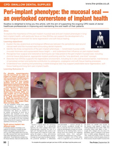
In everyday practice, knowingly or not, patients and clinicians compare the periimplant seal against the periodontium — and it is worth being explicit about how the two differ, because their mere visual similarity hides deep histological and functional distinctions. Those distinctions dictate how the tissue behaves, heals and defends itself.
50 50-51_Probe-Sep_Swallow CPD.indd 50
Around a natural tooth, the soft-tissue collar is anchored by a periodontal ligament, into which collagen fibres insert perpendicularly, encircling the root in a tight cuff. That ligament also carries a rich blood supply and a reservoir of stem cells, giving the gingiva a living, self-renewing attachment with true regenerative capacity. Crucially, the seal is also an effective barrier: the junctional epithelium is deliberately
semi-permeable — porous enough to permit immune surveillance through the flow of crevicular fluid and neutrophils — while the dentogingival complex as a whole, reinforced by its inserting fibres, forms a strong bond that protects the underlying submucosal tissue from pathogen penetration (Bosshardt and Lang, 2005). Around an implant, none of this exists. There is no ligament and no cementum, so
To complete the questions and gain one hour of CPD, visit https://cpd.the-probe.co.uk/
the fibres run parallel and circular to the titanium, lying against it rather than inserting (Berglundh and Lindhe, 1996). The connective tissue itself differs in kind — roughly 85% collagen and only about 3% fibroblasts, closer in character to scar tissue than to healthy gingiva (Berglundh et al., 1991; Araújo and Lindhe, 2018). The blood supply is scarce: where the gingiva draws on three sources, the peri-implant mucosa is fed
The Probe | September 26
01/09/2026 15:57:05