Residency Competition Winners Announced The 108th Annual Foot & Ankle Scientific Seminar featured the annual Gerard V. Yu Scientific Paper Competition and the 9th annual Scientific Poster Competition on May 16, 2024. A special thank you to Dr. Robert Brarens who chaired the competitions. This year’s paper competition featured 6 total submissions. The initial papers were distributed to a panel of judges, who evaluated the papers blindly and selected the finalists: • Aaron Abraham, DPM – The Jewish Hospital • Joseph Brown, DPM – Grant Medical Center • Ross Groeschl, DPM – Grant Medical Center • Damini Patel, DPM – Dayton VA • Alex Pilkington, DPM – University of Cincinnati Medical Center The five finalists presented to a five-judge panel of physicians, who scored the presentations. The winning paper was determined based on a combination of the blind judges’ scores of the papers and the judges’ scores. The competition results are as follows (bolded name represents the presenter): First Place: Joseph Brown, DPM; Zachary Hill, DPM; Ross Groeschl, DPM; Robert Mendicino, DPM; Brian Steginsky, DPM - Effects of Parallax and Distortion in Total Ankle Arthroplasty (Grant Medical Center) Second Place: Joseph Brown, DPM; Zachary Hill, DPM; Ross Groeschl, DPM; Robert Mendicino, DPM; Brian Steginsky, DPM; Joshua Houser, DPM - Parallax and Distortion in Fluoroscopy Units (Grant Medical Center) Third Place: Aaron Abraham, DPM; Robert Brarens, DPM - Surgical Management of Pes Planus in Children with Hypermobile Ehlers-Danlos: A Case Report and Review of Literature (The Jewish Hospital) Honorable Mentions: • Damini Patel, DPM; Wendy Culp, DPM; Judy Kleman, DPM Squamous Cell Carcinoma Presenting as a Non-Healing Wound (Dayton VA) • Alex Pilkinton, DPM; Suhail Masadeh, DPM - Potato Ray: A Model for Understanding Ray Osteotomies and Fixation Techniques for the Surgical Trainee (University of Cincinnati Medical Center) Thank you to our 2024 Resident Paper Competition Judges: Blind Paper Judges: • Dr. Tatyana Hamilton • Dr. Timothy Holmes • Dr. Amy Masowick
• Dr. Richard Weiner • Dr. Sadie Wylie
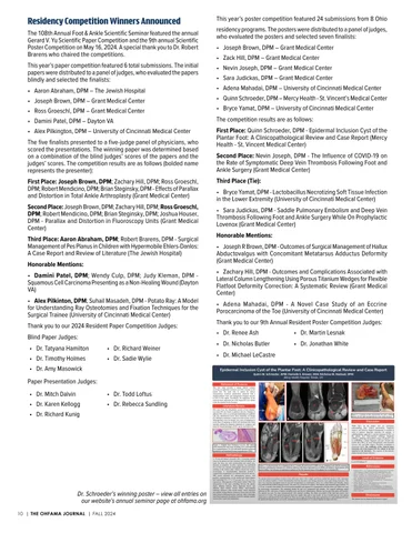
residency programs. The posters were distributed to a panel of judges, who evaluated the posters and selected seven finalists: • Joseph Brown, DPM – Grant Medical Center • Zack Hill, DPM – Grant Medical Center • Nevin Joseph, DPM – Grant Medical Center • Sara Judickas, DPM – Grant Medical Center • Adena Mahadai, DPM – University of Cincinnati Medical Center • Quinn Schroeder, DPM – Mercy Health - St. Vincent’s Medical Center • Bryce Yamat, DPM – University of Cincinnati Medical Center The competition results are as follows: First Place: Quinn Schroeder, DPM - Epidermal Inclusion Cyst of the Plantar Foot: A Clinicopathological Review and Case Report (Mercy Health - St. Vincent Medical Center) Second Place: Nevin Joseph, DPM - The Influence of COVID-19 on the Rate of Symptomatic Deep Vein Thrombosis Following Foot and Ankle Surgery (Grant Medical Center) Third Place (Tie): • Bryce Yamat, DPM - Lactobacillus Necrotizing Soft Tissue Infection in the Lower Extremity (University of Cincinnati Medical Center) • Sara Judickas, DPM - Saddle Pulmonary Embolism and Deep Vein Thrombosis Following Foot and Ankle Surgery While On Prophylactic Lovenox (Grant Medical Center) Honorable Mentions: • Joseph R Brown, DPM - Outcomes of Surgical Management of Hallux Abductovalgus with Concomitant Metatarsus Adductus Deformity (Grant Medical Center) • Zachary Hill, DPM - Outcomes and Complications Associated with Lateral Column Lengthening Using Porous Titanium Wedges for Flexible Flatfoot Deformity Correction: A Systematic Review (Grant Medical Center) • Adena Mahadai, DPM - A Novel Case Study of an Eccrine Porocarcinoma of the Toe (University of Cincinnati Medical Center) Thank you to our 9th Annual Resident Poster Competition Judges: • Dr. Renee Ash
• Dr. Martin Lesnak
• Dr. Nicholas Butler
• Dr. Jonathan White
• Dr. Michael LeCastre Epidermal Inclusion Cyst of the Plantar Foot: A Clinicopathological Review and Case Report Quinn M. Schroeder, DPM; Danielle E. Brewer, MS4; Nicholas M. Nadaud, DPM Mercy Health Hospital, Toledo, OH
Paper Presentation Judges: • Dr. Mitch Dalvin • Dr. Karen Kellogg • Dr. Richard Kunig
This year’s poster competition featured 24 submissions from 8 Ohio
Statement of Purpose
• Dr. Todd Loftus • Dr. Rebecca Sundling
This case study provides a unique clinical scenario involving an epidermal inclusion cyst (EIC) located on the plantar foot. The study aims to explore the clinical characteristics, treatment optimization enhanced with a multidisciplinary team, and management strategies specific to EICs in this region. Secondarily, the confluency of the removed tissue represents one of the largest EICs by volume in the foot reported in the literature.
Literature Review EICs arise from the infundibular portion of hair follicles. Less than 10% are found on glabrous skin [1]. Etiology of glabrous EICs often occur through implantation of epithelial cells into subcutaneous tissue, most commonly via trauma [2]. Definitive treatment is surgical excision of the cyst [3]. Although these cysts are benign, rare cases of malignancies in the form of squamous or basal cell carcinoma have been reported, making the diagnosis important to recognize and treat [4]. General histology of EICs can be seen below [1].
B
A
AH
10 | THE OHFAMA JOURNAL | FALL 2024
E
F
Figure 3: A portion of the excised EIC (E) and a clinical picture (F) five days postoperatively with a drain in place.
Discussion
FDB
AD
While EICs on the plantar foot are infrequent, understanding their clinical presentation, diagnostic nuances, and effective surgical management is essential in order to improve long-term outcomes for patients. A multidisciplinary approach is vital for treatment of EICs on the plantar foot. Interventional radiology, diagnostic radiology, pathology and podiatric surgery all played a role in establishing a proper diagnosis and care. Recent case reports show EICs can occur iatrogenically with minimally invasive foot and ankle surgery. Therefore recognition is paramount [1][5]. The confluency of the removed tissue represents one of the largest EICs by volume in the foot reported in the literature. The response of the patient’s plantar fat pad remains unknown.
FDB
AD
AD
Methodology
Dr. Schroeder’s winning poster – view all entries on our website’s annual seminar page at ohfama.org
AH
AD
Figure 1: Adapted from Posthuma et al [1]. EIC stained with hematoxylin and eosin revealing (A) a well-capsuled cyst (magnification × 5), (B) with lined stratified squamous epithelium and lamellated keratin (magnification x100).
A 37-year-old female presented with a worsening painful mass on the plantar foot that interfered with ambulation. On exam, the mass was well circumscribed and raised; nonfluctuant on palpation. In-office aspiration was performed but no fluid was retained. Radiographs showed soft tissue swelling at the medial ankle without concern for osseous involvement. A musculoskeletal ultrasound showed a compressible encapsulated cyst without concern for abscess. MRI results indicated two subcutaneous masses, with possible communication, on the medial and lateral plantar heel measuring 5.0 x 5.7 x 2.2 cm and 2.0 x 1.0 x 2.6 cm, respectively. Heterogeneity of the masses varied. There was interposition of the mass extending between the plantar fascia, abductor hallucis, flexor digitorum brevis, and abductor digiti minimi muscle bellies. Interventional radiology obtained a fine needle biopsy which displayed non-specific, benign fibrous connective tissue and synovium with extensive fibrinous/necrotic material. After a thorough, team-based approach, the patient elected to undergo surgical excision due to pain with ambulation.
C
C
C
C C
D
Figure 2: A clinical picture (A) is displayed above. Coronal (B) and Axial (C) MRI cuts are displayed with labeled anatomical structures including the calcaneus (C), abductor hallucis (AH), flexor digitorum brevis (FDB), and abductor digiti minimi (AD) muscle bellies. For reference an anatomical rendition of the first muscular layer of the plantar foot is attached (D).
Results The patient underwent radical resection of the soft tissue tumor with a single curvilinear medial heel incision with exploration across the plantar hindfoot. Careful discrimination between the native and foreign structures were made during dissection. The mass was avascular throughout removal and well encapsulated on visualization. There was a fluid filled sack medially, which was penetrated and deflated during removal. The mass was excised in its entirety. The drained sack measured approximately 7.0 x 5.0 cm. A second, firm nodule was excised posteromedially and appeared to communicate with the entire cyst. Both were sent for microscopic examination. The remaining plantar fat pad was compressed and atrophic. The surgical pathology report indicated an EIC with areas of rupture including acute and chronic inflammation, fibrosis, and foreign body giant cell reaction. The patient was seen five days postoperatively with minimal swelling. The drain was pulled at this time and sutures were removed at 3 weeks. The patient progressed back into regular shoe gear at one month. At her 6 month post-op visit, patient relates pain is still present but much improved. There are no signs of return of the mass and the heel is well cushioned. Of note, the patient developed another cyst on the contralateral foot (confirmed on CT) which impedes her daily activities.
Level of Evidence Level of Evidence: IV
References 1. Posthuma, Jelle J., et al. "Traumatic epidermal inclusion cyst after minimally invasive surgery of a displaced intra-articular calcaneal fracture: a case report." The Journal of Foot and Ankle Surgery 57.6 (2018): 1253-1255. 2. Shimizu, Yoshihiko, et al. "Clinicopathologic features of epidermal cysts of the sole: comparison with traditional epidermal cysts and trichilemmal cysts." Journal of cutaneous pathology 32.4 (2005): 280-285. 3. Trinh, Cong Thao, et al. "Overview of epidermoid cyst." European journal of radiology open 6 (2019): 291-301. 4. Houdek, Matthew T., et al. "Giant epidermal cyst of the gluteal region." Radiology case reports 5.4 (2010): 476. 5. Skolnik, Jennifer, Jane Pontious, and Todd Hasenstein. "Development of multiple epidermal inclusion cysts after radiofrequency microtenotomy for plantar fasciitis: a case report." The Journal of Foot and Ankle Surgery 60.5 (2021): 1088-1093.
Disclosures The authors have no financial disclosures to report.