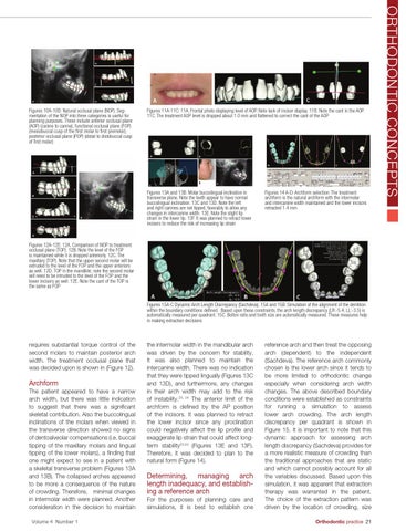
Figures 11A-11C: 11A. Frontal photo displaying level of AOP. Note lack of incisor display. 11B. Note the cant in the AOP. 11C. The treatment AOP level is dropped about 1.0 mm and flattened to correct the cant of the AOP
Figures 13A and 13B: Molar buccolingual inclination in transverse plane. Note the teeth appear to have normal buccolingual inclination. 13C and 13D. Note the left and right canines are not tipped, favorably to allow any changes in intercanine width. 13E. Note the slight lip strain in the lower lip. 13F. It was planned to retract lower incisors to reduce the risk of increasing lip strain
Figures 14 A-D: Archform selection: The treatment archform is the natural archform with the intermolar and intercanine width maintained and the lower incisors retracted 1.4 mm
Figures 12A-12E: 12A. Comparison of NOP to treatment occlusal plane (TOP). 12B. Note the level of the FOP is maintained while it is dropped anteriorly. 12C. The maxillary (TOP). Note that the upper second molar will be extruded to the level of the FOP and the upper anteriors as well. 12D. TOP in the mandible; note the second molar will need to be intruded to the level of the FOP and the lower incisors as well. 12E. Note the cant of the TOP is the same as FOP
Figures 15A-C Dynamic Arch Length Discrepancy (Sachdeva). 15A and 15B. Simulation of the alignment of the dentition within the boundary conditions defined. Based upon these constraints, the arch length discrepancy (LR:-5.4, LL:-3.5) is automatically measured per quadrant. 15C. Bolton ratio and tooth size are automatically measured. These measures help in making extraction decisions
requires substantial torque control of the second molars to maintain posterior arch width. The treatment occlusal plane that was decided upon is shown in (Figure 12).
Archform The patient appeared to have a narrow arch width, but there was little indication to suggest that there was a significant skeletal contribution. Also the buccolingual inclinations of the molars when viewed in the transverse direction showed no signs of dentoalveolar compensations (i.e. buccal tipping of the maxillary molars and lingual tipping of the lower molars), a finding that one might expect to see in a patient with a skeletal transverse problem (Figures 13A and 13B). The collapsed arches appeared to be more a consequence of the nature of crowding. Therefore, minimal changes in intermolar width were planned. Another consideration in the decision to maintain Volume 4 Number 1
the intermolar width in the mandibular arch was driven by the concern for stability. It was also planned to maintain the intercanine width. There was no indication that they were tipped lingually (Figures 13C and 13D), and furthermore, any changes in their arch width may add to the risk of instability.23, 24 The anterior limit of the archform is defined by the AP position of the incisors. It was planned to retract the lower incisor since any proclination could negatively affect the lip profile and exaggerate lip strain that could affect longterm stability23,24 (Figures 13E and 13F). Therefore, it was decided to plan to the natural form (Figure 14).
Determining, managing arch length inadequacy, and establishing a reference arch For the purposes of planning care and simulations, it is best to establish one
reference arch and then treat the opposing arch (dependent) to the independent (Sachdeva). The reference arch commonly chosen is the lower arch since it tends to be more limited to orthodontic change especially when considering arch width changes. The above described boundary conditions were established as constraints for running a simulation to assess lower arch crowding. The arch length discrepancy per quadrant is shown in Figure 15. It is important to note that this dynamic approach for assessing arch length discrepancy (Sachdeva) provides for a more realistic measure of crowding than the traditional approaches that are static and which cannot possibly account for all the variables discussed. Based upon this simulation, it was apparent that extraction therapy was warranted in the patient. The choice of the extraction pattern was driven by the location of crowding, size Orthodontic practice 21
ORTHODONTIC CONCEPTS
Figures 10A-10D: Natural occlusal plane (NOP). Segmentation of the NOP into three categories is useful for planning purposes. These include anterior occlusal plane (AOP) (canine to canine), functional occlusal plane (FOP) (mesiobuccal cusp of the first molar to first premolar), posterior occlusal plane (POP) (distal to distobuccal cusp of first molar)