CONNECTED

F A L L I
S
H E R E

CONNECTED
Welcome to the fall edition of Connected! Hard to believe the leaves are already changing and I am officially 0 and 7 in my elk bow hunting career. I guess I better stick to doing surgery to feed the family! The few grouse and pheasants I manage to find don't fill the freezer for the winter.
We had a busy summer and have added a few new faces and services to the BVS team. We hope you are all doing well and want to make sure we stay Connected with our referral community ~
Josh
CONNECTED
R T U P D A
TE S
We had an unfortunate flood in the linear accelerator last month which shut down the radiation service for a few weeks, but thanks to the quick response of our team, we are now back up and fully functional.
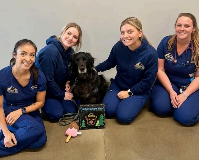
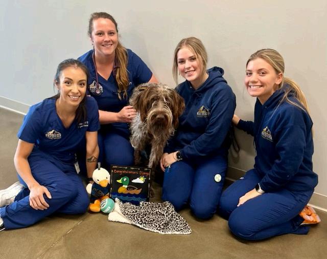
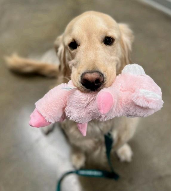
In the last 3 months we have had 18 patients complete their RT protocols. The owners report that their pets did very well during the protocols and they even enjoyed coming in to see us. We do try to make the experience as comfortable as possible. The dogs get toys to play with and a lot of one on one time with the RT team. Here are a few of our recent RT graduates:
E M E R G E
NC Y
CONNECTED
We are also officially open 24/7 for emergency care. This includes transfers, referrals, as well as walk ins on nights and weekends. We will also see overflow cases during the day if needed. We remain committed to being your partner in case management and make sure to avoid direct transfer of cases from ER to Specialty without your involvement. On our end, this means communicating with you in the mornings to see if you would prefer stable cases be transferred back to your hospital or remain at BVS. This is often a slippery slope to manage ER cases to do what is best for the client, the patient, and our referral partners, so be sure to contact us if we are not communicating this effectively or living up to you or your clients' expectations. ER cases can often be the most challenging and owners are often stressed with the least clear picture of what to expect. This includes unfamiliar faces, finances, wait times and prognosis. While I have always considered ER to be a challenging specialty, I think it can be the most rewarding and where we can have the greatest impact in the community. We will continue to strive to improve as we expand the ER service at BVS and will rely on your feedback to help us.
N
E U R O L
O G Y
Bridger Veterinary Specialists is pleased to announce that the Neurology and Neurosurgery service is now offering care on a full time basis. We are excited Dr. Carlos Valerio has joined the team and is available for neurology appointments each week. The neurology and neurosurgery service provides consultation, diagnostics and care for various canine and feline neurological diseases. Advanced diagnostic capabilities include on site MRI and CT. We have extensive experience in the use and interpretation of these imaging modalities. Commonly treated medical neurological conditions including epilepsy, inflammatory brain diseases and brain tumors. We also have extensive neurosurgical experience and perform routine surgery for intervertebral disc disease in the cervical and thoracolumbar spine. We also provide state of the art care for more complicated diseases of the brain and spine, such as lumbosacral stenosis and thoracic spinal malformations. We are on the forefront of development of advanced spinal stabilization systems.
Meet Carlos:
N
E U R O L
O G Y
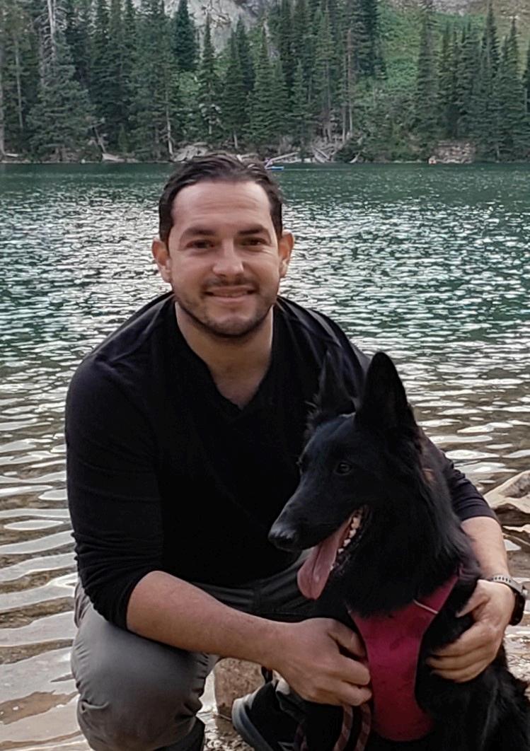
Carlos joined our team in September as our fulltime neurologist. Dr. Robert Bergman is still going to continue to be in Bozeman part-time as well. Raised in Costa Rica, Carlos grew up surrounded by family and pets, fostering a deep connection with animals from an early age. He earned his veterinary degree from the National University of Costa Rica. At Washington State University, he completed an emergency and critical care internship, followed by intensive training in neurology and neurosurgery, completing an internship and a residency. His experience at WSU exposed him to the acute need for neurological services in Montana, where patients often traveled extensive distances for care at WSU. Driven by a desire to address this gap and a personal draw to Montana's beauty, Carlos is thrilled to be joining the Bridger Veterinary Specialists neurology service and looks forward to making a meaningful impact.
O N C O L O G Y
Meet Melanie:
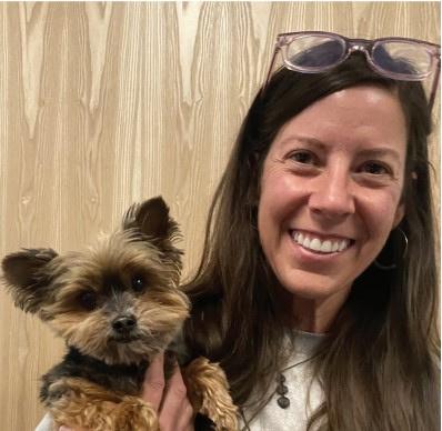
Dr. Melanie McMahon DVM, DACVIM- Oncology joined the BVS team in 2024 as our second medical oncologist. Melanie became interested in the human-animal bond during her undergraduate work at University of Texas at Austin where she received her BA in Psychology. After deciding to pursue veterinary medicine, Dr. McMahon moved to California to attend veterinary school at the University of CaliforniaDavis. She completed an internship at Colorado State University and subsequently completed her medical oncology residency and master’s degree at The Ohio State University. Dr. McMahon practiced oncology full time in Portland, Oregon for 12 years during which time she started a successful oncology service at a busy surgical hospital. For the two years prior to joining Bridger Veterinary Specialists, Dr. McMahon worked as a locum tenens filling in for veterinary oncologists in hospitals all over the United States.
D E N T I S
YRTWe are happy to announce that we have added another service at BVS. We now have a dentist to provide specialty dental care.
Meet Matt:
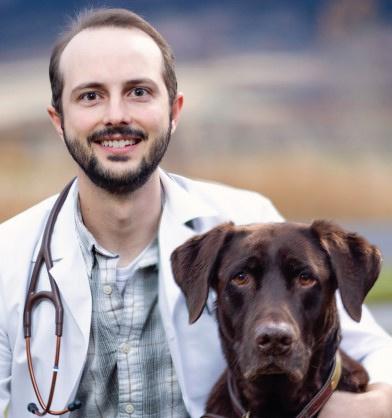
Dr. Evans is a proud graduate of Oregon State University’s College of Veterinary Medicine. He completed a dentistry and oral surgery residency at The Pet Dentist in Tampa Bay and is Board Certified by the American Veterinary Dental College. Prior to pursuing his residency training he practiced in Montana for many years, where his love for both veterinary dentistry and the Big Sky country started. He is excited to be returning to the place he considers home to provide specialty dental care. Every day he sees how improving oral health can improve overall quality-of-life, and he is motivated by a sincere desire to help people by helping their pets. A true “dental nerd” he enjoys all aspects of his job from client education to advanced oral surgery. Dr. Evans is excited to be a part of the BVS team and getting to know his clients and their furry family.
F O R E L I M B L A M E N E S S
Lameness in the Front Limbs of Dogs
Joshua Jackson, DVM, DACVS
Identifying the cause of lameness, especially in the forelimbs of dogs, can be one of the most challenging aspects of veterinary orthopedics. Very often the site of pain can’t be located, even on a thorough orthopedic exam. Stoic and hyperactive dogs will often mask their pain. Diagnosis can be made even more difficult when signs are intermittent, waxing and waning or noticed only after prolonged inactivity. Since diagnosis, prognosis and treatment of forelimb lameness can be so variable, it is important to approach each case methodically and systematically.
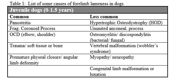
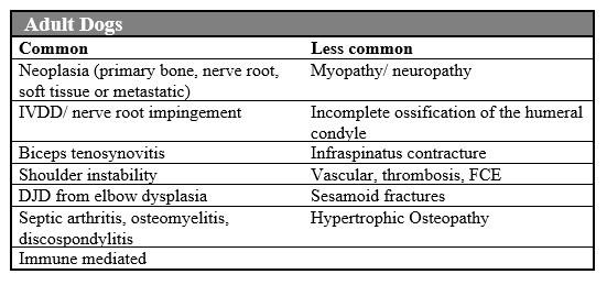
Signalment: a list of some of the potential causes of forelimb lameness is listed in table one. Breed and age can often help guide the search. For instance, young Rottweilers and Labradors have a very high incidence of fragmented coronoid processes.
CONNECTED
Orthopedic/ Neurologic examination: As already stated, physical examination has limitations with some cases of forelimb lameness. This increases the importance of a methodical and complete exam. In my experience, owner assessment of which leg is painful is often incorrect. The patient’s gait should be evaluated at a walk and trot with a loose leash. A trained technician or assistant is often preferable to having the dog led by the owner. Dogs should be palpated while standing at the dogs rear to assess for muscle symmetry and atrophy as well as joint effusion. All long bones should have deep palpation of the entire length to note pain or masses. All joints should be put through a complete range of motion and tested for medial/lateral stability. Hyperflexion and extension of both the elbow and shoulder should be performed, and pain response noted for both forelimbs. The shoulder should have craniomedial palpation of the biceps tendon with the shoulder in flexion. A “drawer” test for shoulder instability should also be performed. In stoic dogs, I will often perform a known painful procedure (toe pinch or ulnar nerve pinch) to assess what the dog’s response to pain would be.
Neurologic assessment should include evaluation of proprioception, withdrawal, neck pain, biceps and triceps reflexes and a deep palpation of the axillary region for masses.
CONNECTED
Diagnostic procedures:
Radiography: The information obtained from signalment and examination guide the first diagnostic steps. Often, radiographs of the limb are a good first step. In dogs with no pain localized, high detail films of the whole limb are often valuable. Elbow radiographs are most useful with hyperflexed lateral views to evaluate the coronoid process, osteophytes on the anconeal process and signs of trochlear sclerosis. Oblique elbow radiographs may enable visualization of a coronoid fragment. Straight AP radiographs are the best way to evaluate for elbow OCD and incomplete ossification of the humeral condyles. Lateral shoulder radiographs with the joint pulled cranially enable the best view to evaluate for OCD and mineralization of the biceps tendon.
Arthrocentesis: Fluid analysis is an easy, non-invasive test the gives significant information about the status of the joints. Differentiating between normal, non-suppurative inflammation and suppurative inflammation is key to identifying the cause of many problems. Arthrocentesis is particularly important when ruling out immune mediated disease or when septic arthritis is possible. Any dog with severe, acute lameness with a chronically arthritic joint, should be screened for septic arthritis.
CONNECTED
Diagnostic procedures (continued):
Ultrasound: as a non-invasive test, can be useful in evaluating biceps tendon disease and evaluating the brachial plexus for masses.
Arthroscopy: the Gold standard for localizing problems within the shoulder and elbow. Most useful for management of elbow dysplasia, OCD, biceps tendon disease and medial glenohumeral ligament desmitis. Can also be useful for obtaining synovial biopsies or performing joint lavage for septic arthritis.
MRI/ CT: MRI is very useful for soft tissue evaluation. Best for localizing lateralized cervical disc disease, brachial plexus tumors and other soft tissue or bone masses. Unfortunately, MRI requires general anesthesia and requires a precise localization. Whole limb MRI is not typically performed. MRI can also be cost prohibitive for many clients trying to localize a source of pain. CT is best for bone and joint detail. It is very sensitive for evaluating incomplete ossification of the humeral condyle and coronoid process disease and picking up calcified lesions in the shoulder. CT can often be performed quickly under sedation and may define pathology missed in survey radiographs.
CONNECTED
EMG/ nerve conduction: often scheduled in conjunction with advanced imaging (MRI/ CT) and can help diagnosis nerve root tumors and primary neuromuscular disorders.
Response to treatment: While listed last, it is not uncommon for us to use response to therapy to guide us on treatment. In particular with shoulder pain in the mature dog where a soft tissue injury (biceps tenosynovitis, medial shoulder instability or supraspinatus tendinopathy) is considered likely. ~60% of shoulder soft tissue injuries will respond to intra articular steroids. We typically use methylpredacetate at 0.5-1mg/kg. Joint/ regional blocks: While used commonly to localize lameness in horses, we don’t often end up going this route and I personally have not had much luck in dogs. An exception might be with distal limb pathology like sesamoid fractures or digit pain and trying to rule those out as the underlying cause.
While it was me that wrote this update, I strongly encourage you to refer your vague, intermittent forelimb lameness cases to either Dr. Wheeler or Dr. Owen as they have a preference for these diagnostic dilemmas! : )
Josh
A L L S Y M P O S I U M SIGN UP
We’d like to welcome you to our Fall Symposium on November 16, 2024 where you will have a chance to meet our new specialists and get 6 hours of FREE RACE approved CE and a delicious lunch. Save the date! Click here to sign up:
Our Symposium topics include:
Ophthalmology with Dr. Crystal Eng, DVM, DVCVO
Neurology with Dr. Carlos Valerio, DVM, DACVIMNeurology
Surgery with Dr. Deandra Owen, DVM, DACVS
Dentistry with Dr. Matt Evans, DVM, DAVDC
Oncology with Dr. Melanie McMahon, DVM, DACVIM-Oncology
Surgery with Ryan Boharski, DVM, Surgical Resident and Erik Kleeman, DVM, Surgical Resident
We also have a special guest that will be joining us to share innovative strategies to decrease stress, prevent burnout, and boost happiness at work! Paul Osincup is the author of the book The Humor Habit.

O S T E O S A R C O M A S T U D Y
Osteosarcoma Clinical Trial at BVS
BVS is working with Colorado State University to evaluate expedited chemotherapy administration following limb amputation for dogs with osteosarcoma.
For many years the standard of care for dogs with osteosarcoma of the limb has been surgical removal of the affected limb followed by chemotherapy. Traditionally, chemotherapy is administered 14 days after surgery to allow the normal tissue to heal. A retrospective study evaluating dogs receiving chemotherapy ≤5 days following a limb amputation had an improved median survival time when compared to dogs receiving adjuvant chemotherapy >5 days following limb amputation. The dogs receiving chemotherapy earlier for treatment did not have an increased incidence of complications, all reported postoperative wound complications healed, and there was an improved MST of 445 days versus 293 days for dogs receiving adjuvant chemotherapy sooner (Marconato et al. 2021 JAVMA).
S T E O S A R C O M A S T U D Y
The Goals of the current clinical trial include:
(1) To determine if there is any impact to limb amputation wound healing when chemotherapy is administered within 24 hours of surgery.
(2) To assess if the survival of dogs receiving chemotherapy within 24 hours of surgery is improved from current standard of care practice.
Dogs are eligible for the clinical trial if they have a bone sarcoma (confirmed with cytology of histopathology) that would be treated with limb amputation. They must not have any evidence of metastasis found on exam and thoracic radiographs. Following amputation, patients receive carboplatin every 21 days for 4-6 treatments. The first dose of chemotherapy is administered the day following surgery.
Please reach out to Dr. Kelly Carlsten, DVM, DACVIM-Oncology with any questions about the trial. Kelly@BVSPETS.com or (408)-548-4226