Infective endocarditis: a history of the development of its understanding
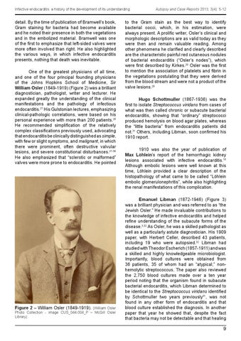
detail. By the time of publication of Bramwell’s book, Gram staining for bacteria had become available and he noted their presence in both the vegetations and in the embolized material. Bramwell was one of the first to emphasize that left-sided valves were more often involved than right. He also highlighted the various ways, in which infective endocarditis presents, nothing that death was inevitable. One of the greatest physicians of all time, and one of the four principal founding physicians of the Johns Hopkins School of Medicine, Sir William Osler (1849-1919) (Figure 2) was a brilliant diagnostician, pathologist, writer and lecturer. He expanded greatly the understanding of the clinical manifestations and the pathology of infectious endocarditis.27 His Gulstonian lectures, emphasizing clinical-pathologic correlations, were based on his personal experience with more than 200 patients.28 He recommended simplification of the relatively complex classifications previously used, advocating that endocarditis be clinically distinguished as simple, with few or slight symptoms, and malignant, in which there were prominent, often destructive valvular lesions, and severe constitutional disturbances.27-30 He also emphasized that “sclerotic or malformed” valves were more prone to endocarditis. He pointed
Figure 2 – William Osler (1849-1919). (William Osler Photo Collection - image CUS_044-004_P – McGill Osler Library).
Autopsy and Case Reports 2013; 3(4): 5-12
to the Gram stain as the best way to identify bacterial cocci, which, in his estimation, were always present. A prolific writer, Osler’s clinical and morphologic descriptions are as valid today as they were then and remain valuable reading. Among other phenomena he clarified and clearly described are the characteristic painful red cutaneous nodules of bacterial endocarditis (“Osler’s nodes”), which were first described by Kirkes.20 Osler was the first to mention the association of platelets and fibrin in the vegetations postulating that they were derived from the blood stream and were not a product of the valve lesions.28 Hugo Schottmuller (1867-1936) was the first to isolate Streptococcus viridans from cases of what was then called chronic or subacute bacterial endocarditis, showing that “ordinary” streptococci produced hemolysis on blood agar plates, whereas the “little bacteria” from endocarditis patients did not.31 Others, including Libman, soon confirmed his 1910 report. 1910 was also the year of publication of Max Löhlein’s report of the hemorrhagic kidney lesions associated with infective endocarditis.16 Although embolic lesions were well known at this time, Löhlein provided a clear description of the histopathology of what came to be called “Löhlein embolic glomerulonephritis”, while also highlighting the renal manifestations of this complication. Emanuel Libman (1872-1946) (Figure 3) was a brilliant physician and was referred to as “the Jewish Osler.” He made invaluable contributions to the knowledge of infective endocarditis and helped refine understanding of the subacute forms of the disease.3,32 As Osler, he was a skilled pathologist as well as a particularly astute diagnostician. His 1909 paper, with Herbert Celler, described 43 patients, including 19 who were autopsied.32 Libman had studied with Theodor Escherich (1857-1911) and was a skilled and highly knowledgeable microbiologist. Importantly, blood cultures were obtained from 36 patients, 35 of whom had an “atypical,” nonhemolytic streptococcus. The paper also reviewed the 2,750 blood cultures made over a ten year period noting that the organism found in subacute bacterial endocarditis, which Libman determined to be identical to the Streptococcus viridans identified by Schottmuller two years previously31, was not found in any other form of endocarditis and that blood culture established the diagnosis. In another paper that year he showed that, despite the fact that bacteria may not be detectable and that healing
9