Chronic meningococcemia: a rare presentation of meningococcal disease: case report
Autopsy and Case Reports 2012;2(1):29-35
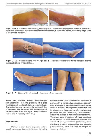
Figure 1 – A – Violaceous maculae suggestive of purpura lesions (arrows) appeared over the swollen and hyperemic lower limbs. Note intense erythema over the knee; B – Necrotic lesions, in the early stage, close to the external malleolus.
Figure 2 – A – Necrotic lesions over the right calf; B – Note also lesions close to the malleolus and the increased volume of the right knee.
Figure 3 – A - Edema of the left ankle; B – Increased left knee volume.
status was favorable allowing corticotherapy with prednisone once the possibility of a post meningococcal reactional status was considered. The patient became afebrile on the eighteenth day of treatment, was discharged with normalization of laboratory tests (CRP = 15 mg/dL) and healed skin lesions and decreased joint swelling.
DISCUSSION Meningococci are diverse organisms and are usually commensal bacteria in humans. According
to some studies, 20–40% of the adult population is permanently or temporarily asymptomatic carriers.1 Only a minority of nasopharyngeal isolates cause invasive disease.2 Meningococci associated with invasive disease elaborate a capsule, which provides protection from desiccation during transmission and aids in the evasion of host immune mechanisms. The major factor of virulence of these organisms is the release of outer-membrane vesicles that consist of lipooligosaccharide (endotoxin), outer membrane proteins, phospholipids, and capsular polysaccharides, which are used as antigen for vaccine production.2,3
31