3 minute read
How Is Atypical Mole Assessment Done With Dermoscopy?

by Ahmad

How is atypical mole assessment done with dermoscopy is a common question for people who want a precise, non-invasive way to evaluate unusual skin lesions. In regions with strong sun exposure, Dermoscopy Mole Evaluation in Dubai has become an essential part of modern skin screening, with clinics such as Dynamic Life Clinic incorporating advanced dermoscopic techniques into routine assessments for clearer, more reliable observations.
Understanding Atypical Moles and Why Assessment Matters
Atypical moles differ from common moles in size, shape, color variation, or border definition. These features do not automatically indicate a serious condition, but they do require closer examination. In Dubai, where sun exposure is frequent year-round, early evaluation helps identify changes that may not be visible to the naked eye and supports long-term skin monitoring.
What Dermoscopy Is and How It Works
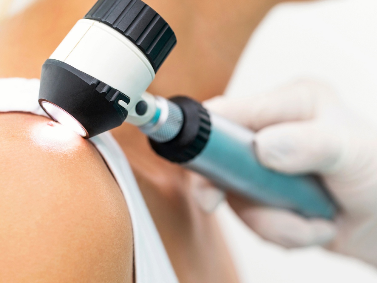
Dermoscopy is a specialized visual examination that uses a handheld device combining magnification and polarized light. This tool allows the examiner to see structures beneath the surface layer of the skin. Unlike basic visual checks, dermoscopy reveals pigment networks, vascular patterns, and symmetry details that provide deeper insight into a mole’s behavior.
Preparing for a Dermoscopic Examination
Preparation is minimal, making dermoscopy practical for routine skin checks. Before the assessment:
The skin is cleaned to remove surface oils or cosmetics
No invasive preparation is required
The examination can be done during a standard skin consultation
This simplicity encourages regular monitoring, especially for individuals with multiple or changing moles.
Step-by-Step Process of Atypical Mole Assessment
During dermoscopic evaluation, the practitioner follows a structured approach to ensure consistency and accuracy:
Visual Inspection – The mole is first assessed with the naked eye to note size, color, and border irregularities
Dermoscope Application – The device is placed gently on the skin to magnify subsurface patterns
Pattern Analysis – Pigment distribution, symmetry, and structural features are examined
Digital Documentation – Images may be recorded for comparison during future visits
This process supports careful tracking of subtle changes over time.
Key Dermoscopic Features Reviewed
Dermoscopy focuses on specific criteria known to help distinguish benign from atypical lesions:
Asymmetry in structure or color
Irregular or disrupted pigment networks
Multiple shades within a single mole
Unusual vascular or dot patterns
Evaluating these features together provides a more reliable assessment than visual inspection alone.
Role of Dermoscopy in Ongoing Monitoring
One of the strongest advantages of dermoscopy is its use in follow-up care. Digital dermoscopic images allow side-by-side comparisons during future assessments. This is particularly valuable for people living in Dubai, where cumulative sun exposure can influence mole appearance gradually rather than suddenly.
When Further Evaluation Is Considered
If dermoscopic findings suggest significant irregularities, additional diagnostic steps may be recommended. Dermoscopy helps reduce unnecessary procedures by identifying which moles truly require closer investigation while reassuring patients when lesions appear stable.
Conclusion
Atypical mole assessment with dermoscopy offers a clear, structured, and non-invasive method for evaluating unusual skin lesions. By revealing details beneath the skin’s surface and supporting ongoing monitoring, dermoscopy plays a vital role in early detection strategies, especially in sun-intensive environments like Dubai. This approach aligns with modern dermatological standards and supports informed, evidence-based skin care decisions.