Aspirational actions
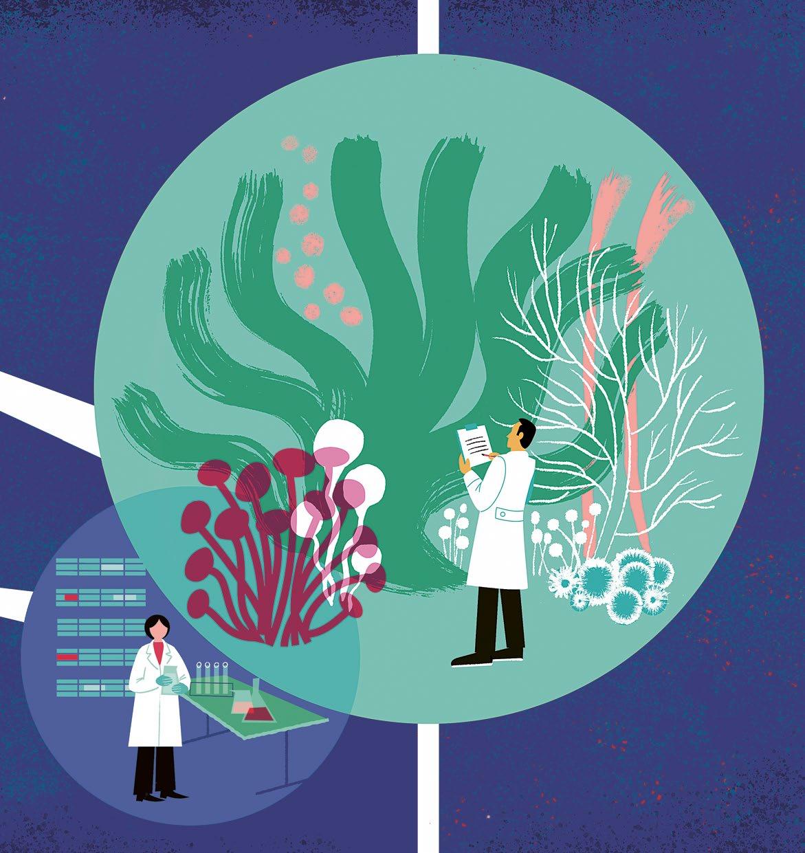
32
12 Revolutionary green hydrogen
22 Disrupting bacterial communication
24 Redesigning maternity and child health clinics
5 Openings Kristiina Kruus and aspirational engineering.
6 Now _ Bits of news, big issues.
10 Oops! Fereshteh Sohrabi and a curious discovery.
OUR THEME Aspirational actions
12 Theme Experts explain how the visions of green hydrogen can become reality.
18 Who _ Arnevi Rautanen and Juha Utriainen run the Torstila manor organic farm.
22 On science _ Researchers are developing new weapons against antibiotic resistance.
24 On science Tomorrow’s maternity clinics will have both avatar and real encounters.
28 Dialogue Laura Arpiainen and Susanna Helke challenge health and social sector with art.
30 On science The Sustainability in Business research group is a pioneer in its field.
32 Collaboration Aalto University Junior offers glimpses into the world of science.
36 On science _ Science news in brief.
38 Collaboration Mentoring programs bring professionals and students together.
42 Partnership AI assisting in dental care and jaw surgery.
44 In-house Outdoor artwork reflects quantum physics.
46 Doctoral theses _ Camilla Andersson and the norm-critical design; Olli Halminen and the health and social services; Henrik Kahanpää and the climate on Mars.
48 Wow! The museum of photography showcased artworks of new graduates.
56 Everyday choices Russel Lai defends information security with cryptography.E
58 Visiting Arto Nurmikko to receive honorary doctorate.
32
Elisabet Cavén's photograph from the series The Home Odyssey, which tells about the time when the whole world stayed at home. This art piece was on display at the Finnish Museum of Photography's exhibition MoA in Photography 23. Read more about the exhibition starting on page 48.
SEDEER EL-SHOWK, INTERNATIONAL EDITOR
‘The more I learn about Aalto, the more I discover unexpected voices here that should be heard and ideas that should be explored. Learning about them –and from them – gives me hope, and being able to amplify them through my work brings me joy.’
‘
KATI NÄRHI, ILLUSTRATOR
In this global situation, hope is a scarce resource. But still, the world is made up of details, some of which are wonderful and delightful, like Charley Harper's illustrations, the scent of jasmine tea, dachshunds, and red and pink combined. Joy, even if it’s temporary, awakens hope.’
PUBLISHER Aalto University, Communications
EDITOR-IN-CHIEF Head of Content and Media Katrina Jurva
MANAGING EDITOR Paula Haikarainen
LAYOUT/PHOTO EDITOR Dog Design
COVER Kati Närhi
CONTRIBUTORS IN THIS ISSUE Matti Ahlgren, Amanda Alvarez, Tiina Aulanko-Jokirinne, Anna Berg, Elisabet Cavén, Richard Fisher, Tiina Forsberg, Terhi Hautamäki, Anna-Katri Hänninen, Minna Hölttä, Jaakko Kahilaniemi, Kalle Kataila, Krista Kinnunen, Alexander Komenda, Marika Lehto, Annika Linna, Juuli Miettilä, Juuso Mäntykivi, Kati Närhi, Samuli Ojala, Lyydia Osara, Aleksi Poutanen, Marjukka Puolakka, Mikko Raskinen, Sedeer el-Showk, Noora Stapleton, Lada Suomenrinne, Niko Tampio, Tiina Toivola, Nita Vera
TRANSLATION Tiina Leivo, Tomi Snellman, Annamari Typpö

ADDRESS PO Box 18 000, FI-00076 Aalto TELEPHONE +358 9 470 01
ONLINE aalto.fi/magazine EMAIL magazine@aalto.fi CHANGE OF ADDRESS crm-support@aalto.fi
PRINTING PunaMusta, 2023 PAPER Maxi Offset 190 g/m2 (covers), Berga Classic Preprint 90 g/m2 (pages)
PRINT RUN 3,000 (English edition), 29,000 (Finnish edition)
SOURCE OF ADDRESSES Aalto University CRM Partnership and alumni data management
PRIVACY NOTICES aalto.fi/services/privacy-notices
ISSN 2489-6772 print ISSN 2489-6780 online
JOB
ON THE
Nita Vera
YMPÄRISTÖMERKKI MILJÖMÄRKT Painotuotteet
The carbon emission of this printed matter is evaluated according to ClimateCalc. www.climatecalc.eu Cert. no. CC-000084/FI
Painotuotteet 1234 5678
4041-0619
4 / AALTO UNIVERSITY MAGAZINE 32
In my research career I’ve focused on how enzymes and microbes break down biomass. Nature’s microbes can decompose plant-based biomass, like wood, into sugars, which pass up the food chain into animals. In this magnificent circular economy, everything is utilised and there is no waste, unlike in many industrial processes. Aspirational engineering is a new way of thinking that seeks to emulate nature’s rational use of resources.
I’m completely convinced that technological development will find solutions for the climate crisis, the loss of biodiversity and the shortage of raw materials. This massive transition is already underway: electric vehicles are taking over, single-use consumption is decreasing, and we are learning to make sustainable choices.
I am particularly encouraged that chemical engineering, which has been seen as a polluting promoter of heavy industry, is now attractive to young people at the same time as the field strives for clean solutions. We are educating the students who will make and apply the aspirational engineering of the future. An example is green hydrogen, which can be produced with Finnish wind energy and used to make, for instance, fertilisers and chemicals, free of emissions.

As a bioengineer, it is amazing to see how renewable biomaterials are now replacing fossil-based packaging like plastic. In addition to replacement, recycling of materials like textiles, fibres and metals is another important way to protect nature. But single-use consumption needs to be cut back, because stopping waste from being created in the first place is the most effective action. Technology can play a role here too, because with judicious and systematic use of raw materials and longer-lasting products, we can reduce waste and promote recycling.
The green transition is happening, and I am convinced that, with the help of technology, we can reach carbonneutrality by 2035, Finland’s goal year.
Kristiina Kruus Dean Aalto University School of Chemical Engineering
Kalle Kataila
Nature invented the circular economy –with aspirational engineering we can follow suit
AALTO UNIVERSITY MAGAZINE 32 \ 5 OPENINGS
I’m completely convinced that technological development will find solutions for the climate crisis, the loss of biodiversity and the shortage of raw materials.
New hydrogen economy training

Hydrogen plays an important role in the green transition from fossil fuels towards renewable energy production. While education in Finland on the hydrogen economy currently falls between different programmes and individual courses, the new project organised by the Finnish Institute of Technology (FITech) network brings together the educational offering that has so far been scattered.
The main target group of the education is the staff of the member companies of Hydrogen Cluster Finland. The cluster involves more than 60 companies and six industry associations. It is open to and suitable for other companies, decision-makers, and students.
An English-language study module of approximately 20-40 ECTS consists of a comprehensive introductory course on hydrogen economy and several optional advanced courses. The education is implemented largely online, and more courses will be started during this year.
The education project is coordinated by Aalto University, and the universities of the FITech network are jointly responsible for the preparation and implementation of the education.
FITech offers selected studies from all of Finland's universities of technology. The studies are open and free of charge for all Finnish citizens and permanent residents of Finland. fitech.io
QS World University Rankings
by Subject 2023
Preliminary simulations of hydrogen flames. The picture shows a swirling, turbulent flame.
in the world.
Aalto’s art and design maintains its position as one of the best.
6th
6 / AALTO UNIVERSITY MAGAZINE 32 NOW
Ilya Morev and Ville Vuorinen
Funding to study hydrogen combustion
Business Finland has granted 1.7 million euros in funding to the HENNES research project, which studies the physics and chemistry of hydrogen combustion. The research is part of Wärtsilä’s ecosystem project, which aims to contribute to the development of zero-emission marine technology, and Aalto is one of the universities involved.
The combustion of hydrogen and its derivative fuels, such as ammonia, produces no carbon dioxide emissions. The EU has set a goal to have clean, low-carbon hydrogen and derivative fuels account for almost a fifth of end-use energy by 2050.
Hydrogen differs from traditional fuels in terms of its thermodynamic and chemical properties and behaves differently in combustion processes. The ignition sensitivity of hydrogen and the small size of the molecule also pose challenges for both combustion control and hydrogen storage.
Accurate modeling of hydrogen combustion is a challenging problem in computational physics and chemistry, the understanding of which is important
when designing efficient combustion processes. Research is also needed on storing liquid hydrogen and developing heat exchangers and vaporizers used in energy transfers.
Combining 3D simulation methods with experiments is at the core of the research. Aalto University Professor Ville Vuorinen’s research team will focus on burner flames and combustion phenomena inside combustion engines, while Professor Simo Hostikka’s team will study the fire safety of hydrogen.
Armin Wehrfritz, Assistant Professor at the University of Turku, and his research group investigate the thermodynamic properties of hydrogen and hydrogen-derived fuels.
With further data, it could become possible to design even more efficient and durable internal combustion engines that run on hydrogen.
In addition to Wärtsilä, the industry partners in the project are AGCO Power, Oilon, Finno Exergy, Vahterus, Auramarine, KK-Palokonsultti Oy and P2X Solutions.
Finland creating common EU model for teaching cybersecurity skills
Aalto University and the Finnish Ministry of Transport and Communications are implementing a large-scale Cyber Citizen project to create a common model for teaching cyber citizenship skills across EU member states. As part of the project, Aalto has published a comprehensive report on the current state of teaching cyber citizenship skills throughout the EU.
The project will create a learning model and a learning portal that includes content tailored to different target groups, such as a game to teach cyber citizen skills. The aim is to improve citizens’ ability to act safely in the digital world.
The project has received five million euros in funding from the EU’s Recovery and Resilience Facility for three years.
Dear readers, thank you for your feedback
According to our readership survey in October–December 2022, the magazine’s content is considered reliable (77% of respondents) and the articles professional (78%).
The magazine’s primary tasks are to keep the reader up to date on Aalto University’s affairs, provide new information and report on recent research results. Creative experiments and new ways of thinking are topics readers would like to see covered more.

An average of 24 minutes was spent reading the magazine. 42% of the respondents want to read the magazine in print and 10% only digitally.
Half of the respondents who have read or browsed the magazine find it very or fairly useful. Only 3% of respondents consider it completely useless.
The primary target group was the alumni, the largest readership group of the magazine. The questionnaire was answered by 669 persons, of which 568 answered in Finnish and 101 in English. 77% of the respondents were alumni.
This survey was carried out by Taloustutkimus.
Aalto University Communications Services thanks all the survey participants for their valuable feedback. The results of the readership survey will be used to develop the magazine's content.
AALTO UNIVERSITY MAGAZINE 32 \ 7
THE DOCUMENTARY FILM ‘Building Ylistaro Church’ (Näin rakennettiin Ylistaron kirkko) depicts the construction phases of this Finnish church between 1847 and 1852. Since there are no photographs from that time, the documentary utilised three-dimensional modeling (3D) created by Aalto University’s Research Institute of Measuring and Modeling for the Built Environment (MeMo).
Laser scanning was used in the modeling process to measure distances and locations. The resulting point cloud depicts the object, and colour values from the object's photographs can be added to create a precise model that is finalised by 3D artists.
The film was directed by Anssi Luoma, filmed and recorded by Mika Koivusalo, and the 3D animations were created by Hannu Hyyppä, Marika Ahlavuo, Sebastian Aho, Hannu Handolin, Matti Kurkela, Toni Rantanen, Matias Ingman, and Risto Känsälä.

THE EXHIBITION ENERGY FUTURES: Critical minerals, climate neutrality and transition pathways explains how the world is moving from fossil fuels to cleaner energy solutions. For example, what will the role of hydrogen and wind power be in the future?
In addition to reducing carbon dioxide emissions, the entire energy system requires a radical change in both energy storage methods and the environmental and economic effects caused by mineral shortages.
The exhibition presents research related to the energy transition carried out at Aalto University. The energy transition means the shift of global energy production and consumption from fossil fuels to renewable energy sources.
The exhibition is on display at the Dipoli gallery (Otakaari 24, Espoo) until September 15, 2023. It can also be viewed online: virtualexhibitions.aalto.fi
 Digitally modelled church roof trusses. The 3D data collected from one of Finland's largest churches can be used to maintain and restore the church.
Aalto University's MeMo Institute
Separated silver-plated copper wire, photovoltaic solar panel waste.
Digitally modelled church roof trusses. The 3D data collected from one of Finland's largest churches can be used to maintain and restore the church.
Aalto University's MeMo Institute
Separated silver-plated copper wire, photovoltaic solar panel waste.
8 / AALTO UNIVERSITY MAGAZINE 29 NOW
Sara Urbanski
THE ROBOBALLET DANCE PERFORMANCE combines technology and classical art.
In February 2023, dance artist Mira Ollila danced with a robot in the historic Tenalji von Fersen Hall at Suomenlinna Sea Fortress. This performance was part of the Infinity Vessel project by glass artists Sini Majuri and Marja Hepo-aho. It combines dance, interactive hologram installation, and a sculpture exhibition.
The robotics were implemented in collaboration with the Aalto University Robotics research group.

AALTO UNIVERSITY MAGAZINE 32 \ 9
Juha-Matti Vahdersalo
A curious discovery
During doctoral researcher

Fereshteh
‘I was doing my master’s thesis when I learned about a summer project that seemed interesting. I wanted to expand my knowledge, so I applied for it.
The idea was to add charge-carrying agents to a ferrofluid – making what we called an electroferrofluid – and then study its behaviour in electric and magnetic fields. We were looking for certain behaviours, like self-assemblies of nanoparticles.
The experiments didn’t seem to be working, and it looked like we had failed. We couldn’t see what we were looking for. Instead, we saw some instabilities in the system that made it difficult to reach our goal.
Some others on the team had worked on other electric field-driven phenomena for another project, which gave us the idea of trying the electrode setup from those experiments. We thought that might simplify the system and generate simpler and more well-defined nanoparticle structures. We already had everything in the lab, so we tried it out of curiosity.
Something interesting happened. We started seeing various instabilities and dynamic pattern formations at a different scale than we were expecting. The first time I saw them, I was really fascinated and had some happy tears in front of the microscope.
It was a new phenomenon – that we had discovered! We decided to study the instabilities and patterns instead of trying to remove them. I looked forward to going to lab and exploring the system every day; I did so many experiments over the following weeks to test out different parameters and try to understand the patterns and how they formed.
We hadn’t expected that adding nanoparticles would lead to new complex behaviour or a whole new pattern formation system in ferrofluids. We’d been trying to do something else, but now this has opened up a whole new area of research. I decided to change my master’s thesis topic and study this system instead, and it has fascinated me ever since.
I think it’s important to always think outside the box and just explore. It’s good that research has certain structures, but when doing experiments, I’m always wondering about other things we could do. I often have a curious and playful approach.
I learned to not get disappointed when an experiment seems to fail and instead change my perspective and ask different questions. This journey has been the most exciting time of my career, and I enjoyed it so much that I’d like to pursue research in the future. Curiosity can go a long way.’
OOPS!
Sohrabi’s master’s project, curiosity turned a failing experiment into a new research avenue.
Text Sedeer el-Showk
Photo Nita Vera
Illustration Juuli Miettilä
Aspirational actions
12 Theme Experts explain how visions of green hydrogen can become reality.
18 Who Arnevi Rautanen and Juha Utriainen run the Torstila manor organic farm.
22 On science _ Researchers are developing new weapons against antibiotic resistance.
24 On science Tomorrow’s maternity clinics will have both avatar and real encounters.
28 Dialogue Laura Arpiainen and Susanna Helke challenge health and social sector with art.
30 On science The Sustainability in Business research group is a pioneer in its field.
32 Collaboration _ Aalto University Junior offers glimpses into the world of science.

Kati Närhi
THEME
AALTO UNIVERSITY MAGAZINE 32 \ 11
Art and science are instruments of hope.
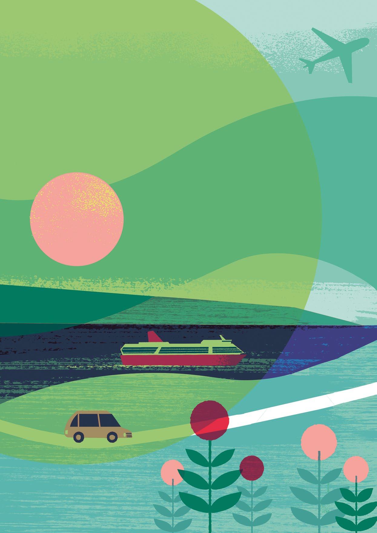
Green hydrogen promises to turn Europe into an energy heavyweight, make traffic emissionfree and replace carbon in steel manufacturing. But can it actually deliver – and how?

THEME Aspirational actions
12 / AALTO UNIVERSITY MAGAZINE 32
The world’s lightest revolution
Translation Tiina Leivo
Illustration Kati Närhi
 Text Minna Hölttä
THEME Aspirational actions
TVhe year 2023 started off well in the small Finnish town of Inkoo, near Helsinki: Norwegian-based Blastr Green Steel published its plan to invest four billion euros to build a plant in the municipality.
Should all go well, the integrated hydrogen production facility will be up and running before the end of this decade, and the plant will produce fossil-free steel and employ 1,200 people. This would mark one of the largest investments in Finland’s industrial history.
This isn’t the only hydrogen-related project in Finland’s future. Major projects are ongoing or being planned in municipalities such as Raahe, Harjavalta, Pori, Lahti and Kokkola. More broadly, the scope of investments in the EU Hydrogen Strategy amounts to hundreds of billions of euros.
Why the sudden fuss about hydrogen?
It all boils down to the energy crisis, the wind power boom and the climate emergency, says Antti Arasto, Vice President of Industrial Energy and Hydrogen at VTT Technical Research Centre of Finland. ‘The carbon neutrality target means that it’s not enough to reduce emissions – we need to get rid of them completely. Green hydrogen alone will not solve the problem, but it will play a major role in the shift.’
Grey, pink or green?
From the climate perspective, hydrogen is an ideal fuel: when it’s burned for energy, the only byproduct is water vapour. Hydrogen has been known to be an energy carrier for two centuries, but it’s had a bumpy ride in powering transport. The crash and burn of the Hindenburg airship in 1937 ended the era of zeppelins, and the 1990s hydrogen car boom quickly cooled down when no mass production or fuelling infrastructure materialised.
But there’s been a lot of interest in industrial uses of hydrogen. The oil industry uses hydrogen to break crude oil into petrol and kerosene, and the chemicals industry uses it to produce ammonia, a key raw material for fertilisers. Fossil-fuel free steel could be produced by using hydrogen to replace coke, a coal-based carbon used for reducing iron ore.
Unfortunately, hydrogen cannot simply be collected. It must be produced. Though there’s no shortage of hydrogen – it’s the most common element in the universe – it does not like solitude. Hydrogen readily binds to other elements in most substances: fossil fuels, water and plant biomass.
Natural gas is by far the most common source material for traditional hydrogen production. In 2020, approximately 90 million tonnes of hydrogen were produced around the world, causing over 800 million tonnes of carbon dioxide emissions – about
equivalent to the annual emissions of Great Britain and Indonesia combined.
The carbon footprint of hydrogen depends on how it’s produced. A colour coding system distinguishes the hydrogen based on its source. Hydrogen derived from coal and oil has the highest emissions and is labelled black; hydrogen extracted from natural gas is called grey. If the carbon dioxide created in the production process is recovered, the hydrogen is labelled blue.
Hydrogen can also be produced by using electricity to break water into hydrogen and oxygen. This method, electrolysis, is simple and emission-free, but it requires energy from electricity. If the electricity is produced with nuclear power, then the emission-free hydrogen is called pink. To be labelled green, hydrogen has to be made using electricity produced from renewable sources, such as wind, solar or hydropower.
A lot of power is needed for electrolysis, so electricity has to be cheap for green hydrogen production and the industries dependent on it profitable. The Inkoo steel plant alone would eat up roughly six terawatt-hours of electricity per year, which is about 7% of Finland’s current total electricity production.
Arasto stresses that green hydrogen production goes hand in hand with increased wind power potential. The price of wind electricity has plummeted in Finland in the 2000s, and onshore wind power has now become the cheapest electricity production method. Finland currently hosts about 1,400 wind power plants, producing around a tenth of the country’s electricity. This share is expected to increase to over a half by 2050.
At the end of this decade, wind power could cover the nation’s entire electricity consumption on a windy day. But what will happen on windless days? Or when the volume of wind power exceeds demand?
Hydrogen cannot simply be collected. It must be produced.
14 / AALTO UNIVERSITY MAGAZINE 31
Road, marine and air traffic powered by hydrogen When wind power production exceeds demand, the extra power can be stored by making hydrogen through electrolysis, which can then be used when wind power is unavailable or insufficient.
In addition to industrial uses, stored hydrogen can serve as fuel for hydrogen engines or in fuel cells that produce electricity out of hydrogen. Toyota plans to release a new hydrogen car later this year, while Airbus is aiming for commercial hydrogen flights in 2035. Stored hydrogen can also be used to produce electricity for the grid with gas turbines, for example, or with Wärtsilä’s engines capable of fast grid balancing.
However, the storage and transport of hydrogen has its challenges. A hydrogen molecule is extremely
small: it only contains two hydrogen atoms, each made of a single proton and a single electron. Hydrogen can thus quickly escape through even the smallest holes. One kilogram of hydrogen contains three times as much energy as the same amount of petrol, but since hydrogen is the world’s lightest substance, a kilogram of hydrogen –even if liquefied – takes up a lot more space than a kilogram of petrol.
Storing and using hydrogen can be made easier by refining it into e-fuels. The only other substance need for this is carbon dioxide, which could be collected directly from the smokestacks of emission-heavy plants. The simplest electric fuel is methane, or natural gas, which could directly replace the fossil natural gas used in heavy road traffic and shipping, for example.

AALTO UNIVERSITY MAGAZINE 32 \ 15
The price of electric fuels is higher than that of oil or natural gas because a lot of electricity is wasted in hydrogen processes, but Arasto finds price comparison to be partially pointless. ‘Considering the climate goals, fossil fuels are no longer an option for the future.’
The EU has set a goal of covering as much as one-fifth of its energy demand with green or lowcarbon hydrogen by 2050. Even before the Ukraine war, the EU Commission had declared that the continent should reduce its energy dependence and that future hydrogen trading should take place in euros instead of dollars.

Realising this plan requires not only massive wind power potential but also an enormous quantity of cheaper and more efficient electrolysers – which means solving the related material challenges.
Platinum and its rivals
Aalto’s Associate Professor Tanja Kallio studies the catalyst materials needed for energy storage and for converting it from one form into another.
During her long career, there’s never been as much demand or need for her research as there is now.
The cause is clear. ‘The International Energy Agency expects the world’s hydrogen production to increase sixfold by 2050. If that happens, we need to find new solutions for the catalysts in electrolysers,’ Kallio says.
The catalysts speed up the reaction in electrolysers, helping store electric power as hydrogen. Precious metals, particularly platinum, are usually used as catalyst materials. The EU has now listed platinum and many other commonly used catalysts as critical raw materials. That designation means that they are either at risk of running out globally –and becoming astronomically expensive as stocks decline – or that they are obtained from geopolitically challenging countries, meaning Europe’s supply is not secure.
That’s why Kallio and her team are developing methods to get as much output from the precious platinum as possible. ‘The ratio of the area of platinum to its volume should be as high as possible.
THEME Aspirational actions
16 / AALTO UNIVERSITY MAGAZINE 32
In other words, the catalyst must have all the platinum on its surface. This isn’t a new idea, but its implementation is challenging: as the area becomes larger, the atoms tend to pile up over time. We’ve tried to solve this by increasing the binding energy of the surface carrying the platinum, and that has stabilised the platinum structures.’
Manufacturing and recycling a catalyst both take a lot of energy, and a part of the material is always lost. The group is therefore also looking for ways to extend the lifespan of catalysts.
In addition to developing catalysts that need less platinum, Kallio’s group has also developed platinum-free catalysts made from cheaper, more common materials, such as iron and carbon. These catalysts are fairly long-lived, but they consume a lot of energy, reducing efficiency.
‘Considering the fact that there’s sometimes almost unlimited energy available, they could still offer a better option than platinum catalysts,’ Kallio says.
Long and crucial chain
The largest source of carbon dioxide emissions in Finland is a steel plant in Raahe, and the demand for steel is rising steeply. Last year, plant owner SSAB announced that it will speed up its investments, and they aim to make production emission-free by 2030, ten years ahead of schedule. That would cut Finland’s cardon dioxide emissions by as much as 7% in one go.
SSAB is leading a research and development project called ‘Fossil Free Steel’ in which the energy company Fortum is investigating the potential of hydrogen production. Mikko Muoniovaara leads hydrogen business development at Fortum and believes steel production provides the most promising business opening. ‘We could directly replace coke fuel with hydrogen in iron ore reduction.’
Fortum is part of a national hydrogen cluster comprising 65 companies that aims to accelerate Finland’s hydrogen industry and hydrogen economy by influencing legislation and investment.
In addition to supportive legislation and financing, the industry needs the latest research results and expertise. ‘The research potential of universities is very important to us,’ says Muonivaara. For example, Fortum and Aalto have jointly studied the option of storing hydrogen in Finnish bedrock.
The cluster was also closely involved in planning the study courses in hydrogen economy offered by the FITech network university. The introductory course was launched at the beginning of this year.
‘We have a lot of expertise here in Finland, but it’s scattered around in different universities. This programme lets us bring it together in one place through remote studies,’ Muoniovaara says.
When VTT Vice President Antti Arasto studied energy technology in Otaniemi two decades ago, green hydrogen was hardly mentioned at all. ‘We’re now moving really quickly towards industrial applications. Many solutions that have been studied at VTT and elsewhere for ten or twenty years are now reality,’ Arasto says.
‘The chain from basic research to applied research, product development and investments in companies, and then to a shift in the whole world’s infrastructure, is tremendously long. The hydrogen revolution is a shining example of the fact that the whole pipeline needs to be invested in, instead of allocating isolated funds at the early and final stages of the process –and that education really is worth the investment.’
‘Price comparison is pointless. Fossil fuels are no longer an option for the future.’
AALTO UNIVERSITY MAGAZINE 32 \ 17

Returning to the roots
Arnevi Rautanen and Juha Utriainen gave up their careers to take up farming in the village of Joroinen in Eastern Finland.
Text Marika Lehto
Translation Annamari Typpö, Sedeer el-Showk Photos Anna-Katri Hänninen
It all started with high-flying dreams of an international life, but in the end their true dreams were realised in the soil of a small village in Northern Savonia.
That’s one way of summing up the journey that Arnevi Rautanen and Juha Utriainen have taken since they started their university studies in the Finnish capital region, where Rautanen studied economics and Utriainen majored in engineering. After several stints abroad, the couple find themselves in the Torstila Manor in the village of Joroinen in Eastern Finland, where they have been farming for the past six years. They grow organic oat and rapeseed on 230 hectares of land. For a family with three children, living in the countryside has been almost entirely positive.
‘The values that brought us to our new home have only grown stronger while living here,’ says Rautanen.
Nowadays, the things the couple holds dear are realised in both their work and their leisure. Twenty years ago, things were different. Back then, two young students felt a conflict brewing between their dreams and reality.
From Paris to Joroinen
Rautanen wasn’t supposed to end up in the countryside. She spent part of her childhood outside of her native Finland and dreamed of living abroad. A year in Hong Kong as an exchange student in her teens increased her interest in international issues, and she chose to major in economic geography at the Helsinki School of Economics (now part of Aalto University), focusing on Southeast Asia.
‘During the first few years, I was quite active in my studies and in student activities, living a happy student life. Towards the end of my university experience, I kept busy with a job and participated in a student exchange programme in Taiwan.’
Rautanen envisioned ‘a future fit for a business major.’ She wanted a global career – and she got one. She worked at Nissan for eight years, spending
the last two in Paris. Her next step might have been Japan, but Rautanen wanted to return to Finland. She had started to question the direction her career was taking.
‘The business world was characterised by callousness, ruthless competition and materialism, which conflicted with my own values.’
Rautanen had often spent her holidays at her family’s farm in Joroinen. Her cousins lived in the Torstila Manor, which had been in the family’s possession since the 1870s. She had warm memories of playing at the farm as a child, going swimming and riding her bike to fetch fresh milk with her aunt. Rautanen found herself wanting to help at the farm.
‘One spring, I had three weeks of holiday and came to Joroinen to do farm work, but it came to nothing because it rained the whole time,’ Rautanen recalls, sitting in the main room of the manor house, built in 1894.
A high-flying first date
Beside Rautanen sits Juha Utriainen, who is also far from where he imagined his student life would take him. Utriainen studied telecommunications systems at the Helsinki University of Technology, one of Aalto’s predecessors. He was determined not to become a programmer, but the job market of the early 2000s steered him towards software engineering anyway – and it turned out that he liked it. Utriainen started working while he was still studying, but that didn’t stop him from also enjoying student life.
‘The Guild of Electrical Engineering almost carried me away,’ Utriainen says, laughing.
After graduating, Utriainen continued working at Accenture. He enjoyed living in Helsinki, but having grown up in the small country town of Orimattila, Utriainen dreamed of a life far away from the hustle and bustle of the city.
An encounter in the summer of 2014 led him to
WHO Aspirational actions
At their family farm, values are more than just empty words — they are principles that the couple lives by every day.
AALTO UNIVERSITY MAGAZINE 32 \ 19
WHO Aspirational actions
love and to a chance to realise that dream. A friend asked Utriainen, an avid aviation enthusiast, whether he had ever taken a blind date to fly a plane. He had not, but being single, he was open to suggestions. The friend gave him his sister’s telephone number. A week later, Utriainen and Rautanen met at the back gate of the Helsinki-Malmi Airport.
Their first date was heavenly. Not long after they had started dating, Utriainen was ‘absolutely, completely smitten.’ They had the same sense of humour and formed a strong team.
Their cooperation was soon put to the test. Rautanen’s aunt was looking for a successor for the Torstila farm. By then, her niece had already done some serious thinking about turning her life around.
After dating for only six months, Rautanen told Utriainen that she would soon own a farm in Joroinen.
‘I would have moved here even without Juha. I have no idea what my life and work here would have looked like in that case,’ she says.
She didn’t have to consider that, because Utriainen knew he would join her. The decision became certain when Utriainen – who likes to hunt – visited Torstila for the first time in early 2015 and saw deer sauntering in the fields and the surrounding landscape at its spring best.
Education eased the career change
Taking over a family farm is a long and complicated affair. The couple’s educational background helped them in the process and in planning their future.
‘We knew how to write a business plan, do the related calculations and test different production line scenarios,’ Rautanen says.
At first, the fresh start meant they were constantly gathering information, applying it and solving problems. Rautanen and Utriainen knew nearly nothing about farming, but the same learning principles apply to any kind of work. And as they succeeded, their lessons reaped rewards.
‘When you sow a field for the first time, which is a technically complex operation, and then something starts growing, it’s like, “What the hell! Damn!”’ Utriainen says excitedly.
‘The first sprouts star in quite a few photos,’ adds Rautanen, laughing.
Modern farming turned out to be a fascinating combination of the basics of life, the village community, science, the global market and advanced technology, including AI.
But every spring the first sprout still feels like a miracle. The annual cycle and weather conditions set the rhythm of the couple’s life.
Traffic jams and commuting to after-school and after-work activities play no part in the family’s life. When the kids, aged 7 and 5 and 2, come home from school and day care, they join their parents in checking fishing traps and touring the fields to see how the crops are developing. The whole family enjoys scouting and going swimming in the neighbouring town.
But from another angle, the life Rautanen and Utriainen lead is far from basic. Farming is also about
running a large-scale global business. It requires an understanding of biology, chemistry and physics. You also have to know how to operate, repair and maintain machinery, understand how the global grain market and food chain works, and master business planning and development.
‘I have a great appreciation for my colleagues who can run and grow their farming businesses profitably,’ Utriainen says.
Perhaps surprisingly, Rautanen and Utriainen lead quite a global life in Joroinen. They sell grain to Germany and are looking for new clients in Central Europe. They have had au pairs and trainees from France. Life in Joroinen also has an international vibe in other ways: there are many Ukrainian immigrants, and a nearby salad manufacturer has many foreign employees.
‘The atmosphere is much more multicultural than where we lived before,’ Utriainen says.
Loneliness and community
Rautanen and Utriainen started farming in Torstila in the spring of 2017. The past six years haven’t been without setbacks. When it comes to machinery, weather and legislation, things sometimes go south.
For Rautanen, the most difficult part has been having no other colleagues besides her spouse. At first, the couple hardly knew anyone in the village.
‘Since then, I’ve made friends, and my hobbies have also led to social contacts.’
Rautanen and Utriainen both have clear responsibilities. Rautanen handles the finances, cropping plans, sales and purchases. Utriainen does more practical field work and is responsible for the machinery. But it’s the wife who operates the combine harvester.
Working together is intensive, and tempers may wear thin, particularly in summer, which is the busiest time of the year.
‘But we get through the tough times, too. We energise each other and respect each other’s decisions and efforts,’ says Rautanen.
The countryside needs people and services
The family wants to continue living in the countryside. Besides running their business, Rautanen and Utriainen want to be involved in developing sustainable farming practices, agricultural automation, and new farming technologies. Soil and water conservation and biodiversity are important to them.
The couple also wants to develop the Finnish countryside. According to Utriainen, keeping rural areas populated is important especially for domestic food production and security of supply.
‘There’s no food production without people to do it. And they won’t do it without other people or services, such as schools and shops.’
A living countryside is also about landscape and tourism value — and first and foremost about life, adds Rautanen.
‘In my experience, the countryside is a good place to live.’
20 / AALTO UNIVERSITY MAGAZINE 32
Arnevi Rautanen
• M.Sc. Economics from the Helsinki School of Business (now part of Aalto University) 2006.
• Nissan Nordic Europe Oy, Section Manager, Accessories 2006–2012.
• Nissan Europe SAS, Sales & Marketing Manager, Accessories 2012–2014.
• Suunto Oy, Global Brand Manager 2014–2017.
• Torstila farm, farmer and entrepreneur 2017–
Is also
• interested in theatre. Was involved in a local theatre group after moving to Joroinen.
• a networker. Organises weekly meetings for immigrants in the area.
• a member of the board in an organic farming co-operative.
Juha Utriainen
• M.Sc. Engineering from the Helsinki University of Technology (now part of Aalto University) 2004.
• worked as a programmer at Accenture and diploma worker at LM Ericsson alongside his studies.
• Accenture, Senior Manager, Financial Services 2004–2013.
• Avanade, Country Director of Delivery 2013–2017.
• Torstila farm, farmer and entrepreneur 2017–
Is also
• an avid participator in woodworking classes at a local adult education centre.
• a shooting sports enthusiast.
• an ice swimmer. The couple like to relax at the Kolppa ice swimming site and sauna.
The hay loft at the Torstila farm has been the venue for many events. Arnevi Rautanen and Juha Utriainen also provide accommodation services.

The path of least resistance
Christopher Jonkergouw is no stranger to languages. His native tongue is Dutch, we are talking in English, and most of his colleagues at Aalto University speak Finnish. Even in his spare time, he plays alongside several nationalities for Espoo rugby club.
However, as a biologist, it’s the ‘language’ and communication of bacteria that he’s particularly interested in.
Bacteria don’t talk, of course, but they do use a form of chemical signalling to pass messages to each other when infecting the human body. ‘Some of the most pathogenic, problematic bacteria utilise signalling molecules very heavily to establish successful infections,’ Jonkergouw explains. ‘A lot of pathogenic processes are directly related to this signalling.’
What if we could disrupt those bacterial communication lines?

It’s a question that Jonkergouw and his colleagues are now exploring. Not only would it make infections less likely to spread, but it could help tackle one of the grandest challenges facing the world: antimicrobial resistance.
The dangers of antimicrobial resistance
In 1945, the Scottish physician Alexander Fleming delivered a lecture in Stockholm to accept his Nobel Prize for the discovery of penicillin. It was a celebratory speech, but
towards the end, he made a prescient forecast about trouble ahead.
‘There may be a danger,’ he warned, ‘in underdosage. It is not difficult to make microbes resistant to penicillin in the laboratory by exposing them to concentrations not sufficient to kill them, and the same thing has occasionally happened in the body.’

In recent years, antimicrobial resistance has caused a ‘stealthy and silent’ pandemic which may not have led the news but has killed millions of people. According to research published in The Lancet in January 2022, it was directly responsible for the deaths of 1.27 million people in 2019, and by some estimates, this number could rise to 10 million deaths per year by 2050.
‘It's kind of an arms race between bacteria and the development of new treatments,’ explains Jonkergouw. But sadly, it’s a race we’re losing. As antibiotic-resistant encounters have risen rapidly, the development of novel therapies has fallen far behind. The number of new antibiotics in trials – as tracked by the World Health Organization – is worryingly small.
In part, this is because ‘it's expensive to make a new drug,’ explains Douglas Häggström of INCATE, the INCubator for Antibacterial Therapies in Europe, which helps universities commercialise research to tackle the antimicrobial resistance problem. But it's more complex than just the cost.
Companies know that even if a new antibiotic makes it through expensive clinical trials, nowadays doctors may use it only as a treatment of last resort. ‘Because resistance develops at a population level, you don't want to prescribe these drugs very often,’ Häggström explains. Ten years later, the patent expires, and the company’s hope of recouping their investment dries up.
In parallel, ‘the science got really hard,’ Häggström continues. The low-hanging fruit have gone. ‘Historically, there have been lots of natural products, but we’ve found most of the things in nature you can find.’
The good news is that alternative approaches are in the works. These include vaccines, immunotherapies, viruses called phages that can infect and kill the bacteria, and therapies that disarm the weapons they use to establish and spread their infection.
It’s this last strategy that Jonkergouw and his colleagues are exploring. What makes their approach novel and interesting is that it doesn’t target the bacteria themselves but instead focuses on their so-called virulence factors.
What are virulence factors?
To understand how the team’s approach works, you first need to know about how bacteria thrive during an infection. The term ‘virulence factors’ collectively includes both offensive and defensive bacterial weapons.
THEME Aspirational actions
22 / AALTO UNIVERSITY MAGAZINE 32
Text Richard Fisher Photos Arivin Therapeutics
Defensive weapons include materials like biofilm, which is a protective matrix that bacteria produce so they can clump together in communities. ‘In practice, what it looks like is just a slimy mess: a whole range of complex polymers from bacterial pathogens,’ explains Jonkergouw. Biofilm helps shield the community from threats, such as our immune defences or antibiotics.
Offensive weapons include bacterial toxins, such as lipopolysaccharides. ‘Bacteria use these toxins to weaken a patient’s defences – your immune system – and create this competitive advantage for themselves,’ says Jonkergouw. ‘In those conditions, they can survive just fine, but you cannot, and this creates huge inflammatory responses and cell death.’
If a therapy could disrupt these weapons, it would pave the way for the immune system or antibiotics to completely wipe out the microbes before they can develop resistance.
Disrupting communication
To deploy virulence factors, bacteria need to communicate.
They do this by producing signalling molecules which can be sensed by other bacteria. If a large enough quorum is reached, the community can collectively spend resources on the energy-intensive production of toxins and biofilms. This group behaviour increases the chances of a successful
infection. Bacteria may be unicellular, but they find strength in numbers.


The way in which bacteria communicate forms the core of Jonkergouw’s approach. ‘A couple of years ago, we explored molecules in the lab that can interfere with this communication,’ he explains. The team realised that, hypothetically, interfering with communication could also help against the World Health Organization's ‘top priority’ resistant pathogens Acinetobacter baumannii and Pseudomonas aeruginosa. ‘However, when you try something like that in research, usually it never ever works,’ he laughs. ‘But here we found that it did work very effectively, and it works against the most problematic pathogens.’
Since then, the research team has been further exploring ways to disrupt bacterial communication and thus mitigate virulence factors. Along with his Aalto colleague Ekaterina Osmekhina, Katarzyna Leskinen of the University of Helsinki, and drug development veteran Tuula Heinonen, Jonkergouw is now developing the findings into a potential therapy through a spin-off company called Arivin Therapeutics.
Arivin is focusing on therapies for cystic fibrosis, chronic obstructive pulmonary disease (COPD) and other respiratory diseases. The team expects that bacteria won’t rapidly develop resistance to the new treatment, unlike antibiotics. Since disrupting the bacteria’s communication doesn’t kill
them, there isn’t strong pressure on the pathogens to evolve.
The treatment is also effective against superbugs – bacteria that are already highly resistant to antibiotics –which are becoming increasingly prevalent due to our heavy dependence on the drugs.
Jonkergouw and the team acknowledge that the company has some way to go before the therapy will be ready as a treatment – but it’s been a strong start, and the science looks promising. Arivin was among the first companies selected by the INCATE incubator, alongside a small group of other promising European companies working on antimicrobial therapies.
Getting clever
In the coming years, the problem of antimicrobial resistance is only going to get more severe, so we’re going to need every new approach we can find.
However, if Alexander Fleming were alive today, the Nobel-winner would no doubt applaud the myriad new ideas and techniques that are emerging to tackle the issue, including efforts to cut off how they communicate.
If Arivin’s therapy can disrupt bacterial virulence, it won't stop the problem of resistance altogether, but it could certainly slow it down. We're in a race with bacteria – a race in which we're falling behind – and techniques like these could help us finally begin to catch up.
Antimicrobial resistance is causing a silent, stealthy pandemic – and the pipeline of new antibiotics is dwindling. The good news is that researchers are turning to other ways to fight bacteria, by targeting the very weapons they deploy during an infection.
Avatars and genuine interaction
Text Tiina Aulanko-Jokirinne
Translation Tiina Leivo, Sedeer el-Showk
Illustration Juuli Miettilä
Nearly all expecting couples and families with small children use maternity and child health clinic services in Finland. The social and healthcare services reform at the turn of the year reassigned the responsibility for organising these services from municipalities to newly founded wellbeing services counties, and the services will undergo reorganisation.
Aalto University’s researchers are helping redesign maternity and child health clinics and positive childbirth experiences. The goal is to take advantage of the opportunities new technologies provide, such as simulating childbirth in a 3-D video conference using an avatar, or a virtual character. The researchers would also like to hold on to best practices from the past, such as the traditional child health clinic card, genuine human interaction and the rotina tradition, visits by family and close friends to meet the newborn and bring foods as a gift.
Heydays and hospitality Maternity and child health clinics started operating in Finland as early as 1922 disseminating information that was previously passed on by the wise older women of the family. Added motivation for launching the activities was concern over child and maternal mortality. The clinics led to clear improvements, and mortality rates are now among the lowest in the world.
In Finland, 1–7 maternal deaths occur annually. The infant mortality rate was 1.8 out of a thousand children born alive in 2020. The deaths of 1–14-year-olds have also become increasingly rare. In 2020, 58 children died in Finland, marking the lowest number in recorded history.
Doctoral student in computer science, doula entrepreneur and childbirth educator Marjaana Siivola says that maternity and child health clinics experienced a kind of heyday at the turn of the millennium.
‘The clinics offered face-to-face encounters, presence, familiar nurses
and birth preparation classes that were based on peer support.’
After the high point, both clinic and childbirth services started to be centralised. Nowadays, the number of public health nurses and physicians at the clinics often falls short of the recommendations.
If there was still doubt before the COVID-19 pandemic that birth preparation classes could be arranged online, the mindset has now changed. A webinar arranged during the COVID period could attract up to a thousand participants.
Marjaana Siivola and Professor of New Media Design and Learning
Teemu Leinonen, together with their colleagues, have studied birth preparation classes arranged as so-called flipped classrooms.
‘The participants are welcome to explore the materials independently. They can watch videos online, read, take a tour in the delivery room, section room and family room through virtual 360° images, play a childbirth game or
ON SCIENCE Aspirational actions
Future maternity and child health clinics may be built on 3D video conferences and avatars but also traditional child health clinic cards and genuine human interaction.
24 / AALTO UNIVERSITY MAGAZINE 32
ON SCIENCE Aspirational actions
watch birth videos through a virtual reality headset. Virtual events provide peer support and answers to puzzling questions, and they also allow you to learn from other participants’ questions,’ Marjaana Siivola says.
Continuity and solutions
Maternity and child health clinic services, including birth preparation classes, have been a part of basic healthcare in Finland, while childbirth has been categorised as specialised medical care. This has led to challenges with the continuity of the care. Maternity clinics have their own nurses, the delivery is handled by a midwife – and there can even be several if the delivery takes a lot of time – and after the delivery, there’s yet another nurse to meet.
In autumn 2022, researchers Hanna Castrén-Niemi and Annika Järvelin, working in the Biodesign Finland programme, observed everyday work at maternity and child health clinics for three weeks. They are now applying for funding for two projects that took shape during that period.
A project led by Castrén-Niemi is looking for ways to optimise the induciton of labour and improve the mother’s birthing experience. Coordinated by Järvelin, another project examines ways to better identify and alleviate a fear of childbirth.
According to the Finnish Institute for Health and Welfare (THL), the share of induced births has been on a clear rise for several years, and the same applies to the number of diagnosed cases of fear of childbirth.
‘The clinics are unable to adequately screen for clients who need help with depression or fear of childbirth, for example. If people are left to cope on their own, they lose trust in the clinics,’ Järvelin notes.
Too much information, too many wires Annika Järvelin says that scattered information is currently one of the challenges at maternity and child health clinics. This may also impact one’s experience of parenthood.
‘As the volume of information increases, parents’ trust in their own capabilities may deteriorate.’
The technological solutions used today do not convince the researchers: they may undermine the childbirth experience. For example, the outdated ultrasound scanners used at the clinics
may even increase unnecessary hospital visits during pregnancy.
‘All the wires and other technology occupy a major role, which may limit movement during childbirth, for example. This, in turn, has a direct impact on the progress of the labour. Discrete technology, such as wireless systems, would respect the person in labour,’ Marjaana Siivola says.
The future shimmers ahead Some clinics have already adopted electronic clinic cards, but Professor Teemu Leinonen is keen to keep the paper format.
‘The card and the notes made in it play a major role in the clinic experience. I also wouldn’t bring much technology into the child–adult interaction.’
In Annika Järvelin’s view, the child’s viewpoint should also be accounted for when planning maternity and child health clinic services. Weighing and taking measurements, being touched by a stranger, cold hands, lifting, unpleasant cushions, vaccination, masks and white coats could all be rethought from a child’s perspective. Tailored services and their timely availability, combined with the right kind of expertise, will be key factors in tomorrow’s maternity and child health clinics.
‘I believe in multi-professional collaboration in which care paths are built systematically together. This allows combining medical, technical and business expertise,’ says Midwife and Research and Testing Manager at Metropolia
University of Applied Sciences
Eija Raussi-Lehto, who completed her doctorate in technology at Aalto in autumn 2022.
From simulated childbirths to baby bubbles
In a few decades, maternity and child health clinics may provide photorealistic 3D-video conferences alongside traditional face-to-face encounters. Future parents could meet the midwife as virtual characters in the space where the delivery will take place.
‘The pregnant avatar’s child would be born in a simulation, which would also allow practising for possible complications,’ Teemu Leinonen explains.
Today’s photorealistic environments are based on still photos. In the future, video footage could be used directly by adding a face from a video to the avatar.
‘The whole body could be modelled, and instead of moving the avatars with a mouse, the simulation could work with the person’s own body movement,’ Leinonen says.
Expressing empathy through video may be difficult, however, and emotions such as fear of childbirth are not only in the mind but also in the body, Leinonen points out.
In her birth preparation classes, Marjaana Siivola emphasises not only the fundamentals of childbirth but also processing the childbirth experience, the postnatal period and supporting the family through the rotina tradition, for example. Negative childbirth experiences often get a lot of publicity, but there is another brighter reality for many: a wonderful baby bubble.
Weighing and taking measurements could be rethought from a child's perspective.
26 / AALTO UNIVERSITY MAGAZINE 32
Art challenges established truths in the care sector
perspective. We’ve both thought a lot about what the system does to people,’ says Arpiainen.
Arpiainen’s project for integrated diverse living solutions for people with memory decline, known in Finnish as MonIA, ended last autumn. The project consortium included government representatives, municipalities, private and public sector developers, and service providers.
Helke’s film Ruthless Times – Songs of Care is a choral documentary about issues in care for the elderly. The film was part of a project called Images of Harmony and Rupture, which explored documentary film as a reflection of fractures in the welfare state.
Text Terhi Hautamäki
Translation Tomi Snellman
Jaakko Kahilaniemi
What does art have to do with health and social services? More than one might expect. Professor Susanna Helke makes documentary films, the latest of which uses cinematic art to highlight the crisis in care work. Professor Laura Arpiainen’s speciality is architecture that promotes health and wellbeing. Archtecture and the design of the built environment have inclusivity and usercenteredness at their very heart, and they aim for equitable design that considers all user groups.
Although Helke and Arpiainen practice different artistic disciplines, they share a desire to change the health and social sector and improve general wellbeing. ‘We’re concerned that the Finnish Health and Social Services Reform has been predominantly planned from an administrative
LAURA ARPIAINEN
is Professor of Practice for Health and Wellbeing Architecture at Aalto University. She is the director of the Sotera research group, which studies health and social services building, accessibility, ageing in society, and urban health and wellbeing.

‘The point was to show the total incompatibility between the logic of money and the production of intangible value,’ says Helke.
An opportunity to ask difficult questions
How can art and artistic research change society? ‘We can do it by telling the truth, asking tough questions and presenting strong opinions. That’s social activism, and that’s what we are doing here at the university – changing the world,’ says Arpiainen.
Her argument is that as artistic disciplines, both architecture and cinema can deeply affect viewers because they communicate something that cannot be described in words or through other means. ‘We can make all the complaints we like about the problems of care for the elderly, but when Susanna creates a film about the subject, it speaks to you on a different level. It’s a powerful way to get the message across.’
Art and artistic research challenge prevailing ways of thinking. Arpiainen and Helke argue that the situation known as a sustainability deficit is, in fact, partly due to the fact that society has succeeded at something: extending people’s lifespans. But this creates a paradox when elderly people are seen exclusively as an economic burden.
According to Helke, the power of documentary film lies in its capacity to make things visible. Instead of presenting an action plan, cinema offers knowledge in an emotional, embodied and experiential form.
Photo
Professor Susanna Helke looks at health and social services through the lens of a filmmaker, Laura Arpiainen as an architect and professor. Both are concerned about the crisis in the care sector in Finland.
28 / AALTO UNIVERSITY MAGAZINE 32 DIALOGUE Aspirational actions
‘By showing a bunch of old people in a remote landscape singing that they are the sustainability deficit, the film carnivalizes political discourse and illustrates the power of language through black humour. Some generations are condemned to live that discourse. Still alive? Oh, you should have died years ago. There’s no room for you in the budget.’
The impact of architecture on wellbeing
Architectural design can also challenge prevailing ideas about care. Arpiainen explains that the design of the built environment rarely takes into account human realities. For example, people with Alzheimer’s shouldn’t be isolated from the rest of society. Instead of an impersonal clinical institution, they need memory anchors around them: familiar buildings, works of art, landmarks and, above all, community.
‘A bench you know from before can bring back a memory of sitting on it with your father when you were five.
You can’t just casually raze the urban environment. You must preserve elements that have been there for a long time,’ she says.
The living environment should be intuitive and accessible so people can live independently for as long as possible. For example, acoustics plays a surprisingly important part in the wellbeing of people with memory disorders: hearing aids can produce echoes, and noise can be distressing and cause an already vulnerable person to withdraw.
Increasing mutual understanding
Professors Helke and Arpiainen have worked extensively with social and health professionals and administrators. Part of this work is bridging the languages used by health and social care professionals and experts of artistic disciplines.
Arpiainen hopes that mutual understanding has increased. Her collaborators have been surprised at the depth of meaning embedded in every detail of architectural design.
She recalls a studio course for designing a huge community health centre. The crucial underlying issue was never expressed: namely, whether there was any point in building such
a massive unit. Care professionals also found it hard to understand some design choices. For instance, white walls are such a well-established ‘clean and sterile’ solution that people are often oblivious to alternatives.
‘It’s easy to talk using their own terminology, like “this allows you to combine procedures more efficiently,” but if you design a purple room, that’s sure to raise eyebrows. We also tried to break up corridors so that the space would be more than just long tunnels with numbered doors,’ she says.
The critical gaze of cinema Helke’s film turns a critical gaze towards a company providing health services and on experts advocating digitalisation.
The criticism in the film is expressed through subtle contrasts, parallels and personal reactions. An encounter between a person with memory decline and a care robot appears absurd. The film also asks whether a poor municipality benefits from outsourcing its health and social services – which are paid for with tax revenues – if in the process it loses control over cost increases.
‘I don’t see why anyone should have the opportunity to make a profit from health care,’ says Helke.
SUSANNA HELKE
is Professor of Research at the Department of Film, Television and Scenography at Aalto University and is director of the Critical Cinema Lab. She is an award-winning filmmaker. Her latest film Ruthless Times – Songs of Care (2022) is about the crisis in elderly care.

But Arpiainen is not quite as critical of the market economy. ‘Competitive bidding or outsourcing may be useful, but outright profiteering certainly isn’t. There are different models of health care, but they must all be humancentred. Nobody wants to be just a number abandoned in a tiny room, wrapped in plastic sheets,’ she says.
Helke has had the opportunity to talk with political influencers and experts. She hopes that experiencing the film has generated a common understanding, even if only for a fleeting moment. Her meetings have given her a sense that something might be moving forward.
Although the film has won awards at festivals, the biggest reward for Helke comes in the form of encounters with the audience. ‘Some people have come to thank me with tears in their eyes or to tell me how they’ve had to provide terminal care for their own mother, and it pains them how things turned out. The film validated their experiences.’
From periphery to business core
Marginaalista liiketoiminnan ytimeen
Sustainability was an unusual, if not odd, research topic in business schools in the early 1990s. But the most radical changes are yet to come, says Professor Minna Halme.
Kauppatieteissä kestävä kehitys oli vielä 1990-luvun alussa outo tutkimusaihe. Radikaaleimmat muutokset ovat kuitenkin vasta edessä, sanoo vastuullisen liiketoiminnan professori Minna Halme.
1992
1992
Rio de Janeirossa järjestetty kestävän kehityksen Earth Summit -huippukokous nosti ympäristöongelmat maailman otsikoihin.
1993
1993
1996
1996
2000–
2000
2005
2005
The UN Conference on Environment and Development in Rio de Janeiro was a major turning point: global environmental problems made headlines. At the time there were only a handful researchers interested in environmental management.
Sustainability in Business -tutkimusryhmä
Tuolloin ympäristöjohtaminen kiinnosti vain muutamia eurooppalaisia ja amerikkalaisia väitöskirjatutkijoita.
”Monet kauppatieteilijät kokivat ammattiidentiteettiä mullistavana sen, että vastuullisuutta on lupa miettiä, sillä on liiketoiminnallistakin merkitystä ja että sitä voi johtaa. Nykyään tämä
Researcher and later professor Raimo Lovio at Helsinki School of Economics and Business Administration established the Sustainability in Business (SUB) research group.
(SUB) Helsingin kauppakorkeakouluun.
Perustajana tutkija, sittemmin professori Raimo Lovio. Aiheina mm. ympäristöjohtaminen ja -raportointi sekä materiaalitehokkuutta lisäävät palveluliiketoimintamallit.
Among the first research topics were environmental management and reporting, followed by service business models that improve material efficiency.
A national contest on social responsibility reporting was established.
Ensimmäinen kansallinen kilpailu yritysten yhteiskuntavastuuraportoinnista. Nykyisin yritysten ja julkishallinnon organisaatioiden Kestävyysraportointikilpailu järjestetään vuosittain.
luvulle tultaessa globaalien toimitusketjujen työolosuhteet nousivat huolenaiheeksi ilmastoja saasteongelmien rinnalle. Alettiin puhua yritysvastuusta, aihe eteni ylimmän johdonkin agendalle.
Human rights in global supply chains emerged in discussions, and climate change got increasing attention. Corporate responsibility made its way as a key concept in the field and began getting more attention from top management, too.
SUB-ryhmän tutkimusaiheeksi nousivat köyhyyttä vähentävät innovaatiot vähävaraisilla markkinoilla. Kohdemaita mm. Afrikassa ja Etelä-Amerikassa.
The SUB group’s research began to focus on innovations solving major sustainability problems and became more global as innovations aimed at alleviating poverty in low-income emerging markets.
ON SCIENCE Aspirational actions
Text Tiina Toivola
Translation Tiina Leivo Illustration Juuli Miettilä
TEEMA
Elämisen tilat
Teksti Tiina Toivola Kuvat Juuli Miettilä
30 / AALTO UNIVERSITY MAGAZINE 32
In 1993, researcher and later professor Raimo Lovio at the Helsinki School of Economics and Business Administration (now Aalto University) established the Sustainability in Business (SUB) research group, one of the first in the world in a business school.
‘We educated a great number of professionals in sustainability management early on, and they now have roles in the industry, public sector, NGOs and consultancy firms, working with business development and corporate responsibility,’ says Minna Halme, Professor of Sustainability Management.
The community built around the SUB research group has changed people’s mindsets throughout its existence.
2010
2010
Helsingin kauppakorkeakoulu, Teknillinen korkeakoulu ja Taideteollinen korkeakoulu yhdistyivät Aalto-yliopistoksi. Monitieteiset How to change the world: Innovating toward sustainability -kurssi ja Creative Sustainability -koulutusohjelma alkoivat.
The Helsinki School of Economics, the Helsinki University of Technology and the University of Art and Design Helsinki merged to form Aalto University. The multidisciplinary ‘How to change the world: Innovating toward sustainability’ course and the Creative Sustainability study programme were launched.
‘At the dawn of this millennium, many business students and professionals still felt it revolutionary for their professional identities to be allowed to think about sustainability as a topic with business relevance, and as something that can be incorporated in business. The world has changed – nowadays all this is self-evident.’
The group’s activities reflect radical creativity. The goal of the ongoing Finix project is to make sustainable fashion a significant business for Finland and Europe.
‘We need new kinds of value creation, design, materials and business models, along with changes in consumption habits and legislation development.’ Nevertheless, production and consumption continue to grow,
aggravating the climate crisis and biodiversity loss.
‘Radical thoughts are starting to be presentable in all contexts, but I still often wonder why natural science knowledge is not respected more when organizations make decisions.’
Halme says we need to engage more deeply in the sustainability transition, from a change of paradigm to implementation, starting from critically examining even ‘holy beliefs’ such as the GDP growth imperative.
‘This calls for risk-taking, which necessitates courage. The success of countries, for example, could be measured with a combined indicator that takes into account the state of the natural environment, wellbeing and long-term economic development – instead of primarily GDP growth.’
2015–2021
2015–2021
2019–2022.
2019–2024
2022
2022
2024
2024
The ‘Smart Energy Transitions’ multidisciplinary research project ran. This project widely impacted the national energy business and policy, accelerating the adoption of renewable energy forms and improving national energy self-sufficiency.
Uusiutuvan energiamurroksen Smart Energy Transitions -tutkimushanke. Hanke käsitti energiajärjestelmän kaikki tasot ja toteutettiin yhdessä muotoilun tutkijoiden kanssa. Tulokset ovat edistäneet uusiutuvan energian käyttöönottoa ja energiaomavaraisuutta Suomessa.
”Radikaalit ajatukset alkavat olla salonki-
Finix – Sustainable textile system. Hanke tutkii ja kehittää kestäviä tekstiilisysteemejä ratkaisuksi pikamuodin aiheuttamiin globaaleihin ympäristö- ja sosiaalisen kestävyyden ongelmiin.
The interdisciplinary 'Finix' project focuses on research into sustainable textile systems to help solve the global environmental and social sustainability problems resulting from fast fashion.
”Tavoitteemme on tehdä kestävästä muodista Suomelle ja Euroopalle merkittävää bisnestä. Tarvitsemme uudenlaista arvonluontia, muotoilua, materiaaleja ja liiketoimintamalleja sekä kuluttajamarkkinoiden ja
Minna Halmeelle Alfred Kordelinin säätiön palkinto (50 000 euroa) työstä, joka on edesauttanut luomaan kestävämpää yhteiskuntaa sekä Suomessa että globaalisti.
Minna Halme was awarded the Alfred Kordelin Foundation prize (€ 50,000) for her work that has created prerequisites for a better future in science, culture and civil society.
ESG- (Environmental, Social, Governance) eli kestävyysraportointi lakisääteiseksi suomalaisille yrityksille.
ESG (Environmental, Social, Governance), or sustainability reporting, will become statutory for Finnish companies
SUB has grown from a team of a few people into a group of some 30 researchers. Around 60 dissertations have been completed, and the group’s alumni work as professors and in managerial positions in companies, the public sector and NGOs in Finland and elsewhere in Europe.
Sustainability in Business on kasvanut muutaman hengen tiimistä noin 30 tutkijan ryhmäksi. Väitöskirjoja on syntynyt noin 60, ja ryhmän alumneja on professuureissa ja johtotehtävissä yrityksissä, julkisella sektorilla ja järjestöissä Suomessa ja Euroopassa.
”Nyt tarvitaan riskinottokykyä ja heittäytymistä – muutos edellyttää rohkeutta. Esimerkiksi valtioiden menestymistä voisi mitata luonnonympäristön tilan, hyvinvoinnin ja pitkän aikavälin taloudellisen kehittymisen yhdistelmämittarilla eikä ensisijaisesti BKT-kasvun kautta.”
AALTO UNIVERSITY MAGAZINE 32 \ 31
Opening up the world of science

COLLABORATION Aspirational actions
Text Juuso Mäntykivi
Translation Tomi Snellman
Photos Kalle Kataila
The laboratory at Aalto University Junior buzzes with excitement as a group of fifth-graders build the cars of the future. Building a scale model of a hydrogen-powered car teaches the kids about solutions to the environmental impacts of transportation – not to mention hydrogen combustion and the operating principle of a fuel cell.
The excited group is led by their teacher, Marianne Wolff. A bus brought them from Westendinpuisto Primary School to the Aalto Junior premises at the School of Chemical Engineering on the Otaniemi campus. Wolff has been an active user of Aalto Junior’s services for years.
‘Learning-by-doing is by far the best kind of activity for primary school children. Children find it inspiring when they’re occasionally taught by someone other than their own teacher,’ says Wolff.
The kids work in a real lab under the guidance of Aalto students. The teaching – which is free for school groups – focuses on STEAM+ subjects: science, technology, engineering, arts, maths, or economics and business.


The highlights Wolff recalls best from her numerous visits to Aalto Junior include running acid-base tests, assembling ‘gizmos’, attending a lecture on the water cycle, and electrical workshops. Optional primary school art studies can also easily be combined with Aalto Junior activities.
There are activities for all age groups, from primary to secondary school students. Aalto Junior also arranges researcher
Aalto University Junior provides free study visits for groups from primary and secondary schools. Aalto students, in turn, earn work experience credits.
AALTO UNIVERSITY MAGAZINE 32 \ 33
COLLABORATION Aspirational actions

plinary and are often run by students from completely different fields,’ she says.
Student instructors have a say in the content of the workshops. For example, a music coding workshop that Hiipakka suggested is currently being established.
Hiipakka says that she’s learned a lot not only from the workshop participants but also from the other instructors, such as art students running art courses. But the most rewarding thing for her is seeing the joy of success when the children discover something new. ‘Pupils may first roll their eyes and say, “I can't do that”. But when they get to try it themselves, they often see that they can.’
The greatest challenge in running workshops is that each group is different. Because the instructors don’t meet the participants in advance, they need to have good situational awareness and be able to handle surprising situations.
A glimpse into studying at Aalto Visiting the Aalto University Junior workshops gives schoolchildren their first glimpse into the university world, and they also get to hear what Aalto students think about their studies.
visits to schools, lectures for secondary school students, camps and other events. Teachers can select and book workshops themselves, but activities get snatched up quickly once they’re published on the programme’s website.
Remote study visits
The Aalto Junior programme can also be delivered remotely, with virtual workshops that can be attended from anywhere in Finland. ‘At the height of the Covid-19 crisis, Junior Lab was a lifesaver,’ says Wolff. ‘Although we have tools for teaching mechanics and electricity at school as well, it’s not the same as the activities at the Junior Lab. Pupils are genuinely excited when they get to attend classes run by Aalto students.’
During the pandemic, Aalto Junior instructors developed activities that could be done at home or school with supplies that could be found in a grocery store. The fun experiments included making a rain cloud in a jar, weaving cloth from recycled materials, building an electric motor from a battery and copper wire, and studying the colours of light with a spectroscope made from paper towels.
Students as teachers
Meri Hiipakka studies acoustics and audio technology at Aalto and has been involved in Aalto Junior since her first semester in 2019. She works up to 20 hours a week as an Aalto Junior instructor. ‘I mostly teach in technology-oriented areas like coding, but many of our workshops are interdisci-
Wolff thinks it’s vital for pupils to have positive experiences with various disciplines from primary school onwards. ‘It’s very educational that children get to see early on that certain fields, such as natural sciences or technology, are not gender specific.’
Aalto Junior can also offer much-needed moments of success to pupils with special needs. ‘Success at school may not be a given for them, but in Junior classes they get to shine.’
When the lights of the miniature cars of the future turn on and the hydrogen vehicles get going, the surprise and joy of the workshop participants is genuine. ‘After a visit to Otaniemi, pupils always ask me when they will next get to attend,’ says Wolff.
Aalto University Junior offers art, science, technology, and economics for children, young people, and teachers – to support learning, for personal joy, and as a source of inspiration.


You can support our activities with a donation.
More information: donor-engagement@aalto.fi
34 / AALTO UNIVERSITY MAGAZINE 32
‘Pupils may first roll their eyes and say, “I can't do that”. But when they get to try it themselves, they often see that they can.’

Smart fabrics respond to changes in temperature
New textiles developed at Aalto University change shape when they heat up, giving designers a wide range of new options. In addition to offering adjustable aesthetics, responsive smart fabrics could also help monitor people’s health, improve thermal insulation, and provide new tools for managing room acoustics and interior design.
The new fabrics weave together old technology and a new approach. Liquid crystalline elastomers (LCEs) were developed in the 1980s. LCEs are a smart material that can respond to heat, light, or other stimuli, and they’ve been used as thin films in soft robotics.
In collaboration with researchers at the University of Cambridge, a team from the research group at Aalto led by Professor Jaana Vapaavuori has now woven fabrics out of LCE yarns using conventional textile crafting techniques and tested how they behave.
The team wove LCE yarn in different patterns to make plain fabric, satin, twill, and a weft rib fabric. They made two versions of each pattern using either a soft or stiff LCE yarn, and then they tested how the different fabrics responded to heat from an infrared lamp.
All of the LCE fabrics contracted as they warmed up, though the exact response differed from pattern to pattern. The changes were reversible –the patterns relaxed back to their original shape as their temperature dropped.
Next, the team combined LCE yarns with linen and nylon in a radial pattern to weave a circle that would lift itself into a cone when heated. Heating the pattern caused the LCE yarn to contract, pulling the cloth up into a cone. As it cooled, the cone relaxed back into a flat circle.

Parliament Sampo sheds light on political debates
Researchers from Aalto University and the University of Helsinki have developed an open data service that contains nearly one million speeches held in the Finnish Parliament since 1907. The entire dataset has been compiled into a semantic web.

For example, Parliament Sampo can show who has mostly frequently interrupted other MPs or what the representatives from different parties have been speaking about.
Parliamentary speeches were digitised and available before Parliament Sampo, but studying the entire corpus was a laborious manual task and lacked the benefits of automation
While the traditional web works with links between webpages, the semantic web works by linking data between webpages, explains Eero Hyvönen, Professor of Computer Science.
‘Data in the semantic web can be enriched with information from several different sources, which a machine can use to associate individual words to larger contexts and offer it to the user.’
Hyvönen leads the semantic computing research group, which has been developing various Sampo services for years with the aim of providing essential national data sets to researchers and citizens. A prominent example is War Sampo, which has already been used by more than a million people.
parlamenttisampo.fi
ON SCIENCE BRIEFLY
The team used radial weaving to make a reactive cone from LCE yarns, linen and nylon.
Pedro Silva / Aalto-yliopisto
Matti Ahlgren
36 / AALTO UNIVERSITY MAGAZINE 32
Pedro Silva
Crop yields reduced by climate extremes

From 1980 to 2009, farmers faced an ever-increasing chance of having to deal with a growing season that was too hot and dry for their crops, according to a new study from an international team led by researchers at Aalto University. Wheat growers saw the biggest change, with the chance of extreme heat and drought during the growing season increasing sixfold over the study period. The risk for maize, rice, and soybean doubled – a smaller increase, but nevertheless considerable. The researchers also investigated the effect of these conditions on crop yields. Their model showed that heat and drought reduced wheat yields by about 4% overall, though some regions saw much greater reductions, notably parts
of Russia and China, both major global producers. Likewise, maize yields were about 3% lower because of hot and dry weather, but the losses were more severe in areas of North America, Eastern Europe and China.
‘As the threat of weather extremes
Better understanding of quantum turbulence
Most people only encounter turbulence as an unpleasant feature of air travel, but it’s also a notoriously complex problem for physicists and engineers. The same forces that rattle planes are swirling in a glass of water and even in the whorl of subatomic particles. Because turbulence involves interactions across a range of distances and timescales, the process is too complicated to be solved through calculation or computational modelling – there’s too much information involved.
Scientists have attempted to tackle the issue by studying the turbulence that occurs in superfluids, which is formed by tiny identical whirls called quantized vortices. Researchers at Aalto University have brought that goal closer with a new study of quantum wave turbulence. Their findings demonstrate a new understanding of how wave-like motion transfers energy
from macroscopic to microscopic length scales, and their results confirm a theoretical prediction about how the energy is dissipated at small scales.
The team studied turbulence in the helium-3 isotope in a unique, rotating ultra-low temperature refrigerator in the Low Temperature Laboratory. They found that at microscopic scales so-called Kelvin waves act on individual vortices by continually pushing energy to smaller and smaller scales – ultimately leading to the scale at which dissipation of energy takes place. This was the first time that the theoretical model of Kelvin waves transferring energy to the dissipative length scales has been demonstrated in the real world.

In the future, an improved understanding of turbulence can help to improve the aerodynamics of vehicles, predict the weather with better accuracy, or control water flow in pipes.
hurting global food production grows, we need to find ways to help farmers adapt to adverse weather conditions, and we also have to reduce the emissions causing these changes in the climate,’ says postdoctoral researcher Matias Heino, who led the study.
The researchers used a unique rotating cryostat in their study.
Mikko Raskinen
Pixabay
AALTO UNIVERSITY MAGAZINE 32 \ 37
Lessons and inspiration from mentorship
Mentors and mentees share their stories.
For many students, university is a time to explore their potential and figure out their future. At Aalto, students can connect with an experienced mentor to guide them through the possibilities and challenges in this journey. The Aalto Mentoring Program offers one-on-one mentoring, while the newer Aalto International Talent Program (AITP) provides mentoring in groups for international students. We asked a mentor and mentee from each about their experience and how it helped them.

COLLABORATION
Text Sedeer el-Showk Photos Aleksi Poutanen
‘After the mentoring, I feel I have more courage and can do the things I actually want to.’
38 / AALTO UNIVERSITY MAGAZINE 32
KHANH VU
Finding a path
KHANH VU, RECENT M.SC. GRADUATE, SCHOOL OF BUSINESS, MENTEE IN AALTO MENTORING PROGRAM:

‘When I started, I didn’t know what career I wanted, so I needed a person to discuss my career path with. I looked for a mentor with a similar background to me, somebody with an international background coming to Finland and working in the same field. I wanted to hear their story and how they made decisions, not just on the professional side but also in their personal path as well.
The discussions and meetings with my mentor made me feel that I had a companion who I could share things with. Of course, you can talk with your friends, but in terms of career, my friends are at the same stage as me. So there were more things to discuss with the mentor, at least at that point.
When we talked about what’s important in life, both of us mentioned time with friends and family. We realised that we need to always remind ourselves what we want to achieve but also compare that with what we consider important in life in the longer term.
I used to think a lot before doing things and maybe consider too many different options. But after the mentoring, I feel I have more courage and can do the things I actually want to.’
Being a co-pilot
JUNHUI WANG, HEAD OF INTEGRATIONS, OUTOKUMPU, THIRD TIME AALTO MENTORING PROGRAM MENTOR:
‘I joined the mentoring programme because I was a mentee many years ago. I had a very inspiring mentor who helped me a lot, and we still keep in touch and meet up sometimes. I wanted to pass that spirit of helpfulness forward and try my best to share my experience. My way of mentoring is to tell the mentee that I’m their co-pilot and they’re in the driver’s seat. In the beginning, I just listen to find out what they want and what their interest area is. Or if they don’t know yet, then I ask what things they don’t like. I always remind them that how I work comes from my history. It’s not the only way, and it might not be right for them. I try to ask the right questions and let them talk about whatever they need to, whether professional development or something more personal. One of my mentees had some personal life challenges and was feeling quite down. At some point during the mentoring, I noticed a difference in her. I could really see a spark in her eyes. She told me that my way of mentoring and the language I used really helped her and gave her support. I was so relieved, and I felt really happy that I was able to help her and see the changes. That was a moment that really touched me. It felt like I did something right.’
‘I try to ask the right questions and let them talk about whatever they need to, whether professional development or something more personal.’
JUNHUI WANG
A chance to learn
MATTI AKSELA, D.SC (TECH), VP, SERVICES, WITHSECURE, THIRD TIME AALTO INTERNATIONAL TALENT PROGRAM MENTOR:
‘Back when I was studying at the Helsinki University of Technology, one of the things I thought was really interesting was getting a glimpse of what life could be after your studies. It wasn’t very common to have interactions with people from industry, but I really valued them when they happened.
I wasn’t specifically looking for an international program but, of course, in Finland we need to have the smartest minds working in the right places, and I’m a big supporter of diversity and happy to do my best to help people who don’t have a Finnish background or know Finnish working culture and hopefully make their transition a little easier. It also gives me the opportunity to speak with students from many different backgrounds and understand things from their perspective.
I’m glad that I’m able to help the students and give them what I honestly would have liked to have even more during my studies, which is exposure to experienced people and a chance to ask questions. And for me, trying to explain something allows me to understand it better. To share something, you need

to structure it in your head and convey it in a way that others understand. It’s good practice for communication in general – so I see the program as a continuous learning opportunity for myself, as well as a chance to give back a little to the Aalto community and get to know many talented students.’
Real life applications
ULVIYYA QULIYEVA, DOCTORAL RESEARCHER, SCHOOL OF ELECTRICAL ENGINEERING, MENTEE IN AALTO INTERNATIONAL TALENT PROGRAM:
‘I was thinking of switching to industry after I finish my PhD, so the International Talent Program was a great opportunity to talk with engineers and HR people from a company. I knew I want to work on products that make it into real life applications, but I didn’t know what kind of skills I should have. Doing a PhD is a bit different from industrial work because you focus on one specific area of research. I realised that to work in industry I should improve my skills in certain areas to be competitive for the types of positions I want to apply to. Now I’m taking specific courses in addition to what I was already doing so my skills will match the requirements of the companies which interest me.
‘I’m a big supporter of diversity and happy to do my best to help people who don’t have a Finnish background.’
COLLABORATION 40 / AALTO UNIVERSITY MAGAZINE 32
MATTI AKSELA
The environment was very relaxed and stressfree. You could ask as many questions as you wanted, and we asked a lot.
As an international student, the mentoring helped me because I realised that in Finland networking is very important. That was kind of a turning point for me. After that, I started going to different events and trying to get to know people
THE AALTO MENTORING PROGRAM started in 2012, though there were predecessor programs at the Helsinki School of Economics and the Helsinki University of Technology. The program connects working-life experts with master’s students at Aalto. It is also multicultural and international, with people of 54 nationalities taking part. So far, 1556 mentoring pairs have gone through the program.
‘Mentoring can promote employability, but that’s not the goal. The goal is to help the students
in different companies instead of being isolated without professional connections in Finland. I haven’t really had very much exposure to industry, so taking part in the mentoring program was really important. Now I’m sure I want to switch to industry.’
develop their working life competencies and broaden their horizons. It’s also a very natural way for alumni to connect with the university and give something back, and it’s a good learning experience for them, too,’ says Kaisa Hölttä, who managed the program until this March.
THE AALTO INTERNATIONAL TALENT PROGRAM began more recently, launching in 2020. Now in its fourth year, the program has connected over 550 master’s and doctoral students with mentors at

26 companies. Many companies have taken part year after year, a testament to the program’s value.
‘We aim to help students learn about Finnish working life and culture and get to know the companies and of course build their networks. Our goal is to encourage students to stay in Finland by enabling them to build a career here over the long term,’ explains program manager Kaisa Paasivirta.
‘The mentoring helped me to realise that in Finland networking is very important. That was kind of a turning point for me.’
AALTO UNIVERSITY MAGAZINE 32 \ 41
ULVIYYA QULIYEVA
Artificial intelligence assists in dental care and jaw surgery
Text Marjukka Puolakka Translation Tiina Leivo Illustration Nature.com (CC BY 4.0)
A dentist inserting a tooth implant must know the exact location of the nerve canal in the patient’s lower jaw to plan the size and position of the implant, along with the overall procedure. This requires X-ray images in which the dentist or radiologist manually specifies the location of the canal point by point. Studying and analysing these images can be arduous and time-consuming.
Dental equipment manufacturer Planmeca, the Finnish Center for Artificial Intelligence (FCAI) and Tampere University Hospital (Tays) joined forces to tackle the problem. The result is an AI-based model that locates the lower jaw nerve canal in 3D X-rays faster than a human and with better precision than other automated methods.
‘The collaboration arose from the needs of experts practising clinical work and from seeking ways to help their everyday work. A lot of time can be saved by using artificial intelligence in patient treatment planning,’ says Vesa Varjonen, Vice President of Research and Technology at Planmeca.
The method is based on training deep neural networks with a mass of clinical data comprised of three-dimensional images rendered with cone beam computed tomography (CBCT).
‘Tampere University Hospital provided us with extensive and versatile clinical materials produced with several 3D-imaging devices. The data was divided at random and part of it used for training the neural networks and part of it isolated for testing and validating the designed method,’ says Aalto University doctoral researcher Jaakko Sahlsten
Artificial intelligence is an efficient and reliable tool Nerves that control the motor functions of the jaw and facial senses run in the nerve canal of the lower jaw, the mandibular canal. In addition to implant placement, its location is crucial in wisdom teeth removal and jaw surgery. The location and route of the canal running inside the jawbone is unique to each person.
‘One of the challenges in training the AI model was that the size of the mandibular canal in a 3D X-ray of the skull is very small compared to the data in the overall image. As a dataset, this type of training material is highly unbalanced,’ Sahlsten notes. Working together with Tays radiologists was key for harnessing the data when training artificial intelligence.
‘When a huge amount of data is fed to the neural network and the location of the mandibular canal is marked in it, it learns to optimise its own internal parameters. The neural network resulting from this learning quickly finds the mandibular canal from the individual 3D data input,’ Varjonen says.

Testing the neural network model with patient data isolated from the research materials demonstrated that the model managed to locate the mandibular canals with high precision: only 1–4% of the cases may be inaccurate.
‘In clinical assessments, experts went through the results produced by the model and discovered that in 96% of the cases they were fully usable in clinical terms. We are highly confident that the model works well,’ Sahlsten says.

PARTNERSHIP
A model locates nerve canals in the lower jaw quickly and precisely, helping radiologists and dentists save time and effort.
42 / AALTO UNIVERSITY MAGAZINE 32
Compared to humans, one of the advantages of artificial intelligence is that it always works with equal efficiency and speed. The AI model speeds up the discovery of the mandibular canal and supports radiologists’ and physicians’ decision-making. Final treatment decisions are always made by a health professional.
Publications verify model functionality
Planmeca is a Finnish family business and one of the world’s leading equipment manufacturers in health technology. Its products are exported to over 120 countries around the world. The company’s business is founded on 3D imaging devices for dental care and the software that supports them. For Planmeca, collaboration with FCAI and Tays means significant new business potential.
‘Digitality and AI used in imaging equipment are important for us. We will integrate the neural network model developed in this research into our imaging software. This will improve the usability and performance of our equipment,’ Varjonen says.
The scientific publications produced in the collaboration are important for all project partners. Some of the results were published in Nature’s Scientific Reports
‘Peer-reviewed publications are solid evidence of the functionality of the model. Deep learning has not previously been used in tasks of this type, which adds to the value of the publications. They also promote doctoral candidates’ thesis work,’ Sahlsten says.
‘The publications will be important for us when applying for a medical device approval for our software. They demonstrate that the software has been designed according to software development processes and scrutinised through all required phases,’ Varjonen notes.
In addition to locating the lower jaw nerve canal, the collaborative project between Planmeca, FCAI and Tays also covered the development of a neural network model for orthognathic surgery, in which anomalies in the lower face area are corrected through surgical measures.
‘The model helps to identify landmarks in the skull area for correcting malocclusion and planning jaw alignment surgery. The same patient data was also used for another AI application,’ Varjonen says.


Going forward, artificial intelligence will have a lot to offer in health applications.
‘I see artificial intelligence as a very powerful tool that physicians and other experts can use when making their first assessments or to get alternative opinions. The challenge with deep learning models is that we cannot give definite grounds as to why the model reaches a specific outcome. Further research is needed to increase the explainability and transparency of the models,’ Sahlsten concludes.

For
Outdoor artwork reflects quantum physics
Near the entrance of the metro station on the Otaniemi campus stands a work of art that, at first glance, is ‘just’ a wooden panel, now slightly green. However, the form language of Quantum Moss conceals enigmas of quantum physics.
The meandering, largely symmetrical pattern of the work tells how the position of a particle behaving like a wave would vary along a vertical, defined axis. The deepest points in the form represent the peak points of the probability distribution at different moments in time.
The artwork was designed by the winners of a campus art competition, student Noora Archer
and researcher Laura Piispanen. ‘We wanted to bring together two world-class fields of expertise at Aalto: quantum technology research and wood construction,’ says Piispanen. Visualising quantum physics through art is a key aspect of both Piispanen’s doctoral thesis and Archer’s master’s thesis.
Quantum Moss is made of spruce plywood. The wind carries spores of moss onto the artwork, causing it to gradually turn green. Over time, the appearance will change again, as part of its decay. The lifespan of this public artwork is intentionally shorter than usual to emphasise the diversity of nature in the campus area.
Aalto University follows the one percent art principle in its building projects. The campus art competition was the university’s fifth project, where approximately one percent of a building project’s allocation is allocated to art purchases. There were two winning entries: Quantum Moss and ‘bio. modules.way’ by Jenna Ahonen and Ayda Grisiute.
 viewing the work, Noora Archer gives a tip from a non-physicist's point of view: ‘If you find the places where the holes in the work can be seen through completely, you know where the particle is most likely to exist.’
Text Noora Stapleton
viewing the work, Noora Archer gives a tip from a non-physicist's point of view: ‘If you find the places where the holes in the work can be seen through completely, you know where the particle is most likely to exist.’
Text Noora Stapleton
AALTO UNIVERSITY MAGAZINE 32 \ 45
Photo Mikko Raskinen
IN-HOUSE
Text Marjukka Puolakka
Translation Tomi Snellman
Photo Camilla Andersson
Design helps dismantle discriminatory norms
Originating in Sweden, norm-critical design is still a new academic discipline. Camilla Andersson’s PhD in design, MArch, is a pioneering work that is strengthening the new field.

‘I examined the norm-critical theory of power. For instance, once gender-discriminatory norms are made visible, we can begin to discuss the power those norms generate. That gave me the basis for laying the foundation for norm-critical design that addresses the dismantling of power structures,’ says Andersson.
Using empirical examples, Andersson shows how norms and power structures can be identified, analysed and challenged in organisations. Therein lies the impetus for change.
The need for physical strength in rescue work
The Swedish Fire and Rescue Service is having difficulty attracting women to join the force. One of the ingrained norms in the sector is the perception that to be competent, a rescue worker must possess masculine physical strength.
‘We looked at working conditions at fire stations and interviewed employees. We also examined public discourses about women being unfit for the job because they don’t have the strength required to ram doors open with an axe in an emergency.’
Andersson wanted to shift the focus from the gender of the person performing tasks that require physical strength to how these tasks are performed. She and her colleagues designed a ‘fire lock’ that connects to a fire alarm and automatically opens a door when it detects smoke and heat. The idea for the digital lock came from a homecare facility for the elderly.
‘The result was a physical object that opened eyes and also launched debate about the fact that by alter-
ing working conditions and practices, we can overcome discriminatory norms against women.’
Client generator for homecare scheduling
Homecare workers for the elderly are often underpaid women with little education. In the project, conducted with SKL (currently SALAR), an organisation representing both public sector workers and employers in Sweden, Andersson sought to point out drawbacks in working conditions and to involve workers in improving them.
‘The care workers reported constant stress and lack of time caused by the route optimisation system they use to schedule home visits. The system did not take into account the fact that a visit cannot be completed in 5–10 minutes if, say, the elderly client has taken a fall. To compensate for this, the workers began to enter imaginary clients into the digital system so as to get extra time for care when needed.’
The managers were aware of the practice and condoned it but left the responsibility for manipulating the system to the care workers themselves.
‘We designed a “client generator” to demonstrate the absurdity of the situation caused by the use in homecare scheduling of a digital system developed for truck routing.’
The physical device made the heart of the matter abundantly clear for all to see. Making the problem visible demonstrated that the issue needed to be addressed at a structural level. For the workers, the materialisation of their innovation allowed their voices to be heard in discussions with management.
Camilla Andersson 10.2.2023: Materializing Norms: Norm-Critical and Speculative Explorations in Design
DOCTORAL THESES
Norms are assumptions about what people should be like –they divide people into ‘us’ and ‘them’. Normcritical design makes discriminatory norms visible, allowing underlying power relations to be renegotiated.
46 / AALTO UNIVERSITY MAGAZINE 32
Approximately 200 doctors of technology, business, arts and philosophy graduate from Aalto University each year. The largest number of doctorates is completed in the technological fields, especially in computer science, engineering physics, and bioproduct technology.
Aalto University doctoral programmes are designed to be completed in four years when studying full time or in eight years if studying part time.
Doctoral theses can be interdisciplinary: they can include parts from other fields of research, for example, on art in a technology thesis or vice versa.
There are some 3 200 doctoral students representing 95 different nationalities. Approximately 1/3 of them are doctoral researchers working at Aalto.
Aalto University doctoral theses online: aaltodoc.aalto.fi shop.aalto.fi
Better care for older people
The management of social and health care services in Finland is often disjointed and piecemeal. For his doctoral thesis, D.Sc. Olli Halminen studied regional services for older people and investigated how their management might be improved. The data in the study consisted of register data on the use of social and health care services by people over age 75 in 65 Finnish municipalities. The thesis is in the field of industrial engineering and management.
In his study, Halminen devised a model of short- and long-term regional social and health service provision, which he then used to examine management practices that may increase cost-effectiveness.
The timely supply of services can introduce an effect of ‘slowing-down efficiency’ in healthcare. For example, high-quality home care and a functioning relationship with the health centre can avoid the need to adopt heavier and more expensive services. It could also prevent overloading of hospitals’ emergency clinics.
The criteria for access to round-the-clock care should be harmonised on the regional level, for both human and economic reasons.
Based on the data, Halminen was also able to calculate which diseases will take up more money and resources in the future. The greatest amount of money and resources will go towards caring for people with dementia. Social services currently account for 68 percent of the total cost of dementia care. As the population ages, resources should be shifted from healthcare to social services.
There are significant regional differences in the structure of social and health services in Finland. The welfare regions should establish unified care practices based on analytics to ensure that patients in similar situations receive similar care.
Olli Halminen 20.1.2023:
Analysing the performance of meso-level care systems including long and short term services
How’s the weather on Mars?
Probes on Mars have collected data on the conditions of the planet since 1976. For his doctoral thesis in space science and technology, M.Sc. Henrik Kahanpää studied measurements of the atmospheric pressure on Mars. Such measurements are important not only for understanding the conditions on Mars but also for modelling climate change on Earth.
At the Finnish Meteorological Institute, Kahanpää participated in the development of a pressure instrument for the Curiosity rover, sent to Mars by NASA in 2011. The Institute had also provided a barometric pressure device for the NASA’s Phoenix probe, landed on Mars in 2008. These instruments are based on technology developed by the Finnish company Vaisala Oyj.
Kahanpää investigated uncertainty factors affecting the pressure measurements and developed correction algorithms to compensate for them. His dissertation showed that the surface atmospheric pressure on Mars can be measured with an accuracy of approximately 0.5 percent by these instruments.
Comparison of the corrected pressure measurements of Phoenix with those of NASA’s Viking landers revealed that no significant changes had taken place in the Martian climate between 1976 and 2008.
Small-scale whirlwinds known as dust devils have been suspected to maintain the dustiness of the Martian atmosphere when no storms occur. By searching for signals caused by dust devils in Curiosity’s weather data, Kahanpää showed that only a few of such vortices are actually strong enough to lift dust from the ground, an observation that challenges previous assumptions. The information about dust devils will help in modelling the planet’s climate and in planning future landing sites for space missions.
Henrik Kahanpää 5.1.2023: On atmospheric pressure measurements and dust devils on planet Mars
AALTO UNIVERSITY MAGAZINE 32 \ 47

WOW! 48 / AALTO UNIVERSITY MAGAZINE 32
This exhibition at the Finnish Museum of Photography showcased nineteen young artists who are new Master of Arts graduates from the Art and Media department. The photographs in the exhibition, held January to March 2023, were part of the graduates’ final thesis works. We present works from four artists.
Niko Tampio: Transitions

‘I don’t look back. And I don’t believe in coincidences. Transitions studies the themes of personal transition and growth through introspection. Displayed for the first time in public, these works have been influenced by the aesthetics of snapshot photography and its immediacy. This immediacy is what grants the photographs a feeling of raw emotion. Nevertheless, the works in the series are well-considered and thoughtfully composed. Photographed in Spain and in Finland between 2016 and 2022, they reflect intimate moments in mundane settings.’
Niko Tampio (b. 1989) works mainly with image and installation. His works are often in dialogue with space, and they shift between the themes of introspection and life. Tampio’s visual language has developed under the influence of Hollywood movies, the ocean and Jackson Pollock. His artistic practice is based on curiosity, observation and a deep interest in the human condition.
MoA in Photography 23

WOW!
Alexander Komenda: Between Eyries

An eagle’s nest is known as an eyrie; a high or inaccessible place from which one can observe what is below them. These are often used for many years, with new material added each breeding season. In some cases, the nests grow large enough to cause structural damage to the tree itself, particularly during bad storms.
‘Alexei is one of many Russians who have fled the mobilization. We both have Polish roots, my family having left Poland in 1987. We both began our migratory journeys at different times, yet ended up in Bishkek, Kyrgyzstan. The encounter with Alexei was a coincidence, at No Name bar in Bishkek, now flooded with young hip Russians. Home could not be defined by either party, nor by myself, a dual citizen born and raised in Canada with Polish heritage.’
Alexander Komenda’s (b. 1992) practice is based in documentary photography, focusing on communities.


MoA in Photography 23 AALTO UNIVERSITY MAGAZINE 32 \ 51
Lyydia Osara: Communications

‘This series explores minds’ hidden languages through gestures of automatic drawing and/or asemic writing and photographic methods. Because mental disorder affects my everyday life, I am overwhelmed daily by various sensations. Most of these sensations feel unexplainable but intense, often feeling as if they are hidden underneath my conscious mind. Attempting to understand these inner sensations and to access this information from my unconscious mind was the inspiration for this series. Through gestures of automatic drawing with little or no control, I feel I can access this information in a form that may look readable, yet it is not.’
Lyydia Osara (b. 1996) is a visual artist working mainly with cameraless experimental photography. Through experimental and non-representative expression Osara works in expressing her inner worlds, hoping to gain further understanding of their often-intense emotions. With this technique they transfer abstract emotion into physical form. Automatic drawing has become an important tool for her because of its cathartic and accessible nature.

MoA in Photography 23
WOW! 52 / AALTO UNIVERSITY MAGAZINE 32
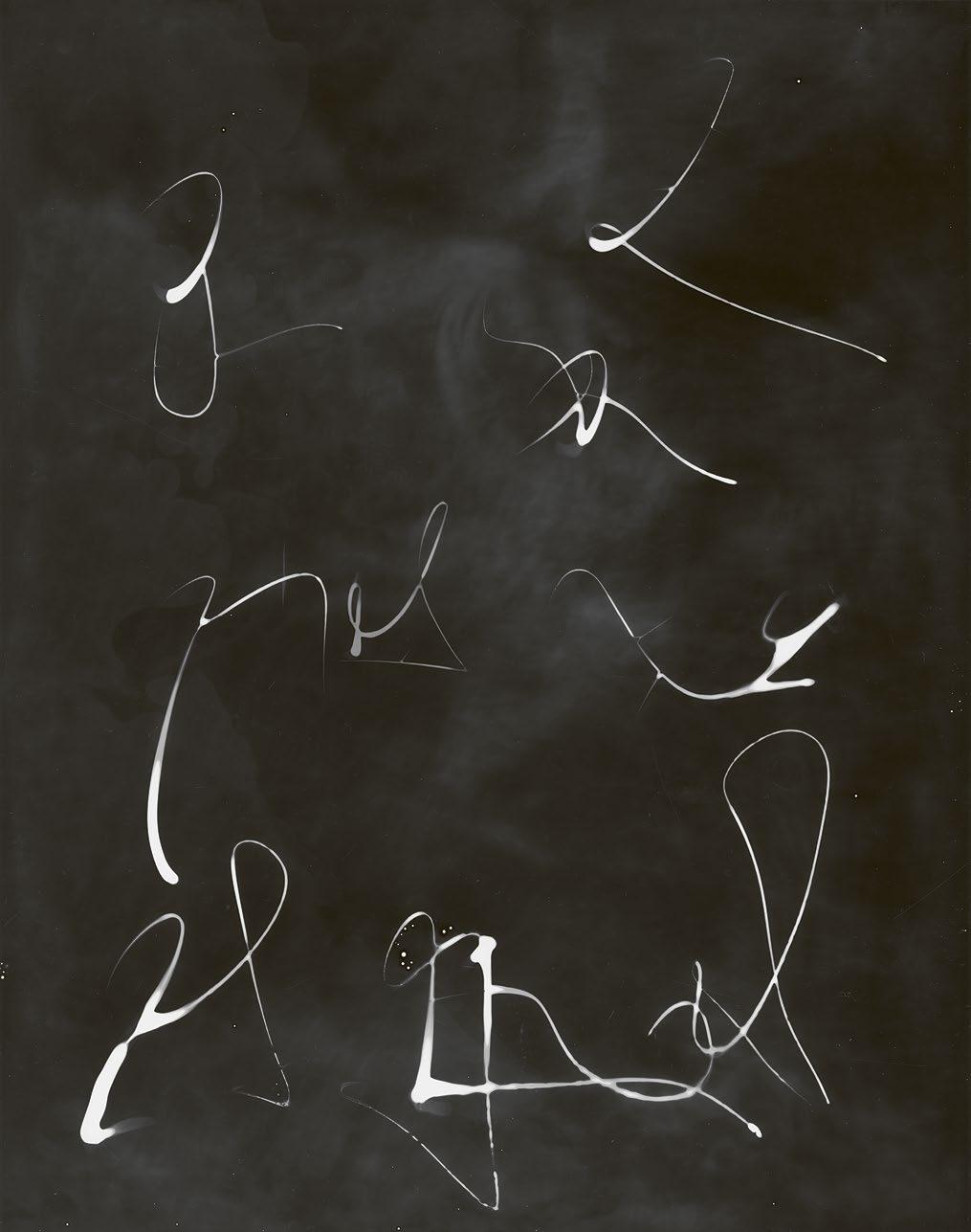
Lada Suomenrinne
‘Hear the wind when I seek for the footprints next to my Máttaráhkku, greatgrandmother. We gather little orbs of lights from the breeze.

“Ádjá gákti mu alde, maid smiehtat?” What would my great-grandmother say when the light orbs hit my grandfather’s regalia on me? Would her footprints walk away from the landscape we share with the same gaze?’
Lada Suomenrinne (b. 1995) was born in Northern Russia but has lived most of their life in the northernmost point of Finland, Sápmi. There Suomenrinne was adopted by their stepfather into his Sámi family. The roots of their work are in cultural identity and belonging; Suomenrinne explores their relationship to the Sámi culture and identity, which they have inherited through adoption. Suomenrinne is in dialogue with nature with whom they search for a place of security as an adopted indigenous person.

MoA in Photography 23
WOW! 54 / AALTO UNIVERSITY MAGAZINE 32

Russell Lai, what do swordsmanship and cryptography have in common?
Assistant Professor Russell Lai wields mathematical symbols and German longswords.
How would you explain cryptography to a five-year-old?
Cryptography is about building systems that are useful to people who do good, but not useful to people who do bad. These systems can be used for things like communicating in a secure way, so that others can’t read or tamper with the messages.
We try to make things secure through mathematics. As scientists, we make sure that if someone wants to break our cryptosystems, they must first solve some very difficult mathematical problems, which we believe to be unsolvable.
How does cryptography touch our everyday lives?
The best kind of security measures are those that you don’t even realise are there. For example, the HTTPS protocol — which you’ll notice in website addresses — is used for encrypting and authenticating web requests.
It protects us every time we visit a webpage. The percentage of HTTPS-protected traffic rose from around 25% in 2016 to around 95% in 2022.
Popular instant messaging apps also use some form of secure messaging protocol, which protects the privacy and integrity of our daily messages, albeit to varying degrees.
What are some common misconceptions about everyday digital security?
One common misconception is to assume that whatever is encrypted is secure. Whenever there is encryption, we should ask who has the decryption key? If you use services like Google Drive or Dropbox, your data is stored in encrypted form but often the service provider has the decryption key. That means that the encryption still allows the service provider to see your data.
In brief, whenever someone is trying to sell us something that is advertised as secure — such as VPN (virtual private network), which many of us use to connect to shared networks when we work remotely — we should question whether the advertised kind of security is the kind that we actually want.
What do you do outside of work?
When I’m not battling with symbols, I’m fighting with swords. I first developed my swordsmanship in the form of iaidō — a Japanese swordbased martial art — in Hong Kong. After moving to Germany, I picked up the art of German longsword, which is nowadays a part of historical European martial arts (HEMA). Recently, I joined the Espoo Association for Historical Fencing, which gives training in German longsword as well as other weapons in HEMA. Apart from being a fun sport, swordsmanship is also a way of life, associated with traits such as righteousness, bravery, and determination.
What does swordsmanship share with cryptography?
As is common to all forms of martial arts, swordsmanship is about defence first and offence second. Similarly, in cryptography, security is by design and never an afterthought. Defence at its best is elegant and effortless in both.
EVERYDAY CHOICES
Text Samuli Ojala
Photo Matti Ahlgren
56 / AALTO UNIVERSITY MAGAZINE 32

Arto Nurmikko to receive honorary doctorate
Text Panu Räty
Brown University
Arto Nurmikko works as a professor of engineering and physics at Brown University. His research covers multiple fields, including microelectronics, nanoscience and photonics, and developing cuttingedge technologies that can be applied to the physical and life sciences. One of Professor Nurmikko’s current areas of interest is neuroengineering, where he works to create devices that could be directly connected to the human brain.
You have been developing, for example, the ability to monitor the function of brain cells using microchips. How would you define your main research areas?
In academia, our lives are determined by intellectual curiosity. I think that’s the driver. And of course, you must try to maintain some focus so that this intellectual curiosity can lead to something concrete that is scientifically meaningful in advancing that particular sector.
If I chronologically look at some of my research, there was an era when I was interested in magnetic semiconductors and a field called semiconductor physics. For the last 15 or 20 years, driven by a curiosity about the brain, I began to interact with colleagues here at Brown University who were experts in neuroscience and brain science.

Out of that came a direction, where the question is how do we get close enough at a single neural cell level, close enough to record electrically, to reconstruct the way that the brain acts as an exceptional computer, with extremely low power consumption.
That led me to work with brain scientists. And I should say that this
is all about collaborations with experts in neuroscience in a field that is loosely called neuro-engineering.
You have received a number of official recognitions during your professional life. What do you consider to be the most valuable achievement of your career? Actually, the answer in my mind is very simple: the reasonably substantial number of PhDs who have graduated from this laboratory and are doing good work all over the world. Numbers don’t mean that much here, but it probably would be on the order of 60 PhDs over the years. For me personally, that’s the achievement.
Scientifically, it’s hard to say. This current project is one of the most interesting ones. It also happens to be one of the hardest ones because we’re trying to compact a lot of electronic capability into a very tiny chip that needs to be also compatible with the biological environment, which is not very friendly toward microchips. And the body may not be that happy with the microchip either. So, there is not a day without encountering a problem.
Nevertheless, if we are successful in elevating this early neurotechnology concept to the level of operating it in a primate, and hopefully then in a human, then I think that would be perhaps considered an accomplishment that’s worth something.
You’ve been studying and working at top US universities for your entire career. What insights can you offer about that environment that might be helpful here in Finland?
I would say that the one thing is the availability of money. The ability to get research funding here, particularly for experimental research –the expensive kind of research that people like me do – is so much easier. Finland could be even better than it is now with enough resources invested.
The ceremonial conferment of the schools of technology at Aalto University will take place on Friday 16 June 2023. In the ceremony eight new honorary doctors will be conferred, one of whom is Arto Nurmikko.
VISITING
Photo
Read more about the celebrations online 58 / AALTO UNIVERSITY MAGAZINE 32
GIVE YOUR IDEAS SPACE
Campus Membership is your ticket to access Aalto University campus with one membership fee. It’s a network of inspiring coworking spaces across campus, which give people and companies a platform to connect, create, rethink and grow.
• Aalto alumni and strategic partners & collaborators
• Companies
• Freelancers
• Organizations in the fields of Aalto’s research themes
• Startups
Who is it for? Become
for a Campus Membership at campusmember.aalto.fi
Apply
a member! Campus
Membership


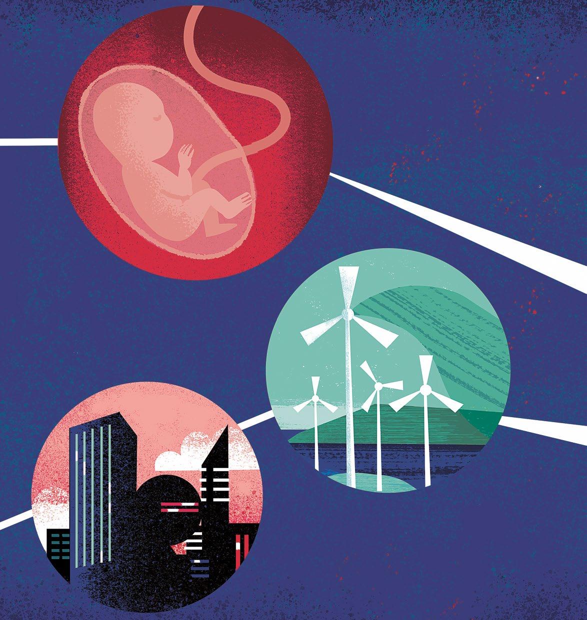
aalto.fi THIS ISSUE LOOKS AT • the hydrogen economy • antimicrobial resistance • art bringing voice to the care sector
