Get Tnm atlas: illustrated guide to the tnm classification of malignant tumours 7th edition james d.
Atlas: Illustrated Guide to the
Visit to download the full and correct content document: https://ebookmass.com/product/tnm-atlas-illustrated-guide-to-the-tnm-classification-of -malignant-tumours-7th-edition-james-d-brierley-editor/
More products digital (pdf, epub, mobi) instant download maybe you interests ...
Andrews’ Diseases of the Skin Clinical Atlas by William James, Dirk Elston, Patrick McMahon Andrews’ Diseases Of The Skin Clinical Atlas By William James
UICC (6e, 2014). Published 2014 by John Wiley & Sons Ltd
All rights reserved. No part of this publication may be reproduced, stored in a retrieval system, or transmitted, in any form or by any means, electronic, mechanical, photocopying, recording or otherwise, except as permitted by law. Advice on how to obtain permission to reuse material from this title is available at http://www.wiley.com/ go/permissions.
The right of James D. Brierley, Hisao Asamura, Elisabeth Van Eycken, Brian Rous to be identified as the authors of the editorial material in this work has been asserted in accordance with law.
Registered Offices
John Wiley & Sons, Inc., 111 River Street, Hoboken, NJ 07030, USA
John Wiley & Sons Ltd, The Atrium, Southern Gate, Chichester, West Sussex, PO19 8SQ, UK
Editorial Office
9600 Garsington Road, Oxford, OX4 2DQ, UK
For details of our global editorial offices, customer services, and more information about Wiley products visit us at www.wiley.com.
Wiley also publishes its books in a variety of electronic formats and by print‐on‐demand. Some content that appears in standard print versions of this book may not be available in other formats.
Limit of Liability/Disclaimer of Warranty
The contents of this work are intended to further general scientific research, understanding, and discussion only and are not intended and should not be relied upon as recommending or promoting scientific method, diagnosis, or treatment by physicians for any particular patient. In view of ongoing research, equipment modifications, changes in governmental regulations, and the constant flow of information relating to the use of medicines, equipment, and devices, the reader is urged to review and evaluate the information provided in the package insert or instructions for each medicine, equipment, or device for, among other things, any changes in the instructions or indication of usage and for added warnings and precautions. While the publisher and authors have used their best efforts in preparing this work, they make no representations or warranties with respect to the accuracy or completeness of the contents of this work and specifically disclaim all warranties, including without limitation any implied warranties of merchantability or fitness for a particular purpose. No warranty may be created or extended by sales representatives, written sales materials or promotional statements for this work. The fact that an organization, website, or product is referred to in this work as a citation and/or potential source of further information does not mean that the publisher and authors endorse the information or services the organization, website, or product may provide or recommendations it may make. This work is sold with the understanding that the publisher is not engaged in rendering professional services. The advice and strategies contained herein may not be suitable for your situation. You should consult with a specialist where appropriate. Further, readers should be aware that websites listed in this work may have changed or disappeared between when this work was written and when it is read. Neither the publisher nor authors shall be liable for any loss of profit or any other commercial damages, including but not limited to special, incidental, consequential, or other damages.
Library of Congress Cataloging‐in‐Publication Data
Names: Brierley, James, editor. | Asamura, H., editor. | Eycken, E. Van, editor. | Rous, Brian Arthur, 1967– editor. | Union for International Cancer Control.
Title: TNM atlas : illustrated guide to the TNM classification of malignant tumours / edited by James D. Brierley, Hisao Asamura, Elisabeth Jozefa Van Eycken, Brian Arthur Rous.
Set in 9/12pt and Frutiger LT Std by SPi Global, Pondy, India
10 9 8 7 6 5 4 3 2 1
CONTENTS
Preface to the Seventh Edition, vii
Acknowledgements, ix
Contributors to the Seventh Edition, xi
Preliminary Note, xiii
HEAD AND NECK TUMOURS,
1
Section Editors Brian O’Sullivan and Shao Hui Huang
Regional Lymph Nodes, 1
Lip and Oral Cavity, 15
Pharynx, 26
Larynx, 47
Nasal Cavity and Paranasal Sinuses, 57
Unknown Primary – Cervical Nodes, 66
Malignant Melanoma of Upper Aerodigestive Tract, 70
Major Salivary Glands, 73
Thyroid Gland, 77
DIGESTIVE SYSTEM TUMOURS, 85
Section Editor James D. Brierley
Oesophagus Including Oesophagogastric Junction, 86
Stomach, 94
Small Intestine, 101
Appendix, 106
Colon and Rectum, 112
Anal Canal, 122
Liver – Hepatocellular Carcinoma, 129
Liver – Intrahepatic Bile Ducts, 135
Gallbladder, 139
Extrahepatic Bile Ducts – Perihilar, 145
Extrahepatic Bile Ducts – Distal, 149
Ampulla of Vater, 152
Pancreas, 159
Well-Differentiated Neuroendocrine Tumours of the Gastrointestinal Tract, 165
Well-Differentiated Neuroendocrine Tumours of the Pancreas, 177
LUNG, PLEURAL AND THYMIC TUMOURS, 181
Section Editor Hisao Asamura
Lung, 182
Pleural Mesothelioma, 193
Thymic Tumours, 200
BONE AND SOFT TISSUE TUMOURS, 209
Section Editor Brian O’Sullivan
Bone, 210
Soft Tissues, 217
Gastrointestinal Stromal Tumours (GIST), 223
SKIN TUMOURS, 227
Section Editor Brian Rous
Carcinoma of the Skin, 233
Carcinoma of Skin of the Head and Neck, 237
Carcinoma of the Skin of the Eyelid, 248
Malignant Melanoma of Skin, 255
Merkel Cell Carcinoma of Skin, 263
BREAST TUMOURS, 267
Section Editor Elisabeth Van Eycken
GYNAECOLOGICAL TUMOURS, 287
Section Editor Brian Rous
Vulva, 288
Vagina, 293
Cervix Uteri, 300
Uterus Endometrium, 310
Uterus – Uterine Sarcomas, 315
Ovarian, Fallopian Tube and Primary Peritoneal Carcinoma, 323
Gestational Trophoblastic Tumours, 331
UROLOGICAL TUMOURS, 335
Section Editor Elisabeth Van Eycken
Penis, 336
Prostate, 345
Testis, 353
Kidney, 371
Renal Pelvis and Ureter, 380
Urinary Bladder, 387
Urethra, 394
Adrenal Cortex Tumours, 404
HODGKIN LYMPHOMA, 409
Section Editor James D. Brierley
NON‐HODGKIN LYMPHOMAS, 424
Section Editor James D. Brierley
Index, 425
PREFACE TO THE SEVENTH EDITION
This new seventh edition of the TNM Atlas incorporates the changes in the TNM System that are in the eighth edition of the TNM Classification of Malignant Tumours 1 In the eighth edition of the TNM Classification staging at many of the tumour sites is unchanged from the seventh edition. However, some tumour entities and anatomical sites have been newly introduced and some tumours contain modifications; this follows the basic philosophy of maintaining stability of the classification over time. The new additions are: p16 oropharyngeal carcinomas, carcinomas of the thymus, neuroendocrine carcinomas of the pancreas and sarcomas of the spine, pelvis, head and neck, retroperitoneum and thoracic and abdominal viscera. In addition, many tumour sites have important updates. The Atlas’s content follows the approach of depicting the Ts, Ns and Ms in graphic terms. The full‐colour artwork is augmented with an increased number of clinical imaging studies illustrating many of the changes between the seventh and eighth editions of the TNM Classification of Malignant Tumours
1 Brierley, J.D., Gospodarowicz, M.K., Wittekind, C. (eds.) (2017) TNM Classification of Malignant Tumours, 8th edn. Chichester: Wiley-Blackwell.
ACKNOWLEDGEMENTS
The editors wish to express their thanks to all who contributed to this seventh edition by comments, questions and their critical interest. In particular we would like to thank Professor Ch. Wittekind, Leipzig, Germany for his enormous contributions as co‐editor of the 4th, 5th and 6th editions of the TNM Atlas, the 5th, 6th, 7th and 8th editions of the TNM Classification of Malignant Tumours and the 2nd, 3rd, 4th and 5th editions of the TNM Supplement He was the driving force behind many of these editions.
The Editors have much pleasure in acknowledging the great help received from the members of the TNM Prognostic Factors Project Committee and the National Staging Committees Global Representatives and International Organizations listed on pages xv–xvi of the TNM Classification of Malignant Tumours, 8th edition. In addition, we would like to thank, Dr Brian O’Sullivan and Dr Shao Hui Huang for providing clinical images for the Head and Neck Tumours chapter, Mr Anthony Griffin, Dr Peter Chung and Dr Ali Hosni for providing clinical images for the Tumours of the Bone and Soft tissues chapter and Dr Richard Tsang for clinical Images in the Lymphoma chapter.
This edition builds on the work by contributors to all previous editions. In particular we would like to thank Dr Leslie Sobin and Dr Christian Wittekind, who along with Dr Hisao Asamura edited the 6th edition. Contributors to all previous editions are listed in the 6th edition pp. x–xiv.
CONTRIBUTORS TO THE SEVENTH
EDITION
Asamura, H. Tokyo, Japan
Brierley, J. Toronto, Canada
Huang, Shao Hui. Toronto, Canada
O‘Sullivan, B. Toronto, Canada
Rous, B. Cambridge, UK
Ruffino, E. Torino. Italy
Van Eycken, E, Brussels, Belgium
Lung
Digestive System, Thyroid, Lymphoma
Head and Neck Tumours
Head and Neck Tumours
Tumours of the Bone and Soft Tissue
Gynaecological Tumours, Skin Tumours
Thymic Tumours
Breast, Urological, Adrenal Cortex Tumours
PRELIMINARY NOTE
The TNM System for describing the anatomical extent of disease is based on assessment of three components:
T – The extent of the primary tumour
N – The absence or presence and extent of regional lymph node metastasis
M – The absence or presence of distant metastasis
The addition of numbers to these three components indicates the extent of the malignant disease, thus:
T0, T1, T2, T3, T4 N0, N1, N2, N3 M0, M1
In effect, the system is a “short‐hand notation“ for describing the extent of a particular malignant tumour.
Each site is described under the following headings:
1) Anatomy
Drawings of the anatomical sites and subsites are presented with the appropriate ICD‐O‐3 topography numbers.1
2) Regional Lymph Nodes
The regional lymph nodes are listed and shown in drawings.
3) T/pT Clinical and Pathological Classification of the Primary Tumour
The definitions for T and pT categories are presented. In the eighth edition (2017) of the TNM Classification the clinical and pathological classifications (T and pT) generally coincide, therefore the same illustrations are valid for the T and pT classifications for most sites.
4) N/pN Clinical and Pathological Classification of Regional Lymph Nodes
The N and pN categories are presented in a fashion similar to the T and pT categories. Differences between N and pN definitions in the seventh edition arise in the case of carcinomas of the breast and penis and germ cell tumours of the testis.
5) M/pM Clinical and Pathological Classification of Distant Metastasis
M localization is given only in selected cases because of its many possible variables.
Please visit the UICC https://www.uicc.org/resources/tnm/publications-resources or Wiley websites for any errata or updates.
1 ICD-O International Classification of Diseases for Oncology, 3rd edn (2000), WHO, Geneva.
HEAD AND NECK TUMOURS
Introductory Notes
The following sites are included:
• Lip, oral cavity
• Pharynx: oropharynx, nasopharynx, hypopharynx
• Larynx: supraglottis, glottis, subglottis
• Nasal cavity and paranasal sinuses
• Malignant melanoma of upper aerodigestive tract
• Major salivary glands
• Thyroid gland
Carcinomas arising in the minor salivary glands of the upper aerodigestive tract are classified according to the rules for tumours of their anatomical site of origin, e.g., oral cavity.
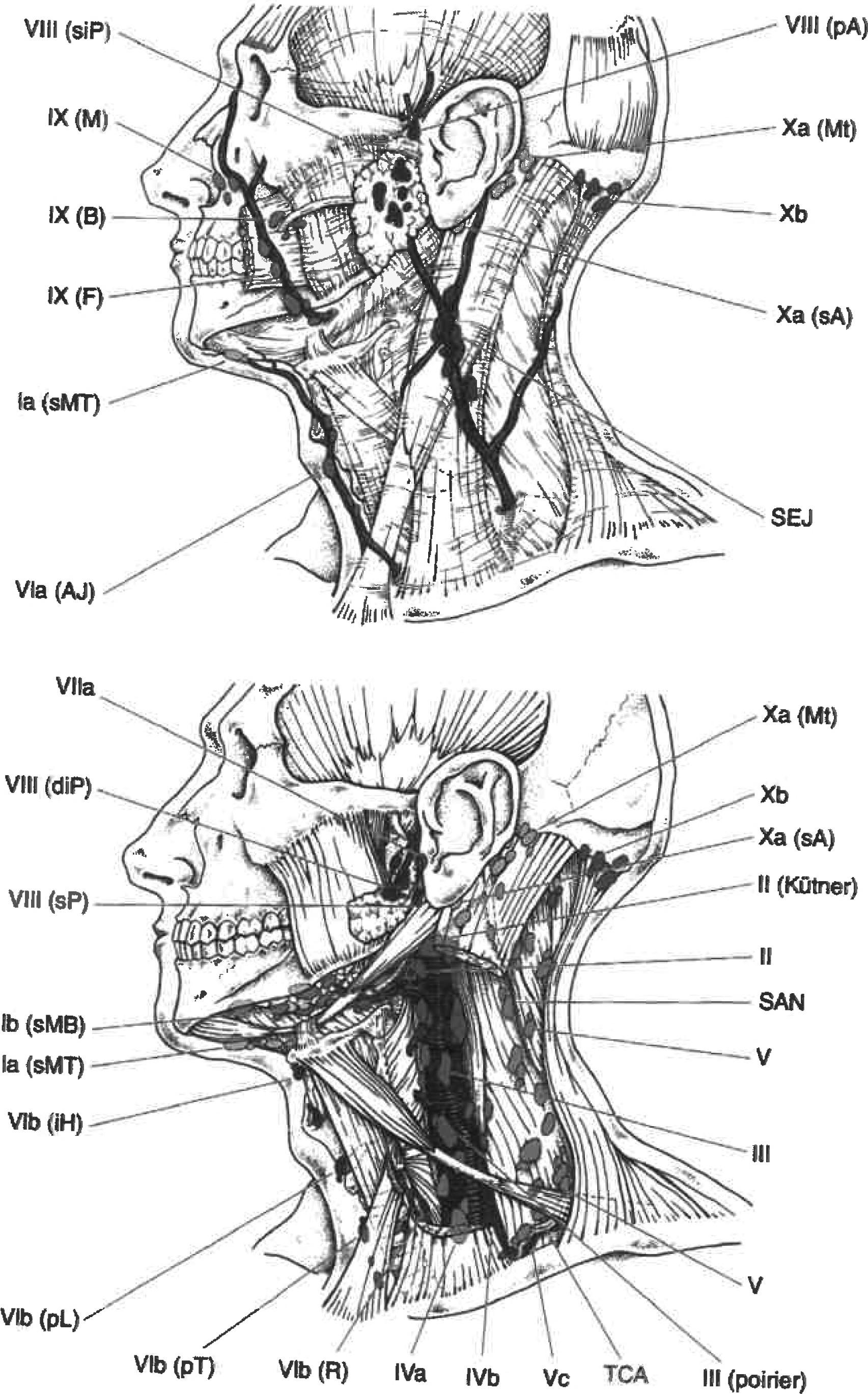
Regional Lymph Nodes (Figs. 1, 2, 3)
The definitions of the N categories for all head and neck sites except p‐16 positive oropharynx, nasopharynx, mucosal malignant melanoma of the upper aerodigestive tract and thyroid are the same.
Midline nodes are considered ipsilateral nodes except in the thyroid.
The status of the regional lymph nodes in head and neck cancer is of considerable prognostic importance. In addition, it is helpful to subdivide the lymph nodes and possible metastasis into specific anatomical subsites and to group these lymph nodes into levels. A consensus guideline from DAHANCA, EORTC, HKNPCSG, NCIC CTG, NCRI, RTOG and TROG has been published and the nodal groups are listed below. However, a number of different classifications exist that use variable level numbers and therefore we recommend the levels be named rather than referred to by number to limit any confusion, although the levels used in the consensus document are given.1 In the consensus classification, the retropharyngeal nodes are classified as Level VII, but in the classification used by the AJCC, Level VII described the upper mediastinal nodes.
1 Grégoire, V., Ang., K., Budach, W., et al. (2013) Delineation of the neck node levels for head and neck tumors: a 2013 update. DAHANCA, EORTC, HKNPCSG, NCIC CTG, NCRI, RTOG, TROG consensus guidelines. Radiother Oncol 2014 110(1):172–181.
TNM Atlas: Illustrated Guide to the TNM Classification of Malignant Tumours, Seventh Edition. Edited by James D. Brierley, Hisao Asamura, Elisabeth Van Eycken, and Brian Rous.
Fig. 1 Source: Modified from Lengele B et al., Radiothor Oncol, 2007; 85(1): 146–155.
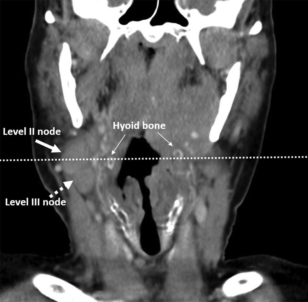
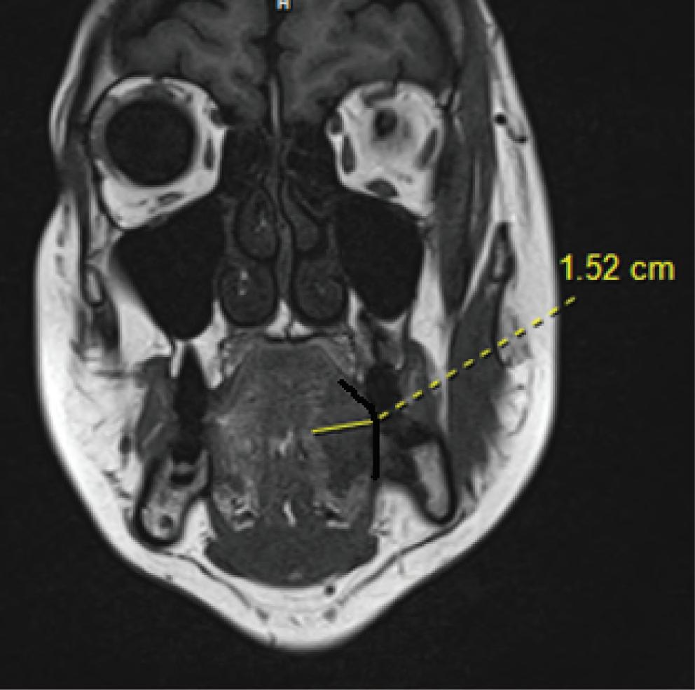
Fig. 2 Axial CT scan showing enlarged right upper jugular (deep cervical) Level II lymph node measuring 2.5 cm in greatest dimension.
Horizontal plane (dotted line) defined by the hyoid bone
Fig. 3 Coronal CT scan showing the same enlarged right upper jugular (deep cervical) Level II lymph node measuring 2.5 cm in greatest dimension, but also an enlarged right medial jugular (deep cervical) Level III lymph node measuring 1.5 cm in greatest dimension. Horizontal plane (dotted line) delineated by the hyoid bone that defines Level II nodes superiorly from Level III nodes inferiorly is marked. This is classified as cN2b: metastasis in multiple ipsilateral lymph nodes, none more than 6 cm in greatest dimension without extranodal extension.
Right sided level II lymph node
1. Submental nodes
2. Submandibular
3. Cranial jugular (deep cervical) nodes
4. Medial jugular (deep cervical) nodes
5. Caudal jugular (deep cervical) nodes
6. Dorsal cervical (superficial cervical) nodes along the spinal accessory nerve
7. Supraclavicular nodes
8. Prelaryngeal and paratracheal (syn. anterior cervical) nodes
9. Retropharyngeal nodes
10. Parotid nodes
11. Buccal nodes (syn. facial nodes)
12. Retroauricular (syn. mastoid, posterior auricular) and occipital nodes
The lymph node groups are defined as follows
1. Submental group
Lymph nodes within the triangular boundary of the anterior belly of the digastric muscle and the hyoid bone.
2. Submandibular group
Lymph nodes within the boundaries of the anterior and posterior bellies of the digastric muscle and the body of the mandible.
3. Upper (cranial) jugular group
Lymph nodes located around the upper third of the internal jugular vein and adjacent spinal accessory nerve, extending from the hyoid bone (clinical landmark) to the skull base. The posterior boundary is the posterior border of the sternocleidomastoid muscle, and the anterior boundary is the lateral border of the sternohyoid muscle. This group includes the jugulodigastric node, which is the most cranial jugular node.
4. Middle (medial) jugular group
Lymph nodes located around the middle third of the internal jugular vein, extending from the carotid bifurcation superiorly to the omohyoid muscle (surgical landmark) or cricothyroid notch (clinical landmark) inferiorly. The posterior boundary is the posterior border of the sternocleidomastoid muscle, and the anterior boundary is the lateral border of the sternohyoid muscle. This group includes the jugulo‐omohyoid lymph node located between the omohyoid muscle and the internal jugular vein.
5. Lower (caudal) jugular group
Lymph nodes located around the lower third of the internal jugular vein, extending from the omohyoid muscle superiorly to the clavicle inferiorly. The posterior boundary is the posterior border of the sternocleidomastoid muscle, and the anterior boundary is the lateral border of the sternohyoid muscle.
6. Dorsal cervical nodes along the spinal accessory chain
This forms the “posterior triangle group”. This comprises predominantly the lymph nodes located along the spinal accessory nerve and the transverse cervical artery.
7. Supraclavicular nodes
The posterior boundary is the anterior border of the trapezius muscle, the anterior boundary is the posterior border of the sternocleidomastoid muscle, and the inferior border is the clavicle.
8. Anterior cervical nodes
Lymph nodes surrounding the midline visceral structures of the neck, extending from the level of the hyoid bone superiorly to the suprasternal notch inferiorly. On each side, the lateral boundary is the medial border of the carotid sheath. Located within
this compartment are the perithyroidal lymph nodes, paratracheal lymph nodes, lymph nodes along the recurrent laryngeal nerves and precricoid lymph nodes. Node group 8 (prelaryngeal and paratracheal nodes) may be further subdivided as follows:
8e: pretracheal near the thyroid isthmus (Delphian)
9. Retropharyngeal nodes
These lie in the buccopharyngeal fascia, behind the upper part of the pharynx and in front of the arch of the atlas.
10. Parotid nodes
These may be subdivided into superficial (in front of the tragus on top of the parotid fascia) and deep parotid nodes. The latter are located underneath the parotid fascia and include intraglandular nodes directly in the parotid gland. The preauricular and infra‐auricular (infra‐ or subparotid) nodes are assigned to the parotid nodes.
11. Buccal (facial) nodes
These include the buccinator nodes located deep on the buccinator muscle, the nasolabial nodes located underneath the nasolabial groove, the molar nodes located in the surface of the cheek and the mandibular nodes located outside the lower jaw.
12. Retroauricular (syn. mastoid, posterior auricular) and occipital nodes
The regional lymph nodes for thyroid include the upper (superior) mediastinal lymph nodes, which may be subdivided into tracheo‐oesophageal (posterior mediastinal) and upper anterior mediastinal nodes. Cervical and mediastinal lymph nodes are not divided by a fascia; the left brachiocephalic vein is considered as the boundary. For the tumour entities listed below, a clinical and a pathological N classification have been introduced in the 8th edition of the UICC TNM Classification of Malignant Tumours:
• Lip and oral cavity
• Oropharynx (p‐16‐negative or oropharyngeal without p‐16‐IH performed)
• Hypopharynx
• Pharynx
• Nasal cavity and paranasal sinuses
• Unknown primary – cervical nodes
• Major salivary glands
• Skin carcinoma of head and neck
N Classification – Regional Lymph Nodes
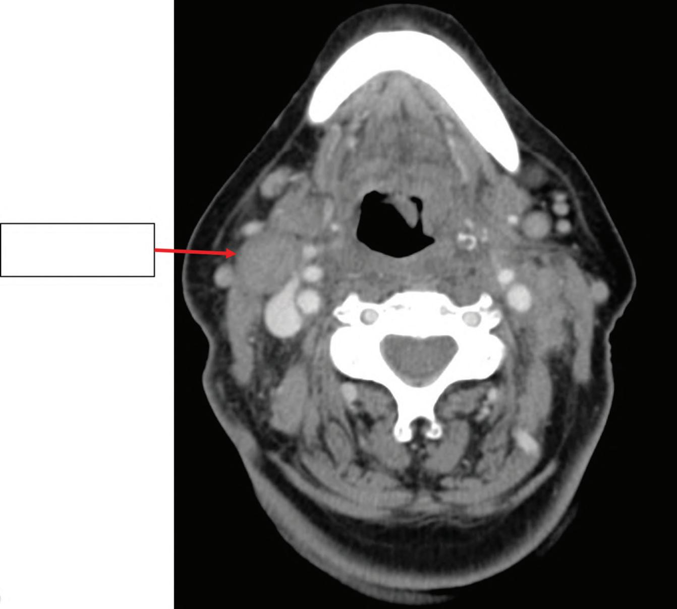
N1 Metastasis in a single ipsilateral lymph node, 3 cm or less in greatest dimension without extranodal extension (Fig. 4)
N2a Metastasis in a single ipsilateral lymph node, more than 3 cm but not more than 6 cm in greatest dimension without extranodal extension (Fig. 5)
N2b Metastasis in multiple ipsilateral lymph nodes, none more than 6 cm in greatest dimension without extranodal extension (Fig. 6)
N1
Ipsilateral
≤ 3 cm
Any head or neck primar y except p16-positive orophar ynx, nasophar ynx, malignant melanoma of upper aerodigestive tract and thyroid gland
> 3 to 6cm
Ipsilateral
Fig. 4 N2a
Fig. 5
Ipsilateral ≤ 6 cm
6
N2c Metastasis in bilateral or contralateral lymph nodes, none more than 6 cm in greatest dimension without extranodal extension (Fig. 7)
≤ 6cm
N2b
Fig.
N2c
Fig. 7
N3a Metastasis in a lymph node more than 6 cm in greatest dimension without extranodal extension (Fig. 8)
> 6cm Without extranodal extension
N3b Metastasis in a single or multiple lymph node(s) with extranodal extension (Figs. 9, 10)
Note
Midline nodes are considered ipsilateral nodes.
N3a
Fig. 8
Fig. 9
Ipsilateral
Any size with extranodal extension
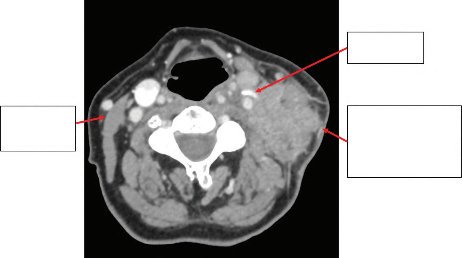
Normal ster nomastoid muscle
Encasement of carotid ar tery
Level II lymph node with subcutaneous tissue invasion (arrow) and engulfment of ster nomastoid muscle
Fig. 10 Axial CT scan showing clinically fixed right upper jugular (deep cervical) Level II lymph node with subcutaneous tissue invasion, engulfment of sternomastoid muscle and encasement of the carotid artery. This is classified as cN3b.
pN Classification – Regional Lymph Nodes
pN0 Histological examination of a selective neck dissection specimen will ordinarily include 6 or more lymph nodes. Histological examination of a radical or modified radical neck dissection specimen will ordinarily include 10 or more lymph nodes. If the lymph nodes are negative, but the number ordinarily examined is not met, classify as pN0. When size is a criterion for pN classification, measurement is made of the metastasis, not of the entire lymph node.
pN1 Metastasis in a single ipsilateral lymph node, 3 cm or less in greatest dimension without extranodal extension (Fig. 11)
Ipsilateral
pN2 Metastasis as described below:
pN2a Metastasis in a single ipsilateral lymph node, 3cm or less in greatest dimension with extranodal extension (Fig. 12), or more than 3 cm but not more than 6 cm in greatest dimension without extranodal extension (Fig. 13)
pN1
Fig. 11
Ipsilateral ≤ 3 cm with extranodal extension
13
Ipsilateral > 3 to 6cm
Fig. 12 pN2a
Fig.
pN2b Metastasis in multiple ipsilateral lymph nodes, none more than 6 cm in greatest dimension without extranodal extension (Fig. 14)
Ipsilateral
6 cm
pN2c Metastasis in bilateral or contralateral lymph nodes, none more than 6 cm in greatest dimension without extranodal extension (Fig. 15)
6cm
pN2b
Fig. 14
pN2c
Fig. 15
16
pN3a
Metastasis in a lymph node more than 6 cm in greatest dimension without extranodal extension (Fig. 16)
> 6cm Without extranodal extension
pN3a
Fig.
pN3b Metastasis in a lymph node more than 3 cm in greatest dimension with extranodal extension, or multiple ipsilateral, contralateral or bilateral with extranodal extension (Figs. 17, 18)
Fig. 18 pN3b
Fig. 17
LIP AND ORAL CAVITY
(ICD‐O
C00, C02–06)
Rules for Classification
The classification applies only to carcinomas of the vermilion surfaces of the lips and of the oral cavity, including those of minor salivary glands. There should be histological confirmation of the disease.
Anatomical Sites and Subsites
Lip* (Fig. 19)
1. External upper lip (vermilion border) (C00.0)
2. External lower lip (vermilion border) (C00.1)
3. Commissures (C00.6)
* In the 9th edition TNM external upper and lower lip C00.0 and C00.1) and commissure (C00.6) will be classified with carcinoma of the skin.
Oral
Cavity (Figs. 20, 21, 22)
1. Buccal mucosa
(i) Mucosa of upper and lower lips (C00.3, 4)
(ii) Cheek mucosa (C06.0)
(iii) Retromolar areas (C06.2)
(iv) Bucco‐alveolar sulci, upper and lower (vestibule of mouth) (C06.1)
2. Upper alveolus and gingiva (upper gum) (C03.0)
3. Lower alveolus and gingiva (lower gum) (C03.1)
4. Hard palate (C05.0)
5. Tongue*
(i) Dorsal surface and lateral borders anterior to vallate papillae (anterior two‐thirds) (C02.0, 1)
(ii) Inferior (ventral) surface (C02.2)
* Note lingual tonsil,C02.4, is classified in the oropharynx
20
C05.0
C05.1 C05.2
C06.2
C03.1
Orophar ynx
Fig.
C00.3
C02.1
C02.1
C02.0
Fig. 21
C02.2
C04.0
Fig. 22
TN Clinical Classification
T – Primary Tumour
TX Primary tumour cannot be assessed
T0 No evidence of primary tumour
Tis Carcinoma in situ
T1 Tumour 2 cm or less in greatest dimension and 5 mm or less depth of invasion (Figs. 23, 24, 25, 26, 27)
≤ 2cm ≤ 5 mm depth of invasion
≤ 2 cm ≤ 5 mm depth of invasion
T1 pT1
Fig. 23
T1
Fig. 24
Fig. 25 Example of measuring depth of invasion on a T1 MRI sequence from the mucosal surface to the deepest point of invasion, perpendicular to the interpreted mucosal plane (in blue), a plane just beneath the closest intact surface of normal mucosa for clinical T category.
“Flat” Tumour DOI = TT
Exophytic Tumour
< TT Ulcerated Tumour
> TT
Tumour
Closest mucosal surface Deepest point of invasion Inter preted submucosal plane
• Black dotted line: IMP (“Inter preted Mucosal Plane”): a plane just beneath the closest intact surface of nor mal mucosa
• Red solid arrow: DOI (“Depth of Invastion”): measured from IMP to deepest point of invasion
• Black dotted arrow: TT (Tumour thickness): measured from centre of tumour surface to the deepest point of invasion
Fig. 26 Schematic Fig.ure depicting the difference between radiologic depth of invasion (DOI) and tumour thickness (TT) for clinical T category.
T2 Tumour 2 cm or less in greatest dimension and more than 5 mm depth of invasion (Figs 28, 29), or Tumour more than 2 cm but not more than 4 cm in greatest dimension and depth of invasion not more than 10 mm (Figs. 30, 31)
T3 Tumour more than 2 cm but not more than 4 cm in greatest dimension and depth of invasion more than 10 mm (Figs. 32, 33) or Tumour more than 4 cm in greatest dimension and not more than 10 mm depth of invasion (Figs. 34, 35)
T4a (lip and oral cavity)
Tumour more than 4 cm in greatest dimension and more than 10 mm depth of invasion (Fig. 36), or (Lip) – Tumour invades through cortical bone, inferior alveolar nerve, floor of mouth or skin (of the chin or the nose) (Figs. 37, 38)