Clinical Practice and Cases in Emergency Medicine


60 The Pectoralis Block: A Case Series of a Novel Modality for Acute Pain Control in the Emergency Department Brewer JS, Sanders N, Ayala A, Nagdev A
64 A Cluster of Neuroinvasive Adenovirus Infections on a College Campus: Case Series Valentini N, Breeden M, Felley LE, Roberts NB, Dinsmore M, Krumheuer A, Grassley C, Montas S, Bassin B, Phillips K
Case Reports


68 Myocardial Bridge of the Left Anterior Descending Artery Causing Pseudo-Wellens’ Syndrome: A Report of Two Cases Guha D, Medoza-Garcia FC, Millen KM, Offenbacher J, Warstadt NM
73 Bilateral Infectious Extensor Tenosynovitis: A Case Report Osipchuk D, Riddell J
77 COVID-19-induced Acute Psychosis Resulting in a Suicide Attempt: A Case Report Piszker A, McManus N


81 Iatrogenic Rhinolith: A Case Report and Review of Literature Mitchell D, Self Q, Orgain C

85 Case Report of Malignant Hyperthermia in the Emergency Department McMurray M, Sowers A, Orthober R, Huecker M

89 A Case Report of a LVAD Driveline Infection Diagnosed by Point-of-care Ultrasound Bielawa N, Cohen A, Patel M, Stankard B, Nelson MJ
Contents continued on page iii
Volume 7, Number 2, May 2023 Open Access at www.cpcem.org ISSN: 2474-252X A Peer-Reviewed, International Professional Journal
Clinicopathological Cases from the University of Maryland 54 44-year-old Man with Hemoptysis and Hypoxemic Respiratory Failure: A Case Report McNeilly BP, Williams DM, Bontempo LJ, Gatz JD
Case Series
In Collaboration with the Western Journal of Emergency Medicine


Please join us for the 2023 Scientific Assembly August 12-16, 2023 Washington Hilton Hotel Washington, DC Save the Date
Penn State Health Emergency Medicine


About Us:
Penn State Health is a multi-hospital health system serving patients and communities across central Pennsylvania. We are the only medical facility in Pennsylvania to be accredited as a Level I pediatric trauma center and Level I adult trauma center. The system includes Penn State Health Milton S. Hershey Medical Center, Penn State Health Children’s Hospital, and Penn State Cancer Institute based in Hershey, Pa.; Penn State Health Hampden Medical Center in Enola, Pa.; Penn State Health Holy Spirit Medical Center in Camp Hill, Pa.; Penn State Health St. Joseph Medical Center in Reading, Pa.; Penn State Health Lancaster Pediatric Center in Lancaster, Pa.; Penn State Health Lancaster Medical Center (opening fall 2022); and more than 3,000 physicians and direct care providers at more than 126 outpatient practices in 94 locations. Additionally, the system jointly operates various health care providers, including Penn State Health Rehabilitation Hospital, Hershey Outpatient Surgery Center, Hershey Endoscopy Center, Horizon Home Healthcare and the Pennsylvania Psychiatric Institute.
We foster a collaborative environment rich with diversity, share a passion for patient care, and have a space for those who share our spark of innovative research interests. Our health system is expanding and we have opportunities in both academic hospital as well community hospital settings.


Benefit highlights include:
• Competitive salary with sign-on bonus

• Comprehensive benefits and retirement package
• Relocation assistance & CME allowance
• Attractive neighborhoods in scenic central Pa.


FOR MORE INFORMATION PLEASE CONTACT: Heather Peffley, PHR CPRP - Penn State Health Lead Physician Recruiter
hpeffley@pennstatehealth.psu.edu
Penn State Health is fundamentally committed to the diversity of our faculty and staff. We believe diversity is unapologetically expressing itself through every person’s perspectives and lived experiences. We are an equal opportunity and affirmative action employer. All qualified applicants will receive consideration for employment without regard to age, color, disability, gender identity or expression, marital status, national or ethnic origin, political affiliation, race, religion, sex (including pregnancy), sexual orientation, veteran status, and family medical or genetic information.
JOIN OUR TEAM EMERGENCY MEDICINE OPPORTUNITIES AVAILABLE
Clinical Practice and Cases in Emergency Medicine
Rick A. McPheeters, DO, Editor-in-Chief
Kern Medical/UCLA- Bakersfield, California
R. Gentry Wilkerson, MD, Deputy Editor University of Maryland School of Medicine
Mark I. Langdorf, MD, MHPE, Senior Associate Editor
University of California, Irvine School of Medicine- Irvine, California
Shahram Lotfipour, MD, MPH, Senior Associate Editor
University of California, Irvine School of Medicine- Irvine, California

Shadi Lahham, MD, MS, Associate Editor
Kaiser Permanente- Orange County, California
Manish Amin, DO, Associate Editor
Kern Medical/UCLA- Bakersfield, California
John Ashurst, DO, Decision Editor/ ACOEP Guest Editor Kingman Regional Health Network, Arizona
Anna McFarlin, MD, Decision Editor
Louisiana State University Health Science Center- New Orleans, Louisiana
Liv Libet, MD, Decision Editor Kern Medical/UCLA- Bakersfield, California
Amin A. Kazzi, MD, MAAEM
The American University of Beirut, Beirut, Lebanon
Anwar Al-Awadhi, MD
Mubarak Al-Kabeer Hospital, Jabriya, Kuwait
Arif A. Cevik, MD
United Arab Emirates University College of Medicine and Health Sciences, Al Ain, United Arab Emirates
Abhinandan A.Desai, MD University of Bombay Grant Medical College, Bombay, India
Bandr Mzahim, MD
King Fahad Medical City, Riyadh, Saudi Arabia
Barry E. Brenner, MD, MPH
Case Western Reserve University
Brent King, MD, MMM University of Texas, Houston
Daniel J. Dire, MD University of Texas Health Sciences Center San Antonio
David F.M. Brown, MD Massachusetts General Hospital/Harvard Medical School
Editorial Board
Edward Michelson, MD Texas Tech University
Edward Panacek, MD, MPH University of South Alabama
Erik D. Barton, MD, MBA Icahn School of Medicine, Mount Sinai, New York
Francesco Dellacorte, MD
Azienda Ospedaliera Universitaria “Maggiore della Carità,” Novara, Italy
Francis Counselman, MD Eastern Virginia Medical School
Gayle Galleta, MD
Sørlandet Sykehus HF, Akershus Universitetssykehus, Lorenskog, Norway
Hjalti Björnsson, MD Icelandic Society of Emergency Medicine
Jacob (Kobi) Peleg, PhD, MPH Tel-Aviv University, Tel-Aviv, Israel
Jonathan Olshaker, MD Boston University
Katsuhiro Kanemaru, MD University of Miyazaki Hospital, Miyazaki, Japan
Christopher Sampson, MD, Decision Editor University of Missouri- Columbia, Missouri
Joel Moll, MD, Decision Editor
Virginia Commonwealth University School of Medicine- Richmond, Virginia
Steven Walsh, MD, Decision Editor Einstein Medical Center Philadelphia-Philadelphia, Pennsylvania
Melanie Heniff, MD, JD, Decision Editor University of Indiana School of Medicine- Indianapolis, Indiana
Austin Smith, MD, Decision Editor Vanderbilt University Medical Center-Nashville, Tennessee
Rachel A. Lindor, MD, JD, Decision Editor Mayo Clinic College of Medicine and Science
Jacqueline K. Le, MD, Decision Editor Desert Regional Medical Center
Christopher San Miguel, MD, Decision Editor Ohio State Univesity Wexner Medical Center
Robert Suter, DO, MHA UT Southwestern Medical Center
Khrongwong Musikatavorn, MD King Chulalongkorn Memorial Hospital, Chulalongkorn University, Bangkok, Thailand
Leslie Zun, MD, MBA Chicago Medical School
Linda S. Murphy, MLIS University of California, Irvine School of Medicine Librarian
Nadeem Qureshi, MD St. Louis University, USA
Emirates Society of Emergency Medicine, United Arab Emirates
Niels K. Rathlev, MD Tufts University School of Medicine
Pablo Aguilera Fuenzalida, MD Pontificia Universidad Catolica de Chile, Región Metropolitana, Chile
Peter A. Bell, DO, MBA Baptist Health Science University
Peter Sokolove, MD University of California, San Francisco
Robert M. Rodriguez, MD University of California, San Francisco
Robert W. Derlet, MD University of California, Davis
Rosidah Ibrahim, MD Hospital Serdang, Selangor, Malaysia
Samuel J. Stratton, MD, MPH Orange County, CA, EMS Agency
Scott Rudkin, MD, MBA
University of California, Irvine
Scott Zeller, MD University of California, Riverside
Steven Gabaeff, MD Clinical Forensic Medicine
Steven H. Lim, MD Changi General Hospital, Simei, Singapore
Terry Mulligan, DO, MPH, FIFEM
ACEP Ambassador to the Netherlands Society of Emergency Physicians
Vijay Gautam, MBBS
University of London, London, England
Wirachin Hoonpongsimanont, MD, MSBATS Siriraj Hospital, Mahidol University, Bangkok, Thailand
Editorial Staff Advisory Board
Amal Khalil, MBA
UC Irvine Health School of Medicine
Elena Lopez-Gusman, JD California ACEP
American College of Emergency Physicians
Adam Levy, BS
American College of Osteopathic Emergency Physicians
John B. Christensen, MD California Chapter Division of AAEM
Randy Young, MD
California ACEP
American College of Emergency Physicians
Mark I. Langdorf, MD, MHPE
UC Irvine Health School of Medicine
Jorge Fernandez, MD
California ACEP
American College of Emergency Physicians University of California, San Diego


Peter A. Bell, DO, MBA
American College of Osteopathic Emergency Physicians
Baptist Health Science University
Robert Suter, DO, MHA
American College of Osteopathic Emergency Physicians UT Southwestern Medical Center
Shahram Lotfipour, MD, MPH
UC Irvine Health School of Medicine
Brian Potts, MD, MBA
California Chapter Division of AAEM

Alta Bates Summit-Berkeley Campus
Isabelle Nepomuceno, BS Executive Editorial Director
Anuki Edirimuni, BS and Visha Bajaria, BS WestJEM Editorial Director
Zaynab Ketana, BS CPC-EM Editorial Director Associate Marketing Director
Stephanie Burmeister, MLIS WestJEM Staff Liaison
June Casey, BA Copy Editor
Cassandra Saucedo, MS Executive Publishing Director
Jordan Lam, BS WestJEM Publishing Director
Rubina Rafi, BS CPC-EM Publishing Director
Avni Agarwal, BS
CPC-EM Associate Publishing Director Associate Marketing Director
Anthony Hoang, BS WestJEM Associate Publishing Director
Volume 7, no. 2: May 2023 i Clinical Practice and Cases in Emergency Medicine
Indexed in PubMed and full text in PubMed Central Available in MEDLINE, PubMed, PubMed Central, Google Scholar, eScholarship, DOAJ, and OASPA. Editorial and Publishing Office: WestJEM/Depatment of Emergency Medicine, UC Irvine Health, 333 City Blvd, West, Rt 128-01, Orange, CA 92866, USA Office: 1-714-456-6389; Email: Editor@westjem.org Official Journal of the California Chapter of the American College of Emergency Physicians, the America College of Osteopathic Emergency Physicians, and the California Chapter of the American Academy of Emergency Medicine
Clinical Practice and Cases in Emergency Medicine
Professional Society Sponsors
American College of Osteopathic Emergency Physicians
California ACEP
Academic Department of Emergency Medicine Subscribers
Albany Medical College
Albany, NY
American University of Beirut
Beirut, Lebanon
Arrowhead Regional Medical Center
Colton, CA
Augusta University
Augusta GA
Baystate Medical Center
Springfield, MA
Beaumont Hospital
Royal Oak, MI
Beth Israel Deaconess Medical Center
Boston, MA
Boston Medical Center
Boston, MA
Brigham and Women’s Hospital
Boston, MA
Brown University
Providence, RI
Carl R. Darnall Army Medical Center
Fort Hood, TX
Conemaugh Memorial Medical Center
Johnstown, PA
Desert Regional Medical Center
Palm Springs, CA
Doctors Hospital/Ohio Health
Columbus, OH
Eastern Virginia Medical School
Norfolk, VA
Einstein Healthcare Network
Philadelphia, PA
Emory University
Atlanta, GA
Genesys Regional Medical Center
Grand Blanc, Michigan
Hartford Hospital
Hartford, CT
Hennepin County Medical Center
Minneapolis, MN
Henry Ford Hospital
Detroit, MI
State Chapter Subscribers
INTEGRIS Health
Oklahoma City, OK
Kaweah Delta Health Care District
Visalia, CA
Kennedy University Hospitals
Turnersville, NJ
Kern Medical Bakersfield, CA
Lakeland HealthCare
St. Joseph, MI
Lehigh Valley Hospital and Health Network
Allentown, PA
Loma Linda University Medical Center
Loma Linda, CA
Louisiana State University Health Sciences Center
New Orleans, LA
Madigan Army Medical Center
Tacoma, WA
Maimonides Medical Center
Brooklyn, NY
Maricopa Medical Center
Phoenix, AZ
Massachusetts General Hospital
Boston, MA
Mayo Clinic College of Medicine
Rochester, MN
Mt. Sinai Medical Center
Miami Beach, FL
North Shore University Hospital
Manhasset, NY
Northwestern Medical Group
Chicago, IL
Ohio State University Medical Center
Columbus, OH
Ohio Valley Medical Center
Wheeling, WV
Oregon Health and Science University
Portland, OR
Penn State Milton S. Hershey Medical Center
Hershey, PA
Presence Resurrection Medical Center
Chicago, IL
California Chapter Division of AmericanAcademy of Emergency Medicine
Robert Wood Johnson University Hospital
New Brunswick, NJ
Rush University Medical Center
Chicago, IL
Southern Illinois University
Carbondale, IL
St. Luke’s University Health Network
Bethlehem, PA
Stanford/Kaiser Emergency Medicine
Residency Program
Stanford, CA
Staten Island University Hospital
Staten Island, NY
SUNY Upstate Medical University
Syracuse, NY
Temple University
Philadelphia, PA
Texas Tech University Health Sciences Center
El Paso, TX
University of Alabama, Birmingham
Birmingham, AL
University of Arkansas for Medical Sciences
Little Rock, AR
University of California, Davis Medical Center
Sacramento, CA
University of California Irvine
Orange, CA
University of California, Los Angeles
Los Angeles, CA
University of California, San Diego
La Jolla, CA
University of California, San Francisco
San Francisco, CA
UCSF Fresno Center
Fresno, CA
University of Chicago, Chicago, IL
University of Colorado, Denver
Denver, CO
University of Florida
Gainesville, FL
University of Florida, Jacksonville
Jacksonville, FL
University of Illinois at Chicago
Chicago, IL
University of Illinois College of Medicine
Peoria, IL
University of Iowa
Iowa City, IA
University of Louisville
Louisville, KY
University of Maryland
Baltimore, MD
University of Michigan
Ann Arbor, MI
University of Missouri, Columbia
Columbia, MO
University of Nebraska Medical Center
Omaha, NE
University of South Alabama
Mobile, AL
University of Southern California/Keck
School of Medicine
Los Angeles, CA
University of Tennessee, Memphis
Memphis, TN
University of Texas, Houston
Houston, TX
University of Texas Health
San Antonio, TX
University of Warwick Library
Coventry, United Kingdom
University of Washington
Seattle, WA
University of Wisconsin Hospitals and Clinics
Madison, WI
Wake Forest University
Winston-Salem, NC
Wright State University
Dayton, OH
Uniformed Services Chapter Division of the American Academy of Emergency Medicine Virginia Chapter Division of the American Academy of Emergency Medicine
Emergency Medicine Association of Turkey Lebanese Academy of Emergency Medicine
MediterraneanAcademyofEmergencyMedicine
Norwegian Society for Emergency Medicine
Sociedad Argentina de Emergencias
Sociedad Chileno Medicina Urgencia ThaiAssociationforEmergencyMedicine
To become a WestJEM departmental sponsor, waive article processing fee, receive print and copies for all faculty and electronic for faculty/residents, and free CME and faculty/fellow position advertisement space, please go to http://westjem.com/subscribe or contact:
Stephanie Burmeister
West
JEM Staff Liaison
Phone: 1-800-884-2236
Email: sales@westjem.org
Clinical Practice and Cases in Emergency Medicine ii Volume 7, no. 2: May 2023
in PubMed and full text in PubMed Central
Chapter Division of the American Academy of Emergency Medicine
Chapter Division of the American Academy of Emergency Medicine
Division of the American Academy of
Medicine
AmericanAcademy
AmericanAcademy
Indexed
International Society Partners Arizona
California
Florida Chapter
Emergency
Great Lakes Chapter Division of the
ofEmergencyMedicine Tennessee Chapter Division of the
ofEmergencyMedicine
This open access publication would not be possible without the generous and continual financial support of our society sponsors, department and chapter subscribers.
Clinical Practice and Cases in Emergency Medicine
Indexed in PubMed and full text in PubMed Central
JOURNAL FOCUS
Clinical Practice and Cases in Emergency Medicine (CPC-EM) is a MEDLINE-indexed internationally recognized journal affiliated with the Western Journal of Emergency Medicine (WestJEM). It offers the latest in patient care case reports, images in the field of emergency medicine and state of the art clinicopathological and medicolegal cases. CPC-EM is fully open-access, peer reviewed, well indexed and available anywhere with an internet connection. CPC-EM encourages submissions from junior authors, established faculty, and residents of established and developing emergency medicine programs throughout the world.
Table of Contents continued
93 Droperidol in the Management of Phantom Limb Pain: Case Report
MC Winstead, KJ Wells, GT Howington
97 Tension Pneumomediastinum and Coronary Artery Thrombosis Following a Motorcycle Accident: A Case Report
SG Rouleau, MAC Manoukian, GX Wong, DK Barnes
101 Emergency-physician Performed, Ultrasound-guided Lateral Femoral Cutaneous Nerve Block for Meralgia Paresthetica: A Report of Two Cases
MM Kongkatong, CD Thom, J Ottenhoff
106 Diagnosing Atypical Flutter in the Post-atrial Fibrillation Ablation Patient: A Case Report
AN Fuher, R Borne, J Cunningham
110 Implanted Penile Prosthetic Visualized During Focused Assessment with Sonography for Trauma
Examination: A Case Report
K Chambers, G Comp
113 Benign Episodic Mydriasis as a Cause of Isolated Anisocoria
A Seibold, J Barnett, L Stack, C Lei
115 Point-of-care Ultrasound Identification of Hepatic Abscess in the Emergency Department
M Blomquist, T Brinkerhoff, D Weech, H Choi
Letters to the Editor
118
An Inexpensive Biomechanical Model to Help Teach and Learn Newer Mandible Reduction Techniques
S Tummings, S Garrett, JG Humphrey, J Haddad, J Roper, DI Bengiamin, TP Young
120 Response to “An Inexpensive Biomechanical Model to Help Teach and Learn Newer Mandible Reduction Techniques”
VWM Lum, J Poh
Policies for peer review, author instructions, conflicts of interest and human and animal subjects protections can be found online at www.cpcem.org.
Volume 7, no. 2: May 2023 iii Clinical Practice and Cases in Emergency Medicine
Images in Emergency Medicine
Education Fellowship at Eisenhower Medical Center, Rancho Mirage, CA


ABOUT THE PROGRAM
SAEM-approved Education Fellowship
Opportunities to learn in both Graduate and Undergraduate Medical Education
Offer “Training to Teach in Medicine” certificate program from Harvard Medical School

One- or two-year fellowship
Competitive salary with full-benefits from Eisenhower Health
ABOUT EISENHOWER MEDICAL CENTER
Rated among the region’s Best Hospitals by U.S. News & World Report
More than 85,000 visits per year
Advanced Primary Stroke Center, STEMI Center, Accredited Geriatric Emergency Department and Level Four Trauma Center
State-of-art medical center
50 private patient rooms
Best EMR: Epic
Three-year Emergency Medicine residency program
LIVING IN THE DESERT
Affordable cost of living
Variety of activities: hiking, shopping, dining, golfing, etc.

Within two hours from many big cities (L.A. and San Diego)
CONTACT
Wirachin Hoonpongsimanont, MD, MS
Cell: 862-216-0466 Email: wirachin@gmail.com
website: gme.eisenhowerhealth.org
39000 Bob Hope Drive, Rancho Mirage, CA 92270 EisenhowerHealth.org
LIVE. WORK. PLAY. PROSPER.
Clinicopathological Cases from the University of Maryland
44-year-old Man with Hemoptysis and Hypoxemic Respiratory Failure: A Case Report
Bryan P. McNeilly, MD*
Dominic M. Williams, DO†
Laura J. Bontempo, MD, MEd‡
J. David Gatz, MD‡
University of Maryland Medical Center, Department of Emergency Medicine, Baltimore, Maryland
CarolinaEast Medical Center, Department of Emergency Medicine, New Bern, North Carolina
University of Maryland School of Medicine, Department of Emergency Medicine, Baltimore, Maryland
Section Editor: Joel Moll, MD
Submission history: Submitted December 6, 2022; Revision received February 2, 2023; Accepted February 14, 2023
Electronically published May 30, 2023
Full text available through open access at http://escholarship.org/uc/uciem_cpcem
DOI: 10.5811/cpcem.1418
Introduction: Hemoptysis can be a highly alarming presentation in the emergency department (ED). Even seemingly minor cases may represent potentially lethal underlying pathology. It requires thorough evaluation and careful consideration of a broad differential diagnosis.
Case Presentation: A 44-year-old man presented to the ED with a concern of hemoptysis in the setting of recent fever and myalgias.
Discussion: This case takes the reader through how to approach the differential diagnosis and diagnostic work-up of hemoptysis in the ED setting and then reveals the surprising final diagnosis. [Clin Pract Cases Emerg Med. 2023;7(2):54–59]
Keywords: Clinicopathological cases; infectious disease; hemoptysis.
CASE PRESENTATION (DR. MCNEILLY)
A 44-year-old man with a history of polysubstance abuse and homelessness presented to an urban emergency department (ED) in Baltimore, Maryland, with hemoptysis. Symptoms started one week prior to arrival, with three days of fevers and myalgias. On day four, he developed nausea, diarrhea, and a dry cough. His cough became progressively worse over the following days, and on day seven he developed hemoptysis, which prompted his visit to the ED. He denied leg swelling, abdominal pain, dysuria, hematuria, arthralgias, rashes, wounds, dizziness, numbness, and headaches. The patient also denied any sick contacts, including tuberculosis or coronavirus disease 2019 (COVID-19) exposures.
The patient had no known past medical or surgical history and did not use any medications. He stated that he had been homeless for several months and had been living on the streets despite the cool weather. He stated that he drank approximately six beers per day and smoked cigarettes.
He occasionally smoked crack cocaine and marijuana. The last time he had used either substance was several weeks prior to his presentation. He also stated that he smoked methylenedioxy-methylamphetamine (MDMA), most recently the morning prior to his arrival.
The patient’s vital signs were as follows: blood pressure of 108/58 millimeters of mercury, heart rate of 138 beats per minute, respiratory rate of 23 breaths per minute, oral temperature of 37.4 ° Celsius, and a room air oxygen saturation of 86%. On physical exam, he appeared distressed, holding a container with approximately 150 milliliters (mL) of bloody sputum. He had bilateral conjunctivitis and was slightly icteric. His heart was tachycardic with a regular rhythm, and his heart sounds were normal. He was speaking in short sentences while actively coughing up blood and was tachypneic with diffuse rhonchi noted in all lung fields. His abdomen was soft and non-tender, with no
Clinical Practice and Cases in Emergency Medicine 54 Volume 7, no. 2: May 2023
* † ‡
McNeilly et al.
44-year-old Man with Hemoptysis and Hypoxemic Respiratory Failure
organomegaly or masses. He had no joint swelling or signs of trauma or injury. His skin was warm and dry without any rashes or lesions. He was alert and oriented to person, place, time, and situation. Although distressed, he was cooperative and able to follow commands. His initial labs (Table 1) sho wed several abnormalities including leukocytosis, anemia, hyponatremia, and elevated transaminases.
K, thousand; mcL, microliter; g, gram; dL, deciliter; mmol, millimole; L, liter; mg, milligram; u, units.
ng, nanogram; mL, milliliter; FEU, fibrinogen equivalent units; mg, milligram; dL, deciliter; hpf, high power field.
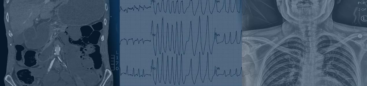
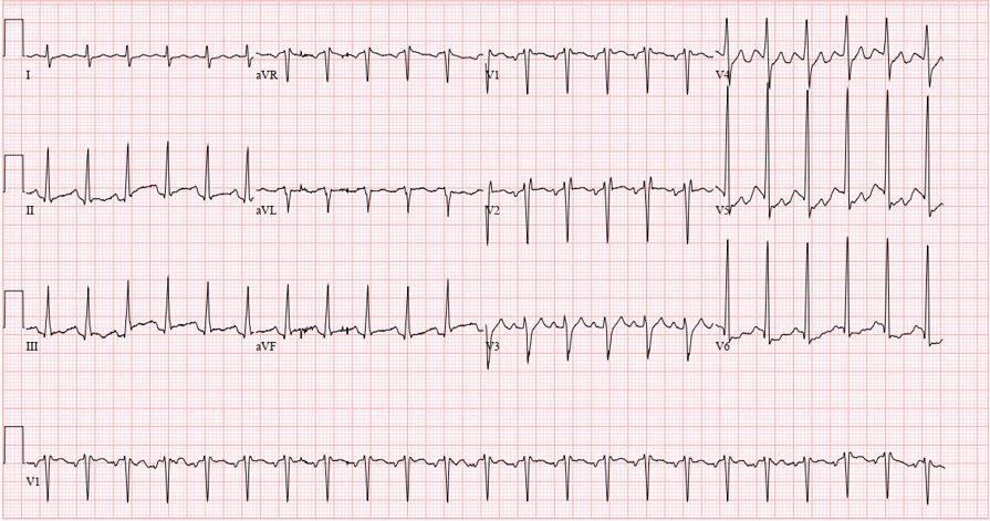
Unfortunately, there were no previous labs available for comparison. An electrocardiogram (ECG) was performed (Image 1), as well as a portable chest radiograph (Image 2). The patient was placed on a non-rebreather mask at a rate of 10 liters per minute.
CASE DISCUSSION (DR. WILLIAMS)
Massive hemoptysis is a worrisome presentation, no matter the demographics of your patient. These patients
Image 1. Electrocardiogram of a 44-year-old man with hemoptysis and hypoxemic respiratory failure showing sinus tachycardia with an incomplete right bundle branch block, left ventricular hypertrophy, possible left atrial enlargement, and marked ST abnormality, concerning for lateral ischemia.
Volume 7, no. 2: May 2023 55 Clinical Practice and Cases in Emergency Medicine
Blood test Patient value Normal range Complete blood count White blood cells 15.3 K/mcL 4.5-11.0 K/mcL Hemoglobin 8.7 g/dL 12.6-17.4 g/dL Hematocrit 25.5% 37.0-50.0% Platelets 147 K/mcL 153-367 K/mcL White blood cell differential Neutrophils 87.0 % 42.6-74.5 % Lymphocytes 3.9 % 20.8-50.5 % Monocytes 3.3 % 2.0-10.3 % Basophils 0.5 % 0.2-1.0 % Eosinophils 0.1 % 0.9-2.9 % Serum chemistries Sodium 128 mmol/L 136-145 mmol/L Potassium 3.7 mmol/L 3.5-5.1 mmol/L Chloride 96 mmol/L 98-107 mmol/L Bicarbonate 19 mmol/L 21-30 mmol/L Blood urea nitrogen 55 mg/dL 9-20 mg/dL Creatinine 1.91 mg/dL 0.66-1.25 mg/dL Glucose 94 mg/dL 70-99 mg/dL Calcium 8.0 mg/dL 8.6-10.2 mg/dL Magnesium 1.4 mg/dL 1.6-2.6 mg/dL Phosphorous 4.9 mg/dL 2.5-4.5 mg/dL Total protein 6.3 g/dL 6.3-8.2 g/dL Albumin 2.8 g/dL 3.5-5.2 g/dL Lactate 1.3 mmol/L 0.5-2.2 mmol/L Hepatic studies Total bilirubin 2.0 mg/dL 0.3-1.2 mg/dL Aspartate aminotransferase 154 u/L 17-59 u/L Alanine aminotransferase 93 u/L 0-49 u/L Alkaline phosphatase 117 u/L 38-126 u/L Cardiac studies N-terminal prohormone of brain natriuretic peptide 1,420 pg/mL <300 pg/mL Blood test Patient value Normal range Troponin <0.02 ng/mL <0.06 ng/mL Coagulation studies Prothrombin time 14.5 seconds 12.1-15 seconds Partial thromboplastin time 37 seconds 25-38 seconds International normalized ratio 1.1 0.8-1.1 D-dimer 1,570 ng/mL FEU <499 ng/mL FEU Urine Studies pH 5.0 5.0-8.0 Protein Negative Negative Ketones Negative Negative Bilirubin Negative Negative Urobilinogen 0.2 mg/dL 0.1-1.8 mg/dL Nitrites Negative Negative White blood cells 3-5 per hpf 0-5 per hpf Red blood cells 0-2 per hpf 0-2 per hpf Bacteria Negative Negative
Table 1. Laboratory results of a 44-year-old man with hemoptysis and hypoxemic respiratory failure.
Table 1. Continued.
often require emergent stabilization, which can limit the opportunity for a detailed history and even prohibit certain imaging options. This patient has a history of homelessness, polysubstance abuse, and likely minimal long-term outpatient medical care, which makes this case uniquely challenging from a diagnostic standpoint. It is a perfect setup for a rare etiology or an uncommon presentation. The starting differential for such a case is broad and includes a range of etiologies such as cardiovascular pathology, infections, malignancy, pulmonary sources, and traumatic injuries.1
Any diagnostic approach should begin with review of the existing information history, exam, and any previous medical records. All of these may point in the direction of an underlying etiology for this patient’s hemoptysis. Summarizing the highlights of the case thus far, this patient has been suffering from fevers and myalgias for one week, followed by the development of a dry cough alongside some nausea and diarrhea. The patient ultimately sought medical attention due to coughing up blood. The exact quantity is poorly characterized, but I would argue this does not matter. While some older definitions of massive hemoptysis focus on rate and total volume of blood loss, the reality is it only takes 150 mL of liquid to fill the entirety of the conducting airways. This patient’s presentation should automatically be considered massive hemoptysis given his presenting respiratory failure and hemodynamic compromise.1

Ultimately, the symptoms detailed in the patient’s history are nonspecific at best and could point toward an infectious, malignant, or even autoimmune etiology. He presented during the COVID-19 pandemic, although he did not have any known exposure to the virus (or to tuberculosis). There is notably no history of trauma reported. This patient unfortunately did not have any previous medical records to offer additional clues.
Physical examination of this patient was concerning, although not necessarily helpful from a diagnostic standpoint. In rare situations, specific findings such as a hemangioma or telangiectasia can suggest a specific etiology such as an underlying vascular malformation. He had a low blood pressure, although not technically hypotensive, and significant tachycardia. This could represent hemorrhagic shock, sepsis, or consequences of his reported MDMA use. Additionally concerning is his room air oxygen saturation of only 86%. His diffuse rhonchi are more suggestive of a systemic or cardiovascular source as opposed to a specific hemorrhagic lesion, although exam alone cannot make this distinction. His noted conjunctivitis could further support an infectious etiology, while his mild icterus might suggest underlying liver disease and an associated coagulopathy.
Laboratory studies are vital in the work-up of hemoptysis but are rarely diagnostic in the ED. This patient’s workup revealed hyponatremia, presumed acute kidney injury, anemia, leukocytosis, and transaminitis. His D-dimer was also elevated. Hyponatremia may suggest a paraneoplastic syndrome, his renal failure could represent a vasculitis, and while a D-dimer is famously nonspecific, one must question the possibility of pulmonary embolism and/or malignancy in the setting of hemoptysis. It would have been potentially helpful to obtain some additional studies, although none of these would have likely led to a definitive diagnosis. (See the complete list of laboratory studies to consider in Table 2)
An ECG may similarly hold suggestive value but is ultimately unlikely to determine the etiology of a patient’s hemoptysis. This patient demonstrated sinus tachycardia with an incomplete right bundle branch block along with left ventricular hypertrophy (LVH) and possible left atrial
Laboratory test
Complete blood cell count
Basic metabolic panel
Renal and liver function
Prothrombin time and international normalized ratio
Blood type
Blood antibody screen
Fibrinogen level
Thromboelastrography (TEG)
Rotational thromboelastogram
Sputum culture
Blood culture
Clinical Practice and Cases in Emergency Medicine 56 Volume 7, no. 2: May 2023 44-year-old Man with Hemoptysis and Hypoxemic Respiratory Failure McNeilly et al.
Image 2. Portable chest radiograph of a 44-year-old man with hemoptysis and hypoxemic respiratory failure showing diffuse bilateral coalescent airspace opacities with air bronchograms (black arrows).
Table 2. Laboratory studies to consider in the work-up of massive hemoptysis.
44-year-old Man with Hemoptysis and Hypoxemic Respiratory Failure
enlargement. Tachycardia and right heart strain again suggest the possibility of pulmonary embolism, while the left atrial enlargement might suggest mitral valve stenosis as the cause of the hemoptysis. However, no murmur was noted on exam, thereby decreasing the probability that he had significant stenosis of the mitral valve, and pulmonary embolism should not cause diffusely abnormal lung sounds as were heard in this patient. Left ventricular hypertrophy could hint at congestive heart failure, although this is a rare cause of hemoptysis and even rarer for it to present so severely.
This leads me to the patient’s imaging, which is often key to reaching a final diagnosis in patients presenting with hemoptysis. As noted earlier, advanced imaging such as computed tomography (CT) is only possible once the patient has been sufficiently stabilized. Therefore, many physicians may have to initially rely on a portable chest radiograph (CXR) alone. A CXR is a bit of a mixed bag, with widely variable rates of diagnostic ability reported in the literature. Sometimes it can identify a localizing lung lesion such as a tumor or cavitation, or potentially a more diffuse process such as pneumonia or diffuse alveolar hemorrhage. This patient’s CXR fits more in the latter category with diffuse bilateral airspace disease and air bronchograms. It is fortunate that he was also able to receive a CT angiography (CTA), as this is often the ideal imaging study to identify the cause of bleeding in hemoptysis, especially if it is from a culprit lesion. As most cases of massive hemoptysis are from the bronchial arterial system, it is worth noting that this CTA should ideally be protocoled differently from the traditional pulmonary artery CTA used when evaluating for pulmonary embolism.1 This patient’s post-intubation CTA demonstrated widespread ground-glass opacities with prominent septal lines, dependent consolidation, and air bronchograms in the bilateral lung bases suggestive of diffuse alveolar hemorrhage and hemorrhagic pneumonia.
Point-of-care bronchoscopy is the only other major diagnostic tool to consider in the evaluation of massive hemoptysis. Availability, however, is inconsistent, and its diagnostic yield is generally lower than that of CT. It would not seem to have much value in this patient, although it could have been considered had the patient been too unstable for transport to a CT scanner.
So, what does this all mean in terms of an underlying diagnosis? There are several ways to categorize the diagnostic possibilities. I previously listed some of the major categories for massive hemoptysis: cardiovascular, infection, malignancy, pulmonary, and traumatic. Others have suggested breaking the differential diagnosis for reported hemoptysis into the following categories: source other than the lower respiratory tract; tracheobronchial source; pulmonary parenchymal source; primary vascular source; and miscellaneous/rare causes.2
At this point, a cardiovascular etiology seems unlikely. The CTA did not identify any vascular malformations or culprit
lesions. The symptoms are too profound for mitral valve stenosis, and there was no significant coagulopathy within the patient’s labs. The CT imaging similarly appears to exclude malignancy and any traumatic injury, and there was no history to suggest the latter. This leaves me focused on pulmonary and infectious etiologies of this patient’s presentation.
This patient unquestionably has diffuse alveolar hemorrhage, but the lingering question of “why” remains. Some possibilities include autoimmune diseases such as granulomatosis with polyangiitis, systemic lupus erythematous, and Goodpasture syndrome. While all three of these can produce an alveolar hemorrhage syndrome, I would have expected more significant and consistent additional features of autoimmune diseases such as arthralgias, rashes, or urinary symptoms. This leaves me predominantly considering an infectious source.
Hemorrhagic pneumonia can be viral (eg, hantavirus, Ebola, Lassa virus), fungal (eg, mycetoma), or bacterial. Bacterial infections may include Mycobacterium (eg, tuberculosis), typical and atypical bacteria, as well as rickettsial and zoonotic disease. This patient presented during the cooler months of the year in the mid-Atlantic, which makes ehrlichiosis, anaplasmosis, or Rocky Mountain spotted fever all unlikely from an epidemiologic standpoint, given the low risk of tick exposure during this window of time. This leads me to consider some of the more infrequently diagnosed bacterial infections and their potential environmental or zoonotic origins.
The patient’s presentation includes a constellation of features that fit with a couple of less common infections. Legionella presents with hyponatremia, alveolar hemorrhage, conjunctivitis, and hemoptysis. Legionella is classically contracted through inhalation or aspiration of contaminated water. There is no such mechanism of exposure reported in the patient’s history. Leptospirosis, however, also has a case to be made here. Classically it has an initial phase from 2-9 days with fever and viral symptoms and produces several physical exam findings exhibited by this patient conjunctival insufflation and scleral icterus. Furthermore, the patient has risk factors secondary to his social determinants of health. He is currently homeless in a city known for its significant rodent population. Brown rats, the most common type in Baltimore, are a known significant natural reservoir for the virus. In the second phase of leptospirosis, patients can demonstrate liver damage and bleeding. The most severe form is referred to as Weil disease and would explain the patient’s acute respiratory distress and altered mental status.
While both diagnoses seem possible, an incidental and unexplained exposure to Legionella simply seems less plausible than exposure to leptospirosis given the known risks from the brown rat population in the patient’s city. Therefore, I think leptospirosis is the most likely diagnosis. It should be confirmed using a urine test for leptospirosis DNA or a serum test for immunoglobulin M antibodies.
Volume 7, no. 2: May 2023 57 Clinical Practice and Cases in Emergency Medicine
McNeilly et al.
CASE OUTCOME (DR. MCNEILLY)
The patient was admitted to the medical intensive care unit (MICU) where he remained intubated for several days. An echocardiogram was performed, which was overall unremarkable, including no sign of mitral valve disease or depressed left ventricular ejection fraction. A bronchoscopy was performed, which showed no signs of inhalation injury, and serial aliquots showed successive clearing, inconsistent with diffuse alveolar hemorrhage. A bronchoscopic culture was obtained that grew methicillin-sensitive Staphylococcus aureus (MSSA). The patient’s blood cultures had been negative up to that point; so his antibiotic regimen was narrowed to cefazolin to cover MSSA pneumonia. Despite continued treatment, the patient’s clinical condition and ventilator requirements did not improve.
Infectious disease (ID) was consulted for a suspected spirochetal infection. Given the time of year (April), ID had a low suspicion for tick-borne illnesses, including ehrlichiosis or Lyme disease. Previous studies had demonstrated a significant reservoir of leptospirosis in the city’s rat population, and there had been numerous rainstorms in the weeks leading up to the patient’s presentation, along with intermittent flooding of the streets. Urine and serum samples were sent for polymerase chain reaction testing and did, in fact, return positive for leptospirosis. He was started on ceftriaxone for severe leptospirosis. After a 10-day stay in the MICU, he was transferred to the medicine floor and then left the hospital against medical advice. He has since presented to the hospital for unrelated complaints, with no apparent sequelae from his bout of leptospirosis.
RESIDENT DISCUSSION
As the most widespread zoonotic disease in the world, leptospirosis is of global importance.3 The incidence of leptospirosis is higher in the tropics and occurs in both industrialized and developing countries. L. interrogans, the causative organism of leptospirosis, is a highly motile obligate anaerobic spirochete with features of both Gram-positive and Gram-negative bacteria.
Leptospirosis typically follows a biphasic pattern, starting with an influenza-like bacteremia phase during which the leptospires are circulating in the blood. The acute bacteremia phase is followed by the immune phase, during which leptospiral toxins result in an immune-mediated response.4 Clinical manifestations of leptospirosis range from an influenza-like illness (anicteric form) to fulminant disease, known as Weil disease.5-7 Weil disease is characterized by a classic triad of jaundice, renal impairment, and hemorrhages. It carries a mortality rate of 5-15%.8-10 Pulmonary hemorrhages are increasingly recognized as a major and potentially lethal complication of leptospirosis. Pulmonary involvement occurs in up to 70% of severe cases and predicts a poor outcome in which death can occur within 48 hours.11,12
Leptospires are carried in the proximal renal tubules of animals, with human infections resulting from exposure to the
urine of carrier animals, either directly or from contaminated soil or water.3 In both rural and urban areas, rats are a major carrier of leptospires. In a study of rats in one American urban center, 65.3% were found to carry antibodies against Leptospira interrogans 13 Given that most cases of leptospirosis are transmitted indirectly via contaminated water,14 the large reservoirs of leptospirosis carried by rats pose a significant threat during floods in both rural and urban settings, particularly to dwellers of lower socioeconomic means.15,16
Indeed, a recent study conducted in 2021 showed that transmission rates of leptospirosis depend on both flooding and temperature. Given the increasing frequency of extreme weather events in the setting of continued global warming, there may be an upsurge in the incidence and magnitude of leptospirosis outbreaks around the world.17 In addition, ongoing climate change may lead to increasing prevalence in regions such as the United States that have thus far experienced fewer cases.
Most cases of leptospirosis are self-limited and resolve without antimicrobial therapy. Prophylactic dosing in some individuals living or traveling in endemic areas may be beneficial.18 Hawaii and Puerto Rico are the most common geographic location in the US for leptospirosis, although cases are also identified throughout the world. It is unclear whether treatment in mild disease limits the progression to severe disease.19-22 A systematic Cochrane review found that antimicrobial therapy did not affect mortality in mild cases; however, there was a nonsignificant trend toward expedited resolution of illness.23 In general, if symptoms are significant enough to come to clinical attention, and the diagnosis is suspected, the patient should receive antimicrobial treatment. For mild disease, a seven-day course of doxycycline, or a three-day course of azithromycin, is recommended. These courses also cover rickettsial infections, which can have a similar presentation. Patients with severe disease should be treated with a seven-day course of intravenous (IV) penicillin, doxycycline, ceftriaxone, or cefotaxime. These patients will likely have some degree of organ failure requiring supportive care, which is generally managed the same as organ failure associated with other etiologies of sepsis. Unique therapies that have been proposed include IV corticosteroids, given the vasculitic nature of the disease process; however, additional studies are needed to support their efficacy.24,25 While plasmapheresis has also been proposed, high-quality data is lacking on its efficacy.26
FINAL DIAGNOSIS
Severe leptospirosis (Weil disease)
KEY TEACHING POINTS
1. Leptospirosis is the most widespread zoonotic disease in the world, and cases may continue to rise as a result of the increased flooding associated with climate change.
Clinical Practice and Cases in Emergency Medicine 58 Volume 7, no. 2: May 2023
McNeilly et al.
44-year-old Man with Hemoptysis and Hypoxemic Respiratory Failure
McNeilly et al.
44-year-old Man with Hemoptysis and Hypoxemic Respiratory Failure
2. Weil disease is a severe manifestation of leptospirosis, and classically presents as a triad of jaundice, renal impairment, and hemorrhage.
3. Pulmonary hemorrhage occurs in up to 70% of patients with Weil disease and portends a poor outcome in which death can occur within 48 hours.
The authors attest that their institution requires neither Institutional Review Board approval nor patient consent for publication of this case report. Documentation on file.
9. Levett PN. Leptospirosis. Clin Microbiol Rev. 2001;14(2):296-326
10. Boertjes E, Hillebrand S, Bins JE, et al. Pulmonary haemorrhage in Weil’s disease. BMJ Case Rep. 2020;13(1):e227570.
11. Carvalho CR and Bethlem EP. Pulmonary complications of leptospirosis. Clin Chest Med. 2002;23(2):469-78.
12. Silva JJ, Dalston MO, Carvalho JE, et al. Clinicopathological and immunohistochemical features of the severe pulmonary form of leptospirosis. Rev Soc Bras Med Trop. 2002;35(4):395-9.
13. Easterbrook JD, Kaplan JB, Vanasco NB, et al. A survey of zoonotic pathogens carried by Norway rats in Baltimore, Maryland, USA. Epidemiol Infect. 2007; 135(7):1192-9.
Address for Correspondence : J. David Gatz, MD, University of Maryland, Department of Emergency Medicine, 110 S Paca Street, 6th Floor, Suite 200, Baltimore, MD 21201. Email: jgatz@som.umaryland.edu.
Conflicts of Interest: By the CPC-EM article submission agreement, all authors are required to disclose all affiliations, funding sources and financial or management relationships that could be perceived as potential sources of bias. The authors disclosed none.
Copyright: © 2023 McNeilly et al. This is an open access article distributed in accordance with the terms of the Creative Commons Attribution (CC BY 4.0) License. See: http://creativecommons.org/ licenses/by/4.0/
14. Sasaki DM, Pang L, Minette HP, et al. Active surveillance and risk factors for leptospirosis in Hawaii. Am J Trop Med Hyg 1993;48(1):35-43.
15. Koizumi N, Muto M, Tanikawa T, et al. Human leptospirosis cases and the prevalence of rats harbouring Leptospira interrogans in urban areas of Tokyo, Japan. J Med Microbiol. 2009;58(Pt 9):1227-30.
16. CDC A. Outbreak of acute febrile illness and pulmonary hemorrhage–Nicaragua, 1995. Morb Mortal Wkly Rep. 1995 Nov 10;44:841-3.
17. Lau CL, Smythe LD, Craig SB, et al. Climate change, flooding, urbanisation and leptospirosis: fuelling the fire? Trans R Soc Trop Med Hyg. 2010;104(10):631-8.
18. Sehgal SC, Sugunan AP, Murhekar MV, et al. Randomized controlled trial of doxycycline prophylaxis against leptospirosis in an endemic area. Int J Antimicrob Agents. 2000;13(4):249-55.
19. McClain JB, Ballou WR, Harrison SM, et al. Doxycycline therapy for leptospirosis. Ann Intern Med. 1984;100(5):696-8.
REFERENCES
1. Atchinson PRA, Hatton CJ, Roginski MA, et al. The emergency department evaluation and management of massive hemoptysis. Am J Emerg Med. 2021;50:148-55.
2. Earwood JS and Thompson TD. Hemoptysis: evaluation and management. Am Fam Physician. 2015;91(4):243-9.
3. Bharti AR, Nally JE, Ricaldi JN, et al. Peru-United States Leptospirosis Consortium. Leptospirosis: a zoonotic disease of global importance. Lancet Infect Dis. 2003;3(12):757-71.
4. Adler B and de la Peña Moctezuma A. Leptospira and leptospirosis. Vet Microbiol. 2010;140(3-4):287-96.
5. Heath CW Jr, Alexander AD, Galton MM. Leptospirosis in the United States. N Engl J Med. 1965;273(16):857-64
6. Vinetz JM, Glass GE, Flexner CE, et al. Sporadic urban leptospirosis. Ann Intern Med. 1996;125(10):794-8.
7. Edwards GA and Domm BM. Human leptospirosis. Medicine (Baltimore). 1960;39:117-56.
8. Smith S, Kennedy BJ, Dermedgoglou A, et al. A simple score to predict severe leptospirosis. PLoS Negl Trop Dis 2019;13(2):e0007205.
20. Watt G, Padre LP, Tuazon ML, et al. Placebo-controlled trial of intravenous penicillin for severe and late leptospirosis. Lancet 1988;1(8583):433-5.
21. Edwards CN, Nicholson GD, Hassell TA, et al. Penicillin therapy in icteric leptospirosis. Am J Trop Med Hyg. 1988;39(4):388-90.
22. Costa E, Lopes AA, Sacramento E, et al. Penicillin at the late stage of leptospirosis: a randomized controlled trial. Rev Inst Med Trop Sao Paulo. 2003;45(3):141-5.
23. Brett-Major DM and Coldren R. Antibiotics for leptospirosis. Cochrane Database Syst Rev. 2012;(2):CD008264.
24. Kularatne SA, Budagoda BD, de Alwis VK, et al. High efficacy of bolus methylprednisolone in severe leptospirosis: a descriptive study in Sri Lanka. Postgrad Med J. 2011;87(1023):13-7.
25. Rodrigo C, Lakshitha de Silva N, Goonaratne R, et al. High dose corticosteroids in severe leptospirosis: a systematic review. Trans R Soc Trop Med Hyg. 2014;108(12):743-50.
26. Fonseka CL and Lekamwasam S. Role of plasmapheresis and extracorporeal membrane oxygenation in the treatment of leptospirosis complicated with pulmonary hemorrhages. J Trop Med 2018;2018:4520185.
Volume 7, no. 2: May 2023 59 Clinical Practice and Cases in Emergency Medicine
The Pectoralis Block: A Case Series of a Novel Modality for Acute Pain Control in the Emergency Department
Jonathan Henry Brewer, MD*
Noah Sanders, MD†
Alexander Ayala, MD†
Arun Nagdev, MD†
Section Editor: Jacqueline Le, MD
Submission history: Submitted December 13, 2022; Revision received March 16, 2023; Accepted March 23, 2023
Electronically published May 27, 2023
Full text available through open access at http://escholarship.org/uc/uciem_cpcem
DOI: 10.5811/cpcem.1408
Introduction: Regional anesthesia has long been used in a perioperative setting for the treatment of both pre- and postoperative pain. Recently, this skill has been brought into the emergency department (ED) as a modality for treating acute pain as the pendulum shifts away from an opioid-based armamentarium and toward a multimodal future. In this case series, we describe a way to use the pectoralis nerve block I and II in the treatment of pain with regard to breast abscesses and/or breast cellulitis managed in the ED.
Case Series: This paper describes three cases, all of which consist of a painful complaint in the thoracic region. The first was a patient diagnosed with a breast abscess. The second patient was diagnosed with breast cellulitis. Finally, the third patient was diagnosed with a large breast abscess that extended into the axilla. All three sustained immense relief with the pectoralis block.
Conclusion: While further research is needed on a larger scale, preliminary data suggests that the ultrasound-guided pectoralis nerve block is an effective and safe modality of acute pain control in regard to breast and axillary abscesses along with breast cellulitis. [Clin Pract Cases Emerg Med. 2023;7(2):60–63]
Keywords: regional anesthesia; nerve block; pocus; procedural ultrasound; case series.
INTRODUCTION
Breast abscesses and cellulitis are common presentations in the emergency department (ED). Adequate analgesia for abscess drainage can be difficult to obtain and is currently limited to local infiltration and/or procedural sedation.1-3 Also, post-procedural pain control with oral and intravenous (IV) medications (often involving opioids) is commonly inadequate. This can be especially concerning in breastfeeding or pregnant women, populations at higher risk for development of breast abscesses and cellulitis.4,6
The pectoralis nerve (Pecs) block I and II, originally developed for analgesia following breast surgery, may be ideal options for pain control in the patient with breast cellulitis or requiring breast abscess drainage in the ED.5 The Pecs I block targets the medial and lateral pectoral nerves to anesthetize the pectoralis major and minor muscles. The Pecs II block targets
the upper intercostal nerves to anesthetize the skin and soft tissue overlying those muscles.5 In this paper, and often in the literature, both blocks together are referred to as the “Pecs block.” It is prudent to know that although there have been newer nomenclature suggestions for these blocks—namely, the “interpectoral plane block” has been suggested to replace Pecs I and the “pectoserratus plane block” has been suggested for Pecs II we will use Pecs I and Pecs II for this case series because of the lack of current consensus regarding nomenclature.7
As emergency physicians become increasingly comfortable with ultrasound-guided nerve blocks, the Pecs I and II block can be an integral part of non-opioid, multimodal pain management for patients with breast cellulitis or abscesses. Herein, we present three cases of breast pain successfully managed by emergency physicians with the Pecs I and II block.
Clinical Practice and Cases in Emergency Medicine 60 Volume 7, no. 2: May 2023 Case Series
* †
Vanderbilt University Medical Center, Department of Emergency Medicine, Division of Emergency Ultrasound, Nashville, Tennessee Highland Hospital, Department of Emergency Medicine, Oakland, California
TECHNIQUE
The Pecs I block consists of an injection into the fascial plane between the pectoralis major and minor muscles. There is debate as to whether a second injection between the pectoralis minor and serratus anterior muscles (Pecs II) is needed, as the former injection provides anesthesia to the medial and lateral pectoral nerves. The Pecs II block, on the other hand, is primarily focused in anesthetizing the upper intercostal nerves, which provides more lateral coverage. In our experience, if there is any pain or swelling to the very lateral aspect of the breast or the axilla region, it may be prudent to add the Pecs II block. However, for most of our usage, the Pecs I block provides adequate anesthesia to the breast tissue.
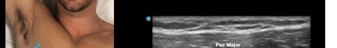
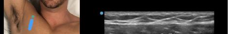
Prior to the start of the procedure, the patient must have IV access and be placed on a cardiac monitor. After informed consent has been obtained, the patient is positioned in the supine position with the head to the contralateral side of the proposed block. The physician stands at the head of the bed above the ipsilateral breast with the ultrasound screen in direct line of sight (commonly at the level of the contralateral hip). The ultrasound probe is initially placed in the sagittal plane at the midclavicular line until the clavicle, pectoralis muscles, and axillary artery and vein are visualized. The transducer is then translated caudally until the third and fourth intercostal spaces are visualized (Image 1).
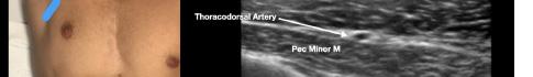
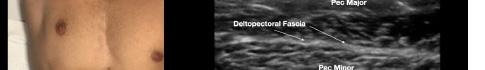
At this point, the pectoralis major and minor muscles can be visualized. By rotating the transducer 45 degrees clockwise, the thoracoacromial artery can be identified between the pectoralis major and minor muscles. Also, the serratus anterior muscle should be identified resting just above the anechoic rib (Image 2).
After appropriate skin disinfection, the block needle is advanced in-plane from the cephalad to caudal aspect of the patient. The needle should be advanced under clear ultrasound visualization during the entire procedure. For the Pecs I block, advance the needle to the fascial plane between the pectoralis

CPC-EM Capsule
What do we already know about this clinical entity? The Pectoralis block has long been used for analgesia following breast surgery. It has been shown to be safe and provides effective pain control to both the skin and soft tissues of the breast.
What makes this presentation of disease reportable? While regional anesthesia is established in the perioperative setting, its use in the emergency department (ED) for acute pain is fairly novel. This block provides the ED with a new approach for pain control.

What is the major learning point?
Due to known complications from opiates and sedation, the Pectoralis block should be considered for patients that present with painful breast complaints.
How might this improve emergency medicine practice?
As we attempt to shift towards a multimodal approach for analgesia, regional anesthesia can provide an effective and safe modality of pain control in the ED when compared to opiates and sedation.
major and minor muscles. Hydrodissection with normal saline will confirm opening of the correct fascial plane (Image 3). Anesthetic can then be gently and slowly deposited in 2-3 milliliters (mL) aliquots to a recommended amount of 15 mL per Pecs block. It is imperative to calculate your weight-based recommended dosage of anesthetic beforehand as to prevent local anesthetic systemic toxicity (LAST). If dilution is needed, the injectate can be mixed with sterile 0.9% normal
Volume 7, no. 2: May 2023 61 Clinical Practice and Cases in Emergency Medicine
Brewer et al. The Pectoralis Block: A Case Series of a Novel Modality for Acute Pain Control in the ED
Image 1. Initial probe placement for the pectoralis nerve block I and II illustrated on a model: the blue line indicates transducer, and the green dot indicates directional marker corresponding to ultrasound image.
Image 2. Final probe placement prior to the pectoralis nerve block I and II: blue line indicates transducer, and green dot indicates directional marker corresponding to ultrasound image.
The
saline. If the Pecs II block is going to be performed for axillary coverage, the needle is then advanced to the plane between the pectoralis minor and serratus anterior muscles (Image 4).


Of note, if both Pecs block I and II are being performed in a single injection, we recommend first depositing anesthetic in the fascial plane between the pectoralis minor muscle and serratus anterior muscle (Pecs II), since this can obscure the superficial structures.

Possible complications of the Pecs block include pneumothorax, vessel or nerve injury, and LAST. A contraindication to either block is cellulitis overlying the site of injection. Calculation of weight-based maximal anesthetic dosing should be performed prior to the block to maximize LAST prevention. For the purpose of this case series, a weight of 70 kilograms (kg) will be assumed. The duration of the block will vary based on the choice of anesthetic. In addition, all patients undergoing the Pecs block require cardiac
monitoring for at least 30 minutes following injection and during the procedure. Clinicians should also be aware of ultrasound-based landmarks and the signs and symptoms of LAST. The symptoms of LAST generally appear as a progression from neurological symptoms (ie, tinnitus, metallic taste, coma, seizures) to cardiovascular symptoms (ie, ventricular dysrhythmias, conduction block, cardiovascular collapse, asystole). As a result, 20% intralipid emulsion should be readily available for any large-volume block, and all clinicians should be clearly aware of dosing protocols.
CASE SERIES

Case One
A 23-year-old pregnant woman presented to the ED with pain and swelling in her left breast. She had a known history of breast cancer and had been seen by her breast surgeon four days prior for mild redness. She had been placed on cephalexin without relief. A point-of-care ultrasound revealed a threecentimeter (cm) abscess on the medial aspect of her breast. The patient’s vital signs were unremarkable other than a mild tachycardia of 110 beats per minute. The surgical service came to the bedside to evaluate the patient and asked for procedural sedation for drainage. Because of ED crowding, the physician opted for a Pecs I block at the bedside with 15 mL of 0.25% ropivacaine. About 30 minutes after completion of the block, five mL of 1% lidocaine with epinephrine was used to anesthetize the skin over the abscess. The surgical team performed a bedside incision and drainage, removing 10 mL of purulent material. The patient was observed in the ED for 24 hours and then discharged with close, outpatient follow-up.
Case Two
A 35-year-old non-pregnant woman presented to the ED with pain and redness in her left medial breast for two days. The patient was ill-appearing with the following vital signs: heart rate 120 beats per minute, respiratory rate 16 breaths per minute, blood pressure 160/85 millimeters of mercury (mm Hg) and temperature 102.6° Fahrenheit. The patient was in moderate distress and complained of 10/10 pain despite receiving 15 milligrams (mg) of IV ketorolac and four mg of IV morphine. A point-of-care ultrasound exam did not show an abscess but rather diffuse cellulitis. An ultrasound-guided Pecs I block was performed with 15 mL of 0.5% bupivacaine. The patient’s pain decreased to 2/10 at 30 minutes post block. The patient was then admitted for IV antibiotics and maintenance oral analgesia.
Case Three
A 26-year-old non-pregnant woman presented to the ED with a six-cm axillary abscess (noted on point-of-care ultrasound). The patient was in severe distress and had vital signs notable for a heart rate of 110 beats per minute but appeared non-toxic. The patient received eight mg of morphine and 15 mg of ketorolac via IV with some relief, but she was unable to tolerate abscess
Clinical Practice and Cases in Emergency Medicine 62 Volume 7, no. 2: May 2023
et al.
Pectoralis Block: A Case Series of a Novel Modality for Acute Pain Control in the ED Brewer
Image 3. Pectoralis nerve block I injection between the pectoralis major and minor muscles: arrow on patient model indicates needle direction; the green dot indicates directional marker, and yellow dots indicate injectate within the fascial plane.
Image 4. Pectoralis nerve block II injection between the pectoralis minor and serratus anterior muscles: the arrow on patient model indicates needle direction; the green dot indicates directional marker; and yellow dots indicate injectate within the fascial plane.
Brewer et al.
The Pectoralis Block: A Case Series of a Novel Modality for Acute Pain Control in the ED
drainage. A Pecs block I and II was performed under ultrasound guidance. Fifteen mL of 0.25% ropivacaine was placed between the pectoralis minor and serratus anterior muscles (Pecs II block) and 10 mL of 0.25% ropivacaine was placed between the pectoralis major and minor muscles (Pecs block I). After 30 minutes the patient’s pain was significantly decreased (3/10), allowing the physicians to place five mL of lidocaine 1% with epinephrine superficial to the abscess site. Incision and drainage was performed successfully with more than 10 mL of purulent material removed. A loop drain was left in place. The patient was discharged with antibiotics, oral pain medicine, and close, outpatient surgery follow-up.
DISCUSSION
In our case series, the Pecs block I and II were shown to be effective methods for controlling pain from breast abscesses and/or cellulitis within the anterior breast and axilla, respectively. Single injections of 10-15 mL of local anesthetic provided excellent analgesia with varying durations based on the type of local anesthetic used. In most cases, we uaed bupivacaine or ropivacaine for longer lasting coverage. Additionally, in all relevant cases, procedural sedation was avoided, with patients tolerating additional local anesthesia as well as incision and drainage at the bedside. These cases demonstrate the utility of the Pecs block for analgesia in patients with pain from breast infections. As such, the Pecs block helps avoid procedural sedation and plays an integral role in non-opioid, multimodal analgesia in the ED.
Especially when the involvement of breast surgeons is warranted (ie, abscesses larger than five cm or adjacent to areola), offering multimodal pain control is vital while determining the ideal location and method for drainage. In settings where access to specialty surgery remains limited, such as many non-academic centers, the Pecs block can be an ideal method for pain control until the patient can be seen by a breast surgeon.
CONCLUSION
Ultrasound-guided pectoralis nerve block I and II can provide safe and rapid analgesia for patients with painful breast abscesses and/or cellulitis. Further research is needed to determine whether both the Pecs block I and II are required to provide adequate analgesia and anesthesia for simple breast abscesses. However, these blocks can be easily performed at the bedside with a portable ultrasound machine with minimal risk to the patient. All in all, we believe that the Pecs block
can serve as an integral component of multimodal, non-opioid, analgesic regimens within the emergency department.
The authors attest that their institution requires neither Institutional Review Board approval, nor patient consent for publication of this case series. Documentation on file.
Address for Correspondence : Jonathan Brewer, MD, Vanderbilt University Medical Center, Department of Emergency Medicine, 1211 Medical Center Dr., Nashville, TN 37232. Email: jonathan.h.brewer@yumc.org.
Conflicts of Interest: By the CPC-EM article submission agreement, all authors are required to disclose all affiliations, funding sources and financial or management relationships that could be perceived as potential sources of bias. The authors disclosed none.
Copyright: © 2023 Brewer et al. This is an open access article distributed in accordance with the terms of the Creative Commons Attribution (CC BY 4.0) License. See: http://creativecommons.org/ licenses/by/4.0/
REFERENCES
1. Downey K and Becker T. Techniques for skin abscess drainage. UpToDate. 2022. Available at: https://www.uptodate.com/contents/ techniques-for-skin-abscess-drainage?search=abscess&topicRef=1105 30&source=see_link#H13. Accessed December 8, 2022.
2. Dixon JM and Khan LR. Treatment of breast infection. BMJ 2011;342(Jun14 2):d3744.
3. Dixon JM. Primary breast abscess. UpToDate. 2020. Available at: https:// www.uptodate.com/contents/primary-breast-abscess?search=breast+ab scess&source=search_result&selectedTitle=1~35&usage_ type=default&display_rank=1#H10. Accessed December 8, 2022.
4. Boakes E, Woods A, Johnson N, et al. Breast infection: a review of diagnosis and management practices. Eur J Breast Health. 2018;14(3).
5. Blanco R and Barrington M. Pectoralis and serratus plane nerve blocks. NYSORA. 2022. Available: https://www.nysora.com/regional-anesthesiafor-specific-surgical-procedures/thorax/pectoralis-serratus-plane-blocks/. Accessed December 8, 2022.
6. Kataria K, Srivastava A, Dhar A. Management of lactational mastitis and breast abscesses: review of current knowledge and practice. Indian J Surg. 2012;75(6):430-5.
7. Sethuraman RM and Narayanan V. Pecs II block: clarifications sought on nomenclature. Reg Anesth Pain. 2022;47(7).
Volume 7, no. 2: May 2023 63 Clinical Practice and Cases in Emergency Medicine
A Cluster of Neuroinvasive Adenovirus Infections on a College Campus: Case Series
Nicholas Valentini, MD*
Madison Breeden, MD†
Laura E. Felley, MD, PhD†
Nathan B. Roberts, MD, PhD*
Maia Dinsmore, MD†
Aaron Krumheuer, MD*
Colleen Grassley, PA-C*
Sacha Montas, MD, JD*
Benjamin Bassin, MD*
Kylee Phillips, MD, MBA*
Section Editor: Melanie Heniff, MD
University of Michigan, Department of Emergency Medicine, Ann Arbor, Michigan University of Michigan, Department of Internal Medicine, Division of Infectious Disease, Ann Arbor, Michigan
Submission history: Submitted December 21, 2022; Revision received March 16, 2023; Accepted March 17, 2023
Electronically published May 27, 2023
Full text available through open access at http://escholarship.org/uc/uciem_cpcem
DOI: 10.5811/cpcem.1406
Introduction: We present six adenovirus cases that emerged from a cluster of respiratory illnesses within a college population. Two patients required intensive care with complicated hospital courses and experienced residual symptoms. Four additional patients were evaluated in the emergency department (ED) with two additional diagnoses of neuroinvasive disease. These cases represent the first known occurrences of neuroinvasive adenovirus infections in healthy adults.
Case Series: An individual presented to the ED with fever, altered mental status, and seizures after being found unresponsive in his apartment. His presentation was concerning for significant central nervous system pathology. Shortly after his arrival, a second individual presented with similar symptoms. Both required intubation and admission to a critical care setting. Over a 24-hour period, four additional individuals presented to the ED with moderate severity symptoms. All six individuals tested positive for adenovirus in their respiratory secretions. A provisional diagnosis of neuroinvasive adenovirus was made after consultation with infectious diseases.
Conclusion: This cluster of cases appears to represent the first known reported diagnosis of neuroinvasive adenovirus in healthy young individuals. Our cases were also unique in demonstrating a significant spectrum of disease severity. Over 80 individuals in the broader college community ultimately tested positive for adenovirus in respiratory samples. As respiratory viruses continue to challenge our healthcare systems, new spectrums of disease are being discovered. We believe clinicians should be aware of the potential severity of neuroinvasive adenovirus disease. [Clin Pract Cases Emerg Med. 2023;7(2):64–67]
Keywords: adenovirus; neuroinvasive; case report; encephalitis.
INTRODUCTION
We present a spectrum of neuroinvasive adenovirus cases that emerged from a cluster of viral respiratory illnesses within a college population. These cases appear to represent the first known occurrences of neuroinvasive adenovirus infections in healthy adults without immunocompromise. Clinicians should be aware of the potential for novel outbreaks of disease among
concentrated populations such as those on a college campus. In addition, clinicians should be aware of the risk of atypical presentations of adenovirus infection.
CASE SERIES
Patient one was a 23-year-old male with a past medical history (PMH) of nephrolithiasis who presented to the
Clinical Practice and Cases in Emergency Medicine 64 Volume 7, no. 2: May 2023 Case Series
* †
emergency department (ED) with altered mental status (AMS) in the setting of one week of infectious symptoms including fever, body aches, chills, headache, and cough. On arrival, the patient was actively seizing. Exam was further notable for hyperreflexia and a petechial rash on the head and upper torso. Signs of trauma were absent. The patient was intubated for airway protection and was empirically treated with dexamethasone, meropenem (due to reported allergies), vancomycin, and acyclovir. Lumbar puncture (LP) was completed with a negative Gram stain and a lymphocytic predominance. Computed tomography (CT) showed no acute pathology.
An electroencephalogram (EEG) was obtained and did not suggest ongoing seizure activity. Blood work showed a mild leukocytosis without additional acute findings. Creatinine kinase (CK) testing was normal. Serum and urine toxicologic studies were negative. A respiratory polymerase chain reaction (PCR) sample was positive for adenovirus. The patient was admitted to the intensive care unit. He underwent expanded testing for immunocompromising and infectious conditions without pertinent findings. Cerebrospinal fluid (CSF) samples were tested for adenovirus and resulted without positive PCR findings. Respiratory samples were sent for expanded genomic testing to the US Centers for Disease Control and Prevention. He was treated with cidofovir. The patient was extubated on day four of his hospital course. His mental status slowly improved after extubation; however, he had persistent deficits in executive functioning and short-term memory. He was discharged on hospital day seven, and at outpatient follow-up two weeks after discharge he continued to have residual deficits in memory and cognition.
Patient two was a 21-year-old male with an unremarkable PMH who presented to the ED due to AMS and seizure in the setting of one week of infectious symptoms including fevers, cough, and congestion. The patient was not following commands but moving all extremities without focal neurological deficits. He had no rash, although there had been reports of rash earlier in the week. The patient was intubated and empirically treated with dexamethasone, cefepime, vancomycin, and acyclovir. An LP showed a lymphocytic predominance and a negative Gram stain. A respiratory PCR was positive for adenovirus. A head CT was initially concerning for a small intracranial hemorrhage in the left parietal lobe, but follow-up magnetic resonance imaging ultimately showed this to be a cavernoma. An EEG did not suggest seizure activity. Toxicologic studies were negative. He was treated with cidofovir. He was extubated on day three of his hospital course, and his mental status slowly improved after extubation. He was discharged home at his mental status baseline after five days in the hospital. Viral cerebrospinal fluid studies including for adenovirus were negative.
Patients three and four presented 24 hours after the initial cases. They were an 18-year-old male and a 23-year-
Population Health Research Capsule
What do we already know about this clinical entity?
Adenovirus is a common respiratory pathogen with varied clinical presentations and the potential to cause severe disease.
What makes this presentation of disease reportable?
This is the first known presentation of neuroinvasive adenovirus in otherwise healthy adults.
What is the major learning point?
There is the potential for severe complications in the presence of adenovirus infections.
How might this improve emergency medicine practice?
Emergency physicians should be aware of emerging pathogens and new presentations of known respiratory infections.
old male, respectively, with no PMH. Presenting symptoms included sore throat, fever, myalgias, cough, and neck pain. Both patients had had close contact with patient one. Physical exam in both cases was notable for nuchal rigidity, no focal neurologic deficits, and no rash or other skin lesions. Blood work was without pertinent findings. A respiratory PCR returned positive for adenovirus in both cases. An LP was performed for both individuals, again notable for a lymphocytic predominance. Both patients became febrile during their ED evaluations. Due to concern for worsening central nervous system (CNS) symptoms of headache and neck stiffness, both patients were admitted to the hospital for observation and ultimately discharged home the following day.
Patient five was a 20-year-old male who presented to the ED for extremity weakness and cough. He was a close contact of three of the previous patients and presented on the advice of family and friends who had knowledge of the disease severity in the prior cases. A physical exam did not reveal any meningismus or focal neurologic abnormalities, and vital signs were within normal limits. His exam was notable for tenderness in the lower extremity muscle groups. His respiratory PCR testing was notable for adenovirus infection. There was concern for viral myositis vs rhabdomyolysis, confounded by a history of recent strenuous exercise. A CK was elevated to greater than 7000 international units per liter; however, renal function and electrolyte testing were normal.
Volume 7, no. 2: May 2023 65 Clinical Practice and Cases in Emergency Medicine Valentini et al.
of Neuroinvasive Adenovirus Infections on a College Campus
A Cluster
A Cluster of Neuroinvasive Adenovirus Infections on
The patient was admitted and discharged two days later. The patient did not develop any signs of meningoencephalitis during admission, and a LP was not performed.
Patient six was a 20-year-old male with a PMH of Crohn’s disease on immunosuppressive therapy who presented shortly after patient five with symptoms of fever, cough, myalgias, and malaise. He was a close contact of several of the prior patients and was aware of the severity of their illnesses. Respiratory PCR testing was positive for adenovirus. Physical exam and vital signs were within normal limits. Lab evaluation was without notable findings; given the absence of CNS symptoms a LP was not performed. The patient was discharged with close outpatient follow-up and did not require admission.
Given the similarity and severity of the above presentations, campus health officials were alerted, and close contacts were notified. Expanded testing ultimately revealed over 80 individuals with confirmed adenovirus infection by respiratory PCR testing. Many additional individuals reported similar symptoms but did not undergo testing. No additional hospitalizations or ED evaluations were reported.
In summary, six patients were evaluated in the ED over three days. Lumbar puncture was performed in four of six cases with a finding of lymphocytic pleocytosis. Adenovirus serum and CSF testing was negative in all patients with suspected neuroinvasive disease. Two critically ill patients received cidofovir. While all patients largely recovered from their illness, patient one had lingering difficulties with executive function and memory.
DISCUSSION
Neuroinvasive adenovirus infections are extraordinarily unusual in healthy patients. Adenovirus is a non-enveloped deoxyribonucleic acid (DNA) virus that is typically spread by inhalation of aerosolized droplets, but fecal-oral spread or contact with exposed surfaces can also produce infection. As the virus is non-enveloped, it is resistant to many disinfectants, although alcohol-based products remain effective.1 Outbreaks are typically seasonal, often occurring in the late fall or early spring. Typical symptoms include fever, pharyngitis, and cough with conjunctivitis and gastrointestinal (GI) symptoms being less common. In immunocompromised patients, adenovirus can cause more severe manifestations including pneumonia, hemorrhagic cystitis, nephritis, and meningoencephalitis.2 Adenovirus infection is a common concern in recipients of both solid organ and hematopoietic stem cell transplants. Depletion of T cells in preparation for transplant is a risk factor for severe infection. Congenital immunodeficiencies are also associated with severe adenovirus infection.
Meningoencephalitis is one of the most uncommon presentations of adenovirus. A single-center review reported meningoencephalitis symptoms in approximately 1% of all adenovirus cases, noting the majority of cases were seen in children age five or less.3 In cases of presumed viral
encephalitis, evaluation for adenovirus is generally only considered once herpes simplex virus, varicella zoster virus, arboviruses, and other common causes of aseptic meningitis have been ruled out.4 Neurologic manifestations of adenovirus infection have been identified, but nearly all cases occurred in pediatric populations or those with predisposing conditions. A review of cases over 21 years identified 48 cases of adenovirus-associated CNS disease in immunocompetent children; the most common manifestations were febrile seizures, encephalitis, aseptic meningitis, and acute disseminated encephalomyelitis. Interestingly, like the patients in this case, 85% had virus detected in the respiratory or GI tract but not in the CSF.5
In a three-year review of adults and children presenting with meningoencephalitis, four adults and one newborn were found to have CSF positive for adenovirus; of the adults, two had HIV, one had sickle cell disease, and one had poorly controlled diabetes.6 A case report in 2006 identified an immunocompetent patient with a history of medulloblastoma status post resection and recent brain irradiation who presented with confusion, difficulty speaking, and increasing somnolence. After the patient developed seizures, a brain biopsy was obtained that was positive for adenovirus. The authors noted that this was the first case of adenovirus encephalitis without prior respiratory involvement. Lung biopsies failed to grow the virus, and the pathology was not typical.7 The isolated encephalitis seen in our two previously healthy young adults is unprecedented.
Treatment decisions in these cases were made more challenging by the fact that there are currently no approved treatments for adenovirus; no randomized controlled trials comparing therapies exist, and most of the literature focuses on pediatric populations or the highly immunocompromised. Ribavirin and ganciclovir have in vitro activity against adenovirus but have not been shown to be effective clinically.2 The most common agent is cidofovir, a cytosine nucleotide analogue that inhibits the viral DNA polymerase, but this treatment is generally reserved for cases of adenovirus in solid organ or hematopoietic transplant patients.2 There is no clear consensus on when treatment is appropriate, as spontaneous viral clearance even in the immunocompromised is not uncommon. Treatment is typically initiated for those with severe disease, disseminated infection, or profound immune deficits.2
In the two severe neuroinvasive cases presented here, the decision to treat was based on the neurologic impairment seen on admission. As nothing is known about the natural history of neuroinvasive adenovirus in healthy individuals, it is unclear how much impact the cidofovir had on their clinical improvement.
CONCLUSION
Adenovirus is a common pathogen in our society often associated with mild respiratory illness. In this case series, we report the development of significant encephalitis from a neuroinvasive strain of adenovirus within a college
Clinical Practice and Cases in Emergency Medicine 66 Volume 7, no. 2: May 2023
College Campus Valentini et al.
a
Valentini et al.
A Cluster of Neuroinvasive Adenovirus Infections on a College Campus
community. As respiratory viruses continue to challenge our healthcare systems, new spectrums of disease are being discovered. We believe clinicians should be aware of the potential for serious neuroinvasive adenovirus disease in their evaluation and management of patients presenting with respiratory and central nervous system symptoms. This series also shows the role of the ED in population health surveillance. Early detection and reporting of these atypical presentations aided public health officials in rapidly expanding surveillance systems and contact tracing. This further emphasizes the multidisciplinary and ever evolving role of the ED within the larger healthcare system.
The authors attest that their institution does not require Institutional Review Board approval, Patient consent has been obtained and filed for publication of this case series. Documentation on file.
Address for Correspondence: Nicholas Valentini, MD, University of Michigan, Department of Emergency Medicine, 1500 East Medical Center Drive, TC-B1-380, Ann Arbor, MI 48109. Email: valentni@med.umich.edu.
Conflicts of Interest: By the CPC-EM article submission agreement, all authors are required to disclose all affiliations, funding sources and financial or management relationships that could be perceived as potential sources of bias. The authors disclosed none.
Copyright: © 2023 Valentini et al. This is an open access article distributed in accordance with the terms of the Creative Commons Attribution (CC BY 4.0) License. See: http://creativecommons.org/ licenses/by/4.0/
with central nervous system dysfunction in children. J Clin Virol. 2013;57:300-4.
4. Tyler KL. Acute viral encephalitis. N Engl J Med. 2018;379:557-66.
REFERENCES
1. Lion T. Adenovirus infections in immunocompetent and immunocompromised patients. Clin Microbiol Rev. 2014;27(3):441-62.
2. Lynch JP 3rd, Fishbein M, Echavarria M. Adenovirus. Semin Respir Crit Care Med. 2011;32:494-511.
3. Huang YC, Huang SL, Chen SP, et. al. Adenovirus infection associated
5. Schwartz KL, Richardson SE, MacGregor D, et al. Adenovirusassociated central nervous systemic disease in children. J Pediatr 2018;205:130-7.
6. Vidal LR, de Almeida SM, Cavalli BM, et. al. Human adenovirus meningoencephalitis: a 3 years’ overview. J Neurovirol 2019;25(4):389-96.
7. Dubberke ER, Tu B, Rivet DJ, et al. Acute meningoencephalitis caused by adenovirus serotype 26. J Neurovirol. 2006;12:235-40.
Volume 7, no. 2: May 2023 67 Clinical Practice and Cases in Emergency Medicine
Myocardial Bridge of the Left Anterior Descending Artery Causing Pseudo-Wellens’ Syndrome: A Report of Two Cases
Debayan Guha, MD*
Franz C. Mendoza-Garcia, MD*
Kathryn M. Millen, MD*
Joseph Offenbacher, MD†
Nicholus M. Warstadt, MD†
Section Editor: Austin Smith, MD
Albert Einstein College of Medicine, Department of Emergency Medicine at the Jacobi and Montefiore Medical Centers, Bronx, New York
New York University Grossman School of Medicine, Ronald O. Perelman Department of Emergency Medicine, New York, New York
Submission history: Submitted September 28, 2022; Revision received February 27, 2023; Accepted March 8, 2023
Electronically published May 26, 2023
Full text available through open access at http://escholarship.org/uc/uciem_cpcem
DOI: 10.5811/cpcem.1404
Introduction: Wellens’ syndrome represents an important, at times overlooked, spectrum of left anterior descending (LAD) coronary artery occlusion, spontaneous reperfusion, and impending reocclusion. Once considered pathognomonic for a thromboembolic coronary event, an increasing number of clinical scenarios have been demonstrated to result in pseudo-Wellens’ syndrome, each requiring unique forms of assessment and management.
Case Report: We describe two clinical presentations in which myocardial bridging (MB) of the LAD led to clinical and electrophysiologic presentations of a pseudo-Wellens’ syndrome.
Conclusion: These reports represent a rare cause of pseudo-Wellens’ syndrome attributed to MB of the LAD. Transient ischemia secondary to myocardial compression of the traversing LAD leads to intermittent angina and electrocardiogram changes that are typical in patients presenting with Wellens’ syndrome secondary to an occlusive coronary event. As with other previously reported pathophysiologic mechanisms that have been shown to mimic Wellens’ syndrome, myocardial bridging should be considered in patients presenting with a pseudo-Wellens’ syndrome. [Clin Pract Cases Emerg Med. 2023;7(2):68–72]
Keywords: emergency medicine; acute coronary syndrome; Wellens’ syndrome; myocardial bridge; case report.
INTRODUCTION
First described in 1982, Wellens’ syndrome represents an important cardiovascular syndrome in the emergency department (ED). Defined by its electrocardiogram (ECG) findings in the context of waxing and waning angina, Wellens’ syndrome was historically taught to herald impending cardiac ischemia due to a left anterior descending (LAD) thromboembolic lesion.1 It is now understood to represent an evolving pathophysiologic pattern of occlusion and spontaneous reperfusion of the LAD. The term pseudo-Wellens’ syndrome is used when the etiology of the patient’s Wellens’-like ECG pattern is not due to an atherosclerotic event.
Myocardial bridging (MB) is a congenital variant in which a coronary artery tunnels through the myocardium and the tunneled artery is constricted on ventricular systole.2 We describe two cases of a pseudo-Wellens’ syndrome attributed to MB of the LAD. We hope to highlight the rapid identification of Wellens’ syndrome via clinical presentation and ECG pattern, educate emergency physicians on MB as a possible anginal cause, and further describe a rare clinical entity.
CASE REPORTS Case One
A 67-year-old female with a past medical history of diabetes, hypertension, hypothyroidism, scoliosis, spinal
Clinical Practice and Cases in Emergency Medicine 68 Volume 7, no. 2: May 2023 Case Report
* †
Guha et al.
Myocardial Bridge of the LAD Artery Causing Pseudo-Wellens’ Syndrome
stenosis, chronic back pain, and cervical radiculopathy presented to the ED complaining of atraumatic neck and back pain radiating to her left shoulder. She denied chest pain, shortness of breath, or any other active cardiopulmonary symptoms. Her pain was not exertional, pleuritic, or positional. Her review of systems was otherwise negative.
On presentation to the ED, her heart rate was 78 beats per minute, blood pressure was 143/62 millimeters of mercury (mm Hg), respiratory rate 14 breaths per minute, oxygen saturation s 99% on room air, and temperature was 36.7° Celsius. She was well-appearing in no acute distress, and there were no notable findings on cardiopulmonary, neurologic, or musculoskeletal exams. Her ECGs are summarized in Images 1A and 1B.
Laboratory studies were significant for mild anemia with a hemoglobin of 11.4 grams per deciliter (g/dL) (reference range: 11.9-15.3 g/dL). Serum troponin and creatinine phosphokinase were within normal limits. Chest radiograph showed a prominent heart size and calcified aorta with clear
CPC-EM Capsule
What do we already know about this clinical entity?
Wellens’ Syndrome is considered an indicator of impending left anterior descending occlusion. Non-thromboembolic mimics have been termed pseudo-Wellens’.
What makes this presentation of disease reportable?
Myocardial bridging is a rare cause of pseudo-Wellens’. Here, two patients with variable presentations are described.
What is the major learning point?
It is important to recognize various pathophysiological mechanisms for pseudoWellens’ as treatment may differ from treatment of an acute coronary syndrome.
How might this improve emergency medicine practice?
This report adds to the knowledge of pseudoWellens’ syndromes, describes physiology of myocardial bridging, and discusses management of patients with known MB.
lungs. Cardiology was consulted for possible Wellens’ syndrome and were concerned for a proximal LAD lesion. The patient received 325 milligrams (mg) aspirin and was started on intravenous (IV) heparin. She was taken for urgent coronary catheterization, which showed a patent coronary lumen. A MB in the mid-LAD was discovered and was thought to explain the patient’s clinical presentation and ECG findings (Images 2A and 2B).
Case Two
A 56-year-old female with a past medical history of hypertension and hyperlipidemia presented to the ED with a two-day history of exertional, left-sided chest pain and dyspnea. Chest pain was non-radiating, described as a tightness, and 10/10 in severity when present. She had associated lightheadedness. She denied symptoms at rest and was asymptomatic while in the ED. Review of systems was otherwise negative. On presentation to the ED, her heart rate was 86 beats per minute, blood pressure was 137/89 mm Hg, respiratory rate 16 breaths per minute, oxygen saturation 98% on room air, and temperature was 36.6° Celsius. Her physical exam was unremarkable.
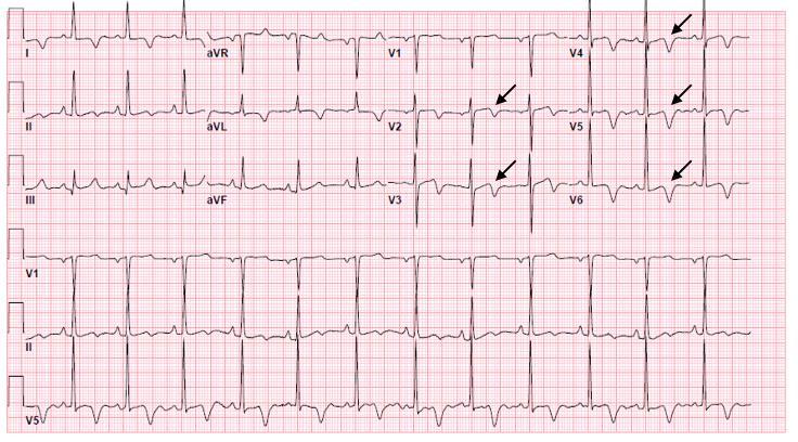
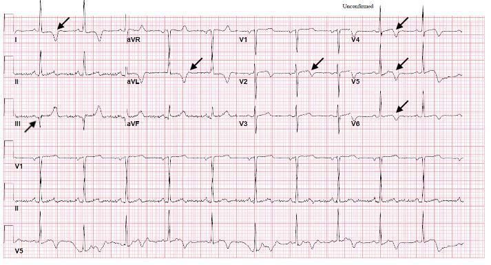
Volume 7, no. 2: May 2023 69 Clinical Practice and Cases in Emergency Medicine
Image 1A. Electrocardiogram on presentation with Q wave in III, deep T-wave inversions in leads I and aVL, and biphasic T-wave morphology in leads V2 and V4-V6; annotated with black arrows.
Image 1B. Follow-up electrocardiogram 20 minutes after presentation with persistent biphasic T-wave inversion in V2, evolving biphasic T-wave morphology in V3, and deeply symmetrically inverted T-waves in V4-V6; annotated with black arrows
Her initial ECG, concerning for Wellens’ syndrome, is shown in Image 3A. An ST-elevation myocardial infarction (STEMI) alert was activated, the patient was given 325 mg of aspirin, started on IV heparin, and taken for cardiac catheterization. Blood laboratory analysis sent prior to the catheterization were remarkable for a troponin I concentration of 0.37 nanograms per milliliter (ng/mL) (reference range: < 0.04 ng/mL). Complete blood count, complete metabolic panel, coagulation studies, lipid panel, and hemoglobin A1c were unremarkable.
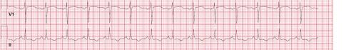
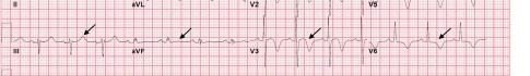
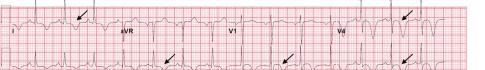
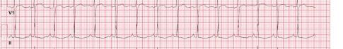
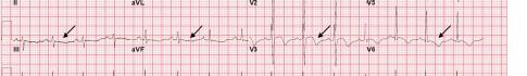
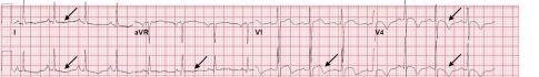
The patient’s left heart catheterization showed mild luminal irregularities in the LAD without a culprit lesion. A mid-LAD MB was identified. No percutaneous coronary intervention (PCI) was performed. Subsequent transthoracic echocardiogram and cardiac magnetic resonance imaging were unremarkable. The patient was started on aspirin and metoprolol, and she was discharged on hospital day two.
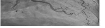
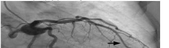
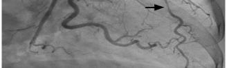
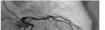
Two weeks after her hospital discharge and while in the cardiology office for follow-up, the patient experienced a
similar episode of 10/10 chest pain. Her ECG is shown in Image 3B. The patient was transferred to the ED, where she reported ongoing chest pain. Her heart rate was 77 beats per minute, blood pressure 121/84 mm Hg, respiratory rate 16 breaths per minute, oxygen saturation 100% on room air, and temperature was 36.6° Celsius. She appeared uncomfortable with an otherwise unremarkable physical exam.
Laboratory analysis was notable for a troponin I concentration of 0.37 ng/mL. Due to ongoing chest pain with new ECG changes, the patient went for coronary angiography, which showed the known MB without new plaque rupture, occlusion, dissection, or alternate explanation for pain. During her hospitalization, metoprolol was discontinued, and verapamil was initiated. She was discharged on hospital day two with cardiology and cardiac surgery follow-up. Despite medical therapy, the patient remained symptomatic at follow-up. She was not interested in surgical intervention.
Image 3A. Presenting electrocardiogram showing T-wave flattening in leads I and aVL; subtle T-wave inversions in II, III, and aVF; new biphasic T-waves in V2 and V3; and new deep symmetric T-wave inversions in V4-V6; annotated with black arrows.
Clinical Practice and Cases in Emergency Medicine 70 Volume 7, no. 2: May 2023 Myocardial
of the LAD
Causing Pseudo-Wellens’ Syndrome Guha et al.
Bridge
Artery
Image 2A. Coronary angiogram demonstrating mid-left anterior descending coronary artery occlusion during systole (arrow).
Image 2B. Coronary angiogram demonstrating mid-left anterior descending patency and distal coronary artery reperfusion during diastole (arrow).
Image 3B. Electrocardiogram on second presentation showing new T-wave inversions in leads I and aVL; pseudo-normalization of T-waves in III and aVF; and deepening of previously noted Twave inversions in V2-V6; annotated with black arrows.
DISCUSSION
Wellens’ syndrome is an important clinical entity in the ED suggesting a dynamic occlusion, reperfusion, and impending reocclusion of the LAD. Once considered to be a STEMI equivalent, evolving understanding of this syndrome calls for aggressive medical management and urgent coronary catheterization.
Traditionally, the syndrome was thought to consist of two distinct ECG patterns found in patients with a history of unstable angina. Type A (25% of patients) presents with biphasic T-waves in V2-V3. Type B (75% of patients) presents with deep symmetric T-wave inversions in V2-V3.3 These findings represent critically high grade LAD stenosis with a high specificity of 96.2%.4 While patients are often pain-free at the time the ECG is taken, subsequent anterior wall myocardial infarction is likely.
While Wellens’ syndrome is associated with thromboembolic occlusion of the LAD, several other physiologic mechanisms have been shown to mimic this electrocardiographic presentation due to transient obstruction of coronary flow and are categorized as pseudo-Wellens’ syndrome. Coronary vasospasm from cocaine use,5 pulmonary embolism,6 stress cardiomyopathy,7 and uncontrolled hypertension8 have been reported to cause pseudoWellens’ syndrome.
Myocardial bridging describes a congenital variant characterized by an intramyocardial route of a segment of one of the major coronary arteries, generally the LAD. The rate of MB in the general population ranges from 5-40%.2 Often discovered incidentally, prevalence is impacted by the diagnostic modality used (coronary angiography, computed tomography, etc). Patients with hypertrophic cardiomyopathy have exponentially higher incidence.9 Myocardial bridging of the LAD is associated with myocardial ischemia, the development of dysrhythmias, and sudden cardiac death.2 Limited evidence suggests the use of beta-blockers (BB) or non-dihydropyridine calcium-channel blockers (CCB) as first-line medical therapy. Stenting is controversial due to high rates of revascularization. If medical management fails, myotomy and coronary artery bypass grafting are viable surgical options. Nitroglycerin accentuates systolic compression of bridged segments and is contraindicated.2 Exercise may induce symptoms and has even been reported to cause fatal arrythmia,10 although no consensus guidelines exist for exercise abstinence.
The typical angiographic feature of a MB is systolic narrowing of a coronary artery, which often resolves completely during the diastolic phase of the cardiac cycle. Because only 15% of coronary flow normally occurs during systole and MB is a systolic angiographic event, clinically significant ischemia has only been demonstrated in specific clinical scenarios. Most commonly, tachycardia can provoke an ischemic event secondary to MB due to the shortening of the diastolic phase and the subsequent increased significance of systolic coronary perfusion.11 If the myocardial muscle is
hypertrophic, the tunneled artery can be compressed during each cycle of systole.12
Rarely, MB has been reported to cause pseudo-Wellens’ syndrome.12-15 In each case previously described, the patient presented with classic anginal symptoms and a Wellens’pattern ECG. Each patient had negative serum troponin concentrations. All patients responded to medical therapy, which primarily included BB, CCB, aspirin, and clopidogrel; and none required PCI or surgery.
In both cases presented here, MB of the mid-LAD, diagnosed via coronary angiography, caused transient LAD stenosis, resulting in pseudo-Wellens’ syndrome. Case one is unique in that the patient had neither chest pain nor classic anginal symptoms. Nevertheless, her dynamic ECG changes were concerning for ischemia, which prompted coronary angiography and led to the diagnosis of MB. Case two is the first in the literature to describe pseudo-Wellens’ from MB leading to a positive troponin and in which the patient had early treatment failure to pharmacological interventions.
CONCLUSION
Myocardial bridging should be recognized as a possible etiology of pseudo-Wellens’ syndrome. Because Wellens’ syndrome often predicts imminent critical ischemia and reports of pseudo-Wellens’ syndromes are rare, acute coronary syndrome should be empirically treated and appropriately ruled out in patients presenting with a concerning history and a Wellens’pattern ECG. Symptomatic worsening following nitroglycerin administration should increase suspicion for MB. Diagnosis is made by coronary angiography or non-invasive advanced coronary imaging techniques. After diagnosis is made, medical management typically includes initiation of beta blockers or calcium-channel blockers, with surgery reserved for refractory cases. Percutaneous coronary intervention is controversial. Patients should be followed by cardiology and cardiac surgery.
The authors attest that their institution does not require Institutional Review Board approval. Patient consent has been obtained and filed for the publication of this case report. Documentation on file.
Address for Correspondence : Nicholus Warstadt, MD, Ronald O. Perelman, Department of Emergency Medicine, 570 First Avenue, New York, New York 10016. Email: nicholus.warstadt@nyulangone.org .
Conflicts of Interest: By the CPC-EM article submission agreement, all authors are required to disclose all affiliations, funding sources and financial or management relationships that could be perceived as potential sources of bias. The authors disclosed none.
Copyright: © 2023 Guha et al. This is an open access article distributed in accordance with the terms of the Creative Commons Attribution (CC BY 4.0) License. See: http://creativecommons.org/ licenses/by/4.0/
Volume 7, no. 2: May 2023 71 Clinical Practice and Cases in Emergency Medicine
Guha et al. Myocardial Bridge of the LAD Artery Causing Pseudo-Wellens’ Syndrome
Myocardial Bridge of the LAD Artery Causing Pseudo-Wellens’ Syndrome
REFERENCES
1. de Zwaan C, Bär FW, Wellens HJ. Characteristic electrocardiographic pattern indicating a critical stenosis high in left anterior descending coronary artery in patients admitted because of impending myocardial infarction. Am Heart J. 1982;103(4 Pt 2):730-6.
2. Lee MS and Chen CH. Myocardial bridging: an up-to-date review. J Invasive Cardiol. 2015;27(11):521-8.
3. Mufti M, Joea R, Sobnosky S, et al. Wellens’ syndrome: an atypical presentation of an already silent killer. J Med Cases. 2018;9(6):173-6.
4. Kobayashi A, Misumida N, Aoi S, et al. Prevalence and clinical implication of Wellens’ sign in patients with non-ST-segment elevation myocardial infarction. Cardiol Res. 2019;10(3):135-41.
5. Lin AN, Lin S, Gokhroo R, et al. Cocaine-induced pseudo-Wellens’ syndrome: a Wellens’ phenocopy. BMJ Case Rep. 2017;2017:1-3.
6. Sedhai YR, Basnyat S, Bhattacharya PT. Pseudo-Wellens’ syndrome in pulmonary embolism. BMJ Case Rep. 2018;11(1):1-4.
7. Ju TR, Yeo I, Pontone G, et al. Pseudo-Wellens syndrome from sepsis-induced cardiomyopathy: a case report and review of the literature. J Med Case Rep. 2021;15(1):151-6.
8. Ola O and Tak T. Pseudo-Wellens syndrome in a patient with
hypertension and left ventricular hypertrophy. Am J Case Rep. 2019;20:1231-4.
9. Mohlenkamp S, Hort W, Ge J, et al. Update on myocardial bridging. Circulation. 2002;106(20):2616-22.
10. Feld H, Guadanino V, Hollander G, et al. Exercise-induced ventricular tachycardia in association with a myocardial bridge. Chest. 1991;99(5):1295-6.
11. Ripa C, Melatini MC, Olivieri F, et al. Myocardial bridging: a ‘forgotten’ cause of acute coronary syndrome - a case report. Int J Angiol. 2007;16(3):115-8.
12. Ambesh P, Sharma D, Kapoor A, et al. Unusual sign from an unusual cause: Wellens’ syndrome due to myocardial bridging. Case Rep Cardiol. 2018;2018 (1):1-3.
13. Milne D, Ramadhin D, Seecheran R, et al. The curious case of pseudo-Wellens’ syndrome and myocardial bridging. J Investig Med High Impact Case Rep. 2022;10(1):1-4.
14. Avram A, Chioncel V, Guberna S, et al. Myocardial bridging-an unusual cause of Wellens’ syndrome: a case report. Medicine (Baltimore). 2020;99(41):1-6.
15. Kaplanis I, Michas G, Arapi S, et al. Myocardial bridge as a cause of pseudo-Wellens’ syndrome. Hellenic J Cardiol. 2017;58(6):453-5.
Clinical Practice and Cases in Emergency Medicine 72 Volume 7, no. 2: May 2023
Guha et al.
Bilateral Infectious Extensor Tenosynovitis: A Case Report
Daria Osipchuk, MD Jeffrey Riddell, MD
Section Editor: Joel Moll, MD
Submission history: Submitted July 27, 2022; Revision received September 29, 2022; Accepted December 27, 2022
Electronically published April 18, 2023
Full text available through open access at http://escholarship.org/uc/uciem_cpcem
DOI: 10.5811/cpcem.1317
Introduction: Infectious extensor tenosynovitis is a rare infection spreading along the extensor tendons of the extremities. It presents a diagnostic challenge in the emergency department (ED) given the nonspecific signs and symptoms, as opposed to the more common flexor tenosynovitis that is diagnosed by the classic Kanavel signs on physical exam.
Case Report: Here we present a case of bilateral extensor tenosynovitis in a 52-year-old female denying past medical history who presented to the ED with two days of bilateral dorsal hand swelling and pain. She denied any risk factors such as direct trauma to the hands or intravenous drug use. The rare diagnosis was suspected in the ED due to a very high complement reactive protein level and a concerning point-of-care ultrasound. Extensor tenosynovitis was ultimately confirmed on computed tomography and by operative irrigation and drainage of the tendon sheaths.
Conclusion: This case demonstrates the importance of keeping extensor tenosynovitis on the differential when seeing a patient with dorsal extremity edema and pain, even if the findings occur bilaterally. [Clin Pract Cases Emerg Med. 2023;7(2):73–76]
Keywords: case report; extensor tenosynovitis.
INTRODUCTION
Purulent tenosynovitis is an infection that tracks along tendons. Most commonly it is seen as a flexor tenosynovitis of the hand and wrist, identified by the Kanavel signs, which include pain with palpation along the flexor tendon, the digit held in flexion, pain with passive extension of the digit, and fusiform swelling of the digit.1 It is much rarer to find in extensor tendons, with only a few case reports and case series documenting infectious extensor tenosynovitis in the literature. The findings in extensor tenosynovitis lack the classic characteristic Kanavel signs of flexor tenosynovitis and, thus, there are frequent delays in diagnosis. As with most hand infections, patients usually report trauma or preceding intravenous (IV) drug use, and the classic dogma is that infections occur unilaterally.2,3 Here we report a case of bilateral extensor tenosynovitis in a patient denying direct trauma or IV drug use.
CASE REPORT
A 52-year-old female with no past medical history presented to the ED with bilateral hand swelling and pain for two days. The patient reported that a few days prior she had strained her left shoulder while holding a rope to help somebody cut down a tree. She presented to an outside hospital, was diagnosed with a rotator cuff tear, and was given IV morphine in the left upper extremity. Subsequently, she developed left hand swelling. She went back to the outside hospital, received IV morphine in the right upper extremity, and developed right hand swelling. The patient denied any direct trauma to her hands. The pain was only alleviated by placing her hands in cold water. In addition, she endorsed atraumatic left knee swelling. The patient endorsed smoking marijuana and a remote history of methamphetamine use but denied any current tobacco, alcohol, or IV drug use.
On arrival, the patient was afebrile with an oral temperature of 36.6° Celsius, tachycardic to 110 beats per
Volume 7, no. 2: May 2023 73 Clinical Practice and Cases in Emergency Medicine Case Report
Keck School of Medicine of University of Southern California, Department of Emergency Medicine, Los Angeles, California
minute, and had a blood pressure of 142/106 millimeters of mercury. Exam revealed puffy edema on the dorsal side of the bilateral hands extending to the wrists with mild erythema bilaterally over the dorsum without overlying induration or fluctuance. Range of motion of fingers and wrists was intact bilaterally but somewhat limited by swelling. There was no pain with micromotion of the wrists. Sensation was intact with strong bilateral radial pulses, brisk capillary refill, and soft compartments. Exam of the left knee revealed mild erythema and edema over the distal thigh with warmth over the knee. There was full range of motion of the knee but pain at extremes of motion. The dorsalis pedis pulse was strong, and the compartments were soft.
Initial differential diagnosis included rheumatologic conditions such as rheumatoid arthritis and systemic lupus erythematosus, infectious etiologies, and allergic reactions to receiving IV morphine. Given the multiple sites of arthralgias without any direct trauma, and minimal erythema with intact range of motion, rheumatologic conditions were at the top of the differential. The appearance of the edema without significant erythema or induration was less consistent with cellulitis. Intact range of motion with arthralgias in multiple locations pointed away from septic arthritis in a patient without a history of IV drug use. Necrotizing soft tissue infection was also unlikely given absence of fever, pain out of proportion, crepitus, or bullae.
Laboratory studies revealed a white blood cell count of 20,000 per millimeter3 (K/ mm3) (reference range: 4.5-10 K/ mm3), 22% bands (0-9%), complement reactive protein (CRP) 631.2 milligrams/liter (mg/L) (0-4.9 mg/L), and rheumatoid factor 45 international units/milliliter (IU/mL) (0-13 IU/mL). Radiographs of the bilateral hands and wrists revealed no acute fracture or dislocation but showed diffuse soft tissue swelling.
Given the laboratory studies of leukocytosis, bandemia, and a very elevated CRP, IV ceftriaxone and vancomycin were started out of concern for bacterial infection. Point-of-care ultrasound was performed, which demonstrated bilateral wrist effusions and fluid surrounding the extensor tendon sheaths.
Orthopedic surgery was consulted and performed bilateral wrist arthrocentesis with aspiration of purulent fluid. Synovial fluid analysis for the left and right wrist demonstrated nucleated cell counts of 37,200 cells/microliter (cells/µL) (reference range: 13-180 cells/µL) and 36,850 cells/µL, and percentage segmented neutrophils of 92% (reference 0-25%) and 98%, respectively. Arthrocentesis of the left knee was also performed, and synovial fluid analysis revealed a nucleated cell count of 24,000 cells/µL and percentage segmented neutrophils of 90%. Computed tomography (CT) of the bilateral upper extremities showed bilateral extensor tenosynovitis and multiple abscesses on the dorsal hands.
The patient was admitted to the inpatient service for extensor tenosynovitis and septic arthritis and ultimately was taken to the operating room by orthopedic surgery. Operative findings showed left extensor tenosynovitis in hand
CPC-EM Capsule
What do we already know about this clinical entity?
Infectious extensor tenosynovitis is usually a unilateral infection that can be identified by pain, edema, and erythema on the dorsum of the extremity.
What makes this presentation of disease reportable?
This rare case of extensor tenosynovitis occurred in the bilateral hands without inciting trauma.
What is the major learning point?
Point-of-care ultrasound can aid in the diagnosis while awaiting computed tomography, magnetic resonance imaging, or the gold standard of surgical debridement.
How might this improve emergency medicine practice?
Extensor tenosynovitis should be on the differential for anyone presenting with soft tissue edema of the dorsum of the hand, even if findings are bilateral.
compartments three and four and right extensor tenosynovitis in compartments two, three, and four with tenosynovectomies performed. The bilateral septic wrist joints and left septic knee joint were irrigated and debrided. Wound cultures and blood cultures showed no growth of either bacteria or fungus. The Neisseria gonorrhea test came back negative. Inpatient broad-spectrum antibiotics were continued, and she was discharged on hospital day 10 with a three-week course of trimethoprim/sulfamethoxazole.
DISCUSSION
This is a rare case of extensor tenosynovitis, even more unusual given it occurred bilaterally without inciting trauma. Tenosynovitis occurs when an infectious or inflammatory fluid fills a potential space in the tendon sheath between the visceral and parietal layers.4 Infection is usually introduced by direct inoculation by penetrating trauma or IV drug use and can spread through the parietal tendon sheaths to surrounding structures.3-5 The extensor tendons are less isolated than flexor tendons; so infection is less likely to be localized exclusively to the extensor tendons.2 Findings of extensor tenosynovitis can be nonspecific but include pain, edema, and erythema on the dorsum of the extremity.2,5
Clinical Practice and Cases in Emergency Medicine 74 Volume 7, no. 2: May 2023 Bilateral Infectious
Osipchuk et al.
Extensor Tenosynovitis
Point-of-care ultrasound (POCUS) may be used to aid in the diagnosis of extensor tenosynovitis by extrapolating from studies on flexor tenosynovitis. Ultrasound has high sensitivity for the diagnosis of flexor tenosynovitis when compared to the gold standard of intraoperative findings during surgical debridement.6 Findings indicating tenosynovitis include hypoechoic or anechoic effusions surrounding the tendons without Doppler flow and thickening of the synovial sheath.7 When performing ultrasound, it can be useful to compare to an unaffected digit for a healthy tissue comparison.6
While bilateral infections are uncommon, polyarthralgia secondary to disseminated bacteremia in conditions such as endocarditis or disseminated gonorrhea does occur.8,9 This patient, however, tested negative for N. gonorrhea. Unfortunately, she did not have an echocardiogram done prior to discharge, but without a history of IV drug use and with negative blood cultures, this would also be unlikely. She did receive IV morphine in her bilateral arms shortly prior to developing hand edema, but it would be unusual for an IV line placed in a hospital setting and removed shortly thereafter to seed an infection significant enough to cause bilateral tenosynovitis and septic arthritis.
Given that this patient’s physical exam findings were nonspecific, the laboratory studies in this case, along with the POCUS, were key in making the diagnosis. Particularly, the very elevated CRP of 631.2 mg/L made it likely that this patient had a bacterial infection. In one study, rheumatologic conditions typically had a CRP level less than 250 mg/L, and 88.9% of cases of high CRP greater than 350mg/L were attributed to infection.10 In another study, 88% of patients with a CRP greater than 500 mg/L were found to have an infection, and their mortality rate was 36%.11
Even with a very high CRP, distinguishing inflammatory versus infectious tenosynovitis can be challenging. Obtaining a good history can be useful, with a history of trauma to the extremity suggesting an infectious etiology, whereas a history of autoimmune disease or prior episodes of joint or extremity swelling would suggest an inflammatory etiology. Inflammatory tenosynovitis may also occur from overuse syndromes, gout, or pseudogout.12-14 In addition to ultrasound, CT and magnetic resonance imaging (MRI) can demonstrate tenosynovitis and often demonstrate abscesses adjacent to the extensor tendons, which would be more suggestive of a septic etiology.3 Operative debridement is the gold standard. In this case, the intraoperative findings of purulent fluid in the synovial sheaths confirmed the diagnosis.
Limitations of this case report include that this patient was treated at an outside hospital prior to her arrival in our ED. She reported receiving IV morphine in her bilateral upper extremities at the outside hospital in the days prior to her bilateral hand swelling, but we cannot confirm where the IVs were placed in relation to where the infections occurred and whether this could have represented a form of direct trauma to inoculate the infection into the dorsum of her hands.
CONCLUSION
Extensor tenosynovitis should be kept high on the differential for anyone presenting with soft tissue edema of the dorsum of the hand. While bilateral infections are rare, they do occur. Point-of-care ultrasound can help with making the diagnosis of extensor tenosynovitis while awaiting CT, MRI, or surgical consult. Any patient with an extremely high CRP greater than 350 mg/L should be presumed to have an infection until proven otherwise and started on antibiotics quickly, particularly as the mortality rate associated with these CRP levels is high. The gold standard for diagnosis and treatment of extensor tenosynovitis is surgical irrigation and debridement.
The authors attest that their institution requires neither Institutional Review Board approval, nor patient consent for publication of this case report. Documentation on file.
Address for Correspondence: Daria Osipchuk, MD, Keck School of Medicine of University of Southern California, 1200 N. State Street GNH 1060, Los Angeles, CA 90033. Email:osipchuk@usc.edu.
Conflicts of Interest: By the CPC-EM article submission agreement, all authors are required to disclose all affiliations, funding sources and financial or management relationships that could be perceived as potential sources of bias. The authors disclosed none.
Copyright: © 2023 Osipchuk et al. This is an open access article distributed in accordance with the terms of the Creative Commons Attribution (CC BY 4.0) License. See: http://creativecommons.org/ licenses/by/4.0/
REFERENCES
1. Kennedy CD, Huang JI, Hanel DP. In Brief: Kanavel’s signs and pyogenic flexor tenosynovitis. Clin Orthop Relat Res 2016;474(1):280-4.
2. Fortier LM, Dasari SP, Gibbs DB. Atraumatic pyogenic extensor tenosynovitis of the extensor digitorum longus. Cureus 2021;13(8):e16952.
3. Reinus WR, De Cotiis D, Schaffer A. Changing patterns of septic tenosynovitis of the distal extremities. Emerg Radiol. 2015;22(2):133-9.
4. Sexton DJ and Leversedge FJ. Infectious tenosynovitis. 2022. Available at: www.uptodate.com/contents/infectious-tenosynovitis. Accessed May 12, 2022.
5. Newman ED, Harrington TM, Torretti D, et al. Suppurative extensor tenosynovitis caused by Staphylococcus aureus J Hand Surg Am 1989;14(5):849-51.
6. Jardin E, Delord M, Aubry S, et al. Usefulness of ultrasound for the diagnosis of pyogenic flexor tenosynovitis: a prospective singlecenter study of 57 cases. Hand Surg Rehabil. 2018;37(2):95-8.
Volume 7, no. 2: May 2023 75 Clinical Practice and Cases in Emergency Medicine
Osipchuk et al. Bilateral Infectious Extensor Tenosynovitis
Bilateral Infectious Extensor Tenosynovitis
7. Padrez K, Bress J, Johnson B, et al. Bedside ultrasound identification of infectious flexor tenosynovitis in the emergency department. West J Emerg Med. 2015;16(2):260-2.
8. Zhou JY, Mittermiller PA, Nishimoto SK, et al. Acute extensor tenosynovitis due to disseminated gonococcal infection. Plast Reconstr Surg Glob Open. 2019;7(9):e2432.
9. Meyers OL and Commerford PJ. Musculoskeletal manifestations of bacterial endocarditis. Ann Rheum Dis. 1977;36(6):517-9.
10. Landry A, Docherty P, Ouellette S, et al. Causes and outcomes of markedly elevated C-reactive protein levels. Can Fam Physician
2017;63(6):e316-23.
11. Vanderschueren S, Deeren D, Knockaert DC, et al. Extremely elevated C-reactive protein. Eur J Intern Med. 2006;17(6):430-3.
12. Wolf JM, Sturdivant RX, Owens BD. Incidence of de Quervain’s tenosynovitis in a young, active population. J Hand Surg Am 2009;34(1):112-5.
13. Weniger FG, Davison SP, Risin M, et al. Gouty flexor tenosynovitis of the digits: report of three cases. J Hand Surg Am. 2003;28(4):669-72.
14. Yoshida H. Tenosynovitis due to pseudogout. N Z Med J 2016;129(1447):84-5.
Clinical Practice and Cases in Emergency Medicine 76 Volume 7, no. 2: May 2023
al.
Osipchuk et
COVID-19-induced Acute Psychosis Resulting in a Suicide Attempt: A Case Report
Alexander Piszker, DO, MS Nicholas McManus, DO
Section Editor: R. Gentry Wilkerson, MD
Submission history: Submitted September 3, 2022; Revision received February 9, 2023; Accepted February 14, 2023
Electronically published May 26, 2023
Full text available through open access at http://escholarship.org/uc/uciem_cpcem
DOI: 10.5811/cpcem.1401
Introduction: Psychosis associated with coronavirus disease 2019 (COVID-19) has been previously, but infrequently, reported in the literature. We present a rare case of COVID-19-associated severe psychosis and suicide attempt in an 80-year-old male with no personal or known family history of psychiatric disease. Our patient’s symptoms appeared to be longer lasting than most other reported cases in the available literature.
Case Report: After a COVID-19 diagnosis, our patient experienced fluctuating, long-lasting psychiatric symptoms over a six-month period. During this time, he was unable to function independently. Suggested mechanisms involve a multifactorial combination of neuroinflammation and increased societal stress due to the direct and indirect effects of the virus, respectively.
Conclusion: More research is needed to help identify risk factors, prognostic indicators, and a standard of care for psychosis associated with COVID-19. [Clin Pract Cases Emerg Med. 2023;7(2):77–80]
Keywords: Suicide attempt; COVID-19; psychosis; emergency; stabbing; case report.
INTRODUCTION
Since the beginning of the global coronavirus disease 2019 (COVID-19) pandemic, adults aged >65 years old have accounted for a disproportionate amount of COVID-19related morbidity and mortiality.1 The severe acute respiratory syndrome coronavirus (SARS-CoV) epidemic in 2003 saw case associations with psychiatric manifestations;2 likewise, the COVID-19 pandemic shares similar manifestations. There is an increased risk (5.8% above baseline) of new psychiatric diagnoses in the 90 days after a COVID-19 infection.3 The risk of new psychiatric symptoms is greater for those patients with a family history of psychiatric conditions.3 We present the rare case of COVID-19-associated severe, prolonged psychosis with suicide attempt in an 80-year-old male with no known personal or family history of psychiatric disease.
CASE REPORT
The patient was an 80-year-old male with relevant past medical history including hypertension, hyperlipidemia,
chronic kidney disease stage IIIa, and he was without documented psychiatric history. He initially presented to the emergency department (ED) via ambulance for three days of generalized weakness. He was given one liter of normal saline en route by paramedics. At that time, the patient’s electrocardiogram showed sinus bradycardia, chest radiograph (CXR) was unremarkable, and the comprehensive metabolic panel (CMP), complete blood count (CBC), troponin, B-type natriuretic peptide, and thyroid studies were all unremarkable as well. Aside from sinus bradycardia, his other vital signs were within normal limits. He did test positive for COVID-19, and he qualified for monoclonal antibody therapy. At that time, he was determined to be stable for discharge home.
Three days later (COVID day 6), he presented to a different ED, again for a chief complaint of generalized weakness. He again received intravenous (IV) fluids, was found to be vitally stable, and had an unremarkable laboratory work-up similar to his previous ED visit. He was discharged home with a prescription for dexamethasone (which he did not fill). Three days later
Volume 7, no. 2: May 2023 77 Clinical Practice and Cases in Emergency Medicine Case Report
Michigan State University College of Osteopathic Medicine, Trinity Health Muskegon Emergency Medicine Residency, Muskegon, Michigan
COVID-19-induced Acute Psychosis Resulting in a Suicide Attempt
(COVID day 9), he presented again to the ED for a chief complaint of generalized weakness and altered mental status. Regarding his altered mental status, he reported intermittent confusion and hallucinations involving seeing wolves coming after him. His vital signs all were within normal limits.
The patient received a broad laboratory and imaging workup including CBC, CMP, troponin, lactate, C-reactive protein, thyroid studies, and blood cultures, all of which were unremarkable. Additionally, a lumbar puncture was performed with cell count, glucose, protein, and Gram stain all returning unremarkable. A computed tomography (CT) of the head and CXR were also performed and resulted without emergent findings. The patient was admitted to the hospital at this visit and observed overnight. He was observed to have waxing and waning mental status. At his best he was fully alert and oriented, while at worst he did have intermittent delusions and hallucinations involving wolves. He did maintain insight into his hallucinations and realized they were not real.
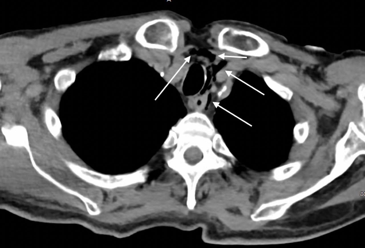
The patient’s daughter flew in from out-of-state and agreed to stay with her father after discharge for a few weeks to ensure primary care and neuropsychology follow-up. The patient was discharged on COVID day 10. Later that same day, he presented to the ED as a priority one trauma activation. The patient had multiple self-inflicted stab wounds to the abdomen and one to the anterior neck. His daughter had called emergency medical services after finding him in the basement with an eight-inch steak knife protruding from his abdomen. The patient had removed the knife from his abdomen and attempted to stab himself in the neck before his daughter was able to take control of the knife. The patient was unable to provide much history on arrival; however, he stated he had done this because he was “a horrible person.”
Initial vital signs showed a blood pressure of 211/116 millimeters of mercury (mm Hg), a temperature of 36.2° C, bradycardia at 53 beats per minute, a respiratory rate of 22 breaths per minute, and an oxygen saturation of 97% on room air. The patient had a stab wound overlying the anterior neck; however, his airway was intact and there was no pulsatile hematoma, no subcutaneous crepitus, or stridor, although his voice was hoarse. There were multiple stab wounds across the abdomen with an expanding hematoma over the right lower quadrant. There was no active external hemorrhage, and the patient was without peritoneal signs. Initial interventions included a tetanus booster, 1 liter of normal saline, 2 grams of ceftriaxone, and 500 milligrams of metronidazole, all administered intravenously. The patient was hemodynamically stable for CT. A CT angiogram of the neck was performed and showed no extravasation of contrast through the great vessels or common carotid arteries. Computed tomography of the chest with IV contrast showed free air consistent with penetrating injury (Image).
An emergent exploratory laparotomy was performed, as well as closure of the abdominal wounds. Eleven stab wounds to the abdomen were noted; six of them were full thickness
CPC-EM Capsule
What do we already know about this clinical entity?
Coronavirus disease of 2019 (COVID-19) has case associations with psychiatric manifestations. The risk of new psychiatric diagnoses within 90 days of a COVID-19 infection is 5.8% above baseline.
What makes this presentation of disease reportable?
Our patient, without history of previous psychological disease, lost six months of functional status secondary to his COVID19-related psychosis.
What is the major learning point?
As seen with our patient, the COVID-19associated psychiatric symptoms can be severe enough to progress to a suicide attempt and their course can be prolonged.
How might this improve emergency medicine practice?
Recognizing that agitation and delirium are associated with COVID-19 infections may help guide testing and treatment in patients with similar clinical pictures.
entering the peritoneal cavity. A thorough exploration of the abdomen revealed no injury to the liver, stomach, small bowel, or colon. The rectus sheath hematomas were decompressed, and the wounds closed.
Clinical Practice and Cases in Emergency Medicine 78 Volume 7, no. 2: May 2023
Piszker et al.
Image. Computed tomography of the chest with contrast showing free air (arrows) adjacent to the trachea contained within the mediastinum secondary to the self-inflicted stab wound to the patient’s neck.
After stabilization by the trauma team, he was evaluated by psychiatry. At that time (COVID day 11), he continued to endorse suicidal ideation, visual hallucinations, and depressive symptoms. Inpatient psychiatric placement was recommended. He was started on sertraline. The patient continued to express suicidal ideation throughout his hospital stay with documented quotes such as “I am nothing but evil and do not deserve to be living.” On COVID day 15 he was discharged from the hospital to inpatient psychiatric care.
On COVID day 18, while at the inpatient psychiatric facility, he was found to have an acute kidney injury and was transferred back to the hospital for medical management. There he was evaluated by neuropsychology and had an unremarkable magnetic resonance image (MRI) of the brain. Over the next 14 days, he had escalating anxiety that was unresponsive to quetiapine and haloperidol. Benzodiazepine administration worsened the patient’s delirium. The patient’s quetiapine was discontinued, and his regimen was changed to olanzapine and hydroxyzine.
On COVID day 29 the patient had an abrupt, significant improvement in mentation where he was calm, cooperative, and appropriately conversive. Over the next seven days, he continued to have waxing and waning mental status. Sodium valproate was briefly initiated; however, he developed a papular, erythematous rash shortly after, and this was discontinued. Mirtazapine was then added to the patient’s regimen, and his sertraline dose was increased. By COVID day 43 the patient had significant improvements in his mental status and depressive symptoms. He took a mini-mental state examination and scored 24 of 25, which is within the normal range. He also took a St. Louis University Mental Status Examination and scored 20 of 30, which is consistent with cognitive impairment and dementia. On COVID day 44 he was discharged to adult foster care with 24-hour supervision on a medication regimen of olanzapine, mirtazapine, and sertraline.
He was moved into an adult foster-care home with close outpatient follow-up with his family medicine physician. After multiple follow-up visits, interval hospitalization, and serial medication adjustments, the patient’s mental status stabilized, and he was discharged from adult foster care. He is maintained on sertraline, olanzapine, and mirtazapine. He now lives safely on his own, plays golf multiple times a week, and can care for his pet dog. Despite the eventual, significant resolution in our patient’s symptoms, he ultimately lost six months of functional status secondary to his COVID-19-associated psychosis.
DISCUSSION
Although this case is similar to existing cases in the literature, the patient demographics and presentation are unique. This case adds to existing literature surrounding the elderly population and COVID-19. The exact mechanism of this patient’s psychosis is unknown. There was no documented personal psychiatric history, history of suicidal ideation, or reported significant family psychiatric history. Furthermore,
our patient did not fill his dexamethasone prescription; so the likelihood of medication-induced psychosis is very low. Our patient’s CT of the brain, MRI of the brain, and cerebral sinus fluid studies did not show any evidence of neuroinflammation or neurologic injury; however, an extended encephalitis panel was not performed.
A review of the available literature revealed common psychiatric symptoms associated with COVID-19 such as anxiety and depression, as well as more severe manifestations such as delusions, paranoia, and suicidal thoughts.4 Similar to what we report regarding our patient, other case reports have documented new-onset psychiatric symptoms in patients without a previous psychiatric history.4–7 While some of the reported cases of newonset psychiatric symptoms did not progress to a suicide attempt, there are reports of progressive psychosis leading to suicide attempts similar to our patient. Special attention is paid to two cases involving attempted suicide by neck laceration, similar to our patient although these patients were much younger (37 and 52 years old, respectively).8,9
SARS-CoV-2 is known to cause large-scale inflammatory responses, sometimes referred to as “cytokine storm,” which studies have implicated in the pathogenesis of psychiatric symptoms in acute COVID-19.10,11 Other authors suggest a more multifactorial mechanism for the pathogenesis of newonset psychiatric symptoms associated with a COVID-19 infection. These authors take into consideration both the neuroinflammation and the psychosocial effects of physical/ social isolation, hospitalization and its associated procedures, and the sociocultural effects of the pandemic.12 Specifically regarding our patient, a possible link to the development of new-onset psychiatric symptoms in the peri-COVID-19 setting could be his chronic kidney disease. Researchers in the United Kingdom suggest a link between kidney disease patients and mental health problems, suggesting a risk increase of 100% compared to the general population.13 Otherwise, there were no other social stressors (eg, no known financial difficulties, loss of a loved one, or illicit substance use) apparent in our patient’s history.
While there certainly are multiple reports of newonset psychiatric symptoms in patients without previous psychiatric history,4–7 there are also reports highlighting the onset of COVID-19-associated psychiatric symptoms in patients with either personal or family history of psychiatric symptoms.7,14 Additionally, our patient had a protracted course of his psychosis and recurrent agitation and delirium. This is incongruent with most cases reported in the literature. In one retrospective study of patients with new-onset psychotic symptoms and confirmed diagnosis of SARS-CoV-2, excluding patients with a previous personal or family history of severe mental disorders, 80% of the patients experienced resolution of their symptoms in less than two weeks.15 Even though the overall number of patients studied was limited, data suggests that in a majority of patients their psychiatric symptoms are not long-lasting. However, due to the recent
Volume 7, no. 2: May 2023 79 Clinical Practice and Cases in Emergency Medicine Piszker et al.
in a
COVID-19-induced Acute Psychosis Resulting
Suicide Attempt
COVID-19-induced Acute Psychosis Resulting in a Suicide Attempt Piszker et al.
onset of the phenomena described in this report, the optimal treatment regimen is still unknown and can include a variety of medications.
CONCLUSION
Additional research is needed to delve into this complication of COVID-19, as reported cases in the literature are still quite rare. It remains unknown whether psychosis associated with COVID-19 requires a different standard of care compared to other causes of delirium and psychosis.
The authors attest that patient consent is not required for publication of this case report. The Institutional Review Board approval has been documented and filed for publication of this case report. Documentation on file.
infection: a case report. Riv Psichiatr. 2022;57(1):40-3.
4. Bakre S, Chugh K, Oke O, et al. COVID-19 induced brief psychotic disorder: a case report and review of literature. Case Rep Psychiatry 2022;2022:9405630.
5. Correa-Palacio AF, Hernandez-Huerta D, Gómez-Arnau J, et al. Affective psychosis after COVID-19 infection in a previously healthy patient: a case report. Psychiatry Res. 2020;290:113115.
6. Smith CM, Gilbert EB, Riordan PA, et al. COVID-19-associated psychosis: a systematic review of case reports. Gen Hosp Psychiatry 2021;73:84-100.
7. Ferrando SJ, Klepacz L, Lynch S, et al. COVID-19 psychosis: a potential new neuropsychiatric condition triggered by novel coronavirus infection and the inflammatory response? Psychosomatics. 2020;61(5):551-5.
8. Gillett G and Jordan I. Severe psychiatric disturbance and attempted suicide in a patient with COVID-19 and no psychiatric history. BMJ Case Rep. 2020;13(10):e239191.
Address for Correspondence: Alexander Piszker, DO, MS, Michigan State University College of Osteopathic Medicine, Department of Emergency Medicine, 1500 E Sherman Blvd, Muskegon, MI 49444. Email: alexander.piszker@mercyhealth.com.
Conflicts of Interest: By the CPC-EM article submission agreement, all authors are required to disclose all affiliations, funding sources and financial or management relationships that could be perceived as potential sources of bias. The authors disclosed none.
Copyright: © 2023 Piszker et al. This is an open access article distributed in accordance with the terms of the Creative Commons Attribution (CC BY 4.0) License. See: http://creativecommons.org/ licenses/by/4.0/
9. Chacko M, Job A, Caston F 3rd, et al. COVID-19-induced psychosis and suicidal behavior: case report. SN Compr Clin Med 2020;2(11):2391-5.
10. Kulaga SS and Miller CWT. Viral respiratory infections and psychosis: a review of the literature and the implications of COVID-19. Neurosci Biobehav Rev. 2021;127:520-30.
11. Corsi-Zuelli F, Loureiro CM, Shuhama R, et al. Cytokine profile in first-episode psychosis, unaffected siblings and community-based controls: the effects of familial liability and childhood maltreatment. Psychol Med. 2020;50(7):1139-47.
12. Wang Z, Chen X, Lu Y, et al. Clinical characteristics and therapeutic procedure for four cases with 2019 novel coronavirus pneumonia receiving combined Chinese and Western medicine treatment. Biosci Trends. 2020;14(1):64-8.
REFERENCES
1. World Health Organization. WHO COVID-19 Dashboard. 2023. Available at: https://covid19.who.int/. Accessed November 27, 2022.
2. Lee DT, Wing YK, Leung HC, et al. Factors associated with psychosis among patients with severe acute respiratory syndrome: a casecontrol study. Clin Infect Dis. 2004;39(8):1247-9.
3. Gallo M, Leone B, Tarallo F, et al. Onset psychosis and PTSS after severe respiratory symptomatology caused by SARS-CoV-2
13. Damery S, Brown C, Sein K, et al. The prevalence of mild-tomoderate distress in patients with end-stage renal disease: results from a patient survey using the emotion thermometers in four hospital Trusts in the West Midlands, UK. BMJ Open. 2019;9(5):e027982.
14. Lanier CG, Lewis SA, Patel PD, et al. An unusual case of COVID-19 presenting as acute psychosis. J Pharm Pract. 2022;35(3):488-91.
15. Parra A, Juanes A, Losada CP, et al. Psychotic symptoms in COVID-19 patients. A retrospective descriptive study. Psychiatry Res 2020;291:113254.
Clinical Practice and Cases in Emergency Medicine 80 Volume 7, no. 2: May 2023
Iatrogenic Rhinolith: A Case Report and Review of Literature
Daniel Mitchell, BS*
Quinn Self, MD† Carolyn Orgain, MD†
Section Editor: Melanie Heniff, MD
The University of Vermont - Larner College of Medicine, Burlington, Vermont The University of Vermont Health Network, Department of Otolaryngology Surgery, Burlington, Vermont
Submission history: Submitted September 12, 2022; Revision received January 12, 2023; Accepted January 26, 2023
Electronically published May 26, 2023
Full text available through open access at http://escholarship.org/uc/uciem_cpcem
DOI: 10.5811/cpcem.1395
Introduction: Unilateral nasal obstruction is a common complaint with a broad differential diagnosis that includes anatomic asymmetry, unilateral infective or inflammatory conditions, and benign and malignant sinonasal masses. A rhinolith is an uncommon foreign body in the nose, which serves as a nidus for calcium salt deposition. The foreign body can be endogenous or exogenous in origin and may remain asymptomatic for many years before incidental discovery. When left untreated, stones may cause unilateral nasal obstruction, rhinorrhea, nasal discharge, epistaxis or, in rare cases, progressive destruction leading to septal/palatal perforation or oro-antral fistula. Surgical removal is an effective intervention with limited complications reported.
Case Report: This article describes a 34-year-old male who presented to the emergency department (ED) with unilateral obstructing nasal mass and epistaxis, which was found to be an iatrogenic rhinolith. Successful surgical removal was performed.
Conclusion: Epistaxis and nasal obstruction are common presentations to the ED. Rhinolith is an uncommon clinical etiology that if left undiagnosed may lead to progressive destructive disease; it should be included in the differential for any unilateral nasal symptoms of unclear origin. Appropriate work-up for any suspected rhinolith includes computed tomography, as biopsy is risky given the broad differential of unilateral nasal mass. When identified, surgical removal has a high success rate with limited complications reported. [Clin Pract Cases Emerg Med. 2023;7(2):81–84]
Keywords: Rhinolith; nasal mass; epistaxis; nasal drainage; case report.
INTRODUCTION
Nasal obstruction and epistaxis are common complaints in acute care medicine. Obstruction may be acute or chronic, bilateral or unilateral, and may be anatomical or inflammatory in origin.1 Common causes of unilateral nasal obstruction include anatomic asymmetries, allergic or infectious sinusitis, and benign or malignant neoplastic processes.2 It is often difficult to differentiate the etiology of nasal masses on physical examination alone. Any unilateral nasal mass should undergo imaging and referral to otolaryngology for biopsy.2-4
A rhinolith is a rare, often overlooked etiology for unilateral nasal obstruction, epistaxis, and nasal drainage
that presents similarly to neoplastic nasal masses on physical examination.5 A rhinolith forms when an endogenous or exogenous foreign body serves as a nidus for calcium deposition over many years.6 Computed tomography (CT) is useful for diagnosis and assessment of size and local mass effect, as well as for surgical approach decision-making.7,8 Complication following stone removal is rare and typically limited to bleeding, local infection, or ipsilateral sinusitis.6
We present a case of iatrogenic rhinolith in a neurocognitively intact adult male likely arising from retained packing after an intranasal operation 2.5 years prior
Volume 7, no. 2: May 2023 81 Clinical Practice and Cases in Emergency Medicine Case Report
* †
in Afghanistan, and we discuss clinical, pathological, and radiological features.
CASE REPORT
A 34-year-old male who had recently immigrated from Afghanistan presented to the emergency department (ED) with several years of right-sided nasal obstruction and several days of intermittent self-limiting right-sided epistaxis. Past medical and surgical history was significant for an intranasal procedure 2.5 years prior while still in Afghanistan. Initial evaluation by the emergency physician revealed a whitish-gray, rockhard mass in the floor of the right nasal passage, which was biopsied for culture. A CT was also obtained. Biopsy revealed aggregates of filamentous, Gram-positive rods with short, modified acid-fast microorganisms. Computed tomography revealed a 3.5-centimeter calcified mass inferior to the right turbinate (Image 1).
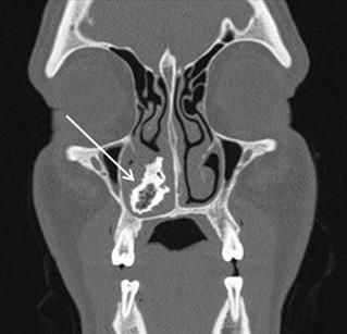
Outpatient referral and subsequent surgical removal was recommended by otolaryngology. Due to physical exam and imaging characteristics, antibiotics were not recommended. The patient ultimately underwent uncomplicated surgical removal. Intraoperatively a large, calcified nasal mass was encountered between the septum and inferior turbinate (Image 2). This was adherent to the surrounding nasal mucosa. With removal, friable mucosa was exposed, and mucosal bleeding was encountered that was controlled with topical vasoconstrictors. The foreign body was removed piecemeal and demonstrated a calcified exterior and a synthetic rubbery center (Image 3). He recovered without complication.
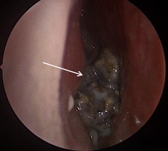
DISCUSSION
A rhinolith is a calcification in the nasal passage that originates when a foreign body serves as a nidus for inorganic
CPC-EM Capsule
What do we already know about this clinical entity?
Rhinoliths are benign intranasal calcified foreign bodies that often present with nonspecific symptoms such as epistaxis or nasal obstruction with an indolent course.
What makes this presentation of disease reportable?
A neurocognitively intact adult presented with epistaxis and intranasal mass. Removal suggested retained surgical material from history of nasal surgery
What is the major learning point?
There is a broad differential for intranasal masses including benign and malignant etiologies. Imaging and subspecialty consultation are often indicated.
How might this improve emergency medicine practice?
Rhinoliths are uncommon, difficult to visualize and are nonspecific in presentation. Definitive management is removal. They should remain on the differential for nasal complaints.
salt deposition.9 Rhinoliths are uncommon and may be overlooked and underdiagnosed, as the estimated prevalence is approximately 1 in 10,000 patients.10 Nasal foreign body is a common finding in children and neurocognitively impaired
Clinical Practice and Cases in Emergency Medicine 82 Volume 7, no. 2: May 2023 Iatrogenic Rhinolith Mitchell et al.
Image 1. Coronal computed tomography demonstrating calcified mass indicated by white arrow in the inferior meatus.
Image 2. Anterior rhinoscopy demonstrating calcified mass indicated by white arrow.
adults when a bead or similar object is manually placed into the nose.5 Rhinoliths, however, are seen across all age groups and mental states. Given the time required for stones to form, they are most often reported in adults between the third and sixth decades of life.11,12
Rhinoliths may be found incidentally on anterior rhinoscopy, nasal endoscopy, or imaging studies when asymptomatic.6 Symptoms of occlusive stones may include unilateral nasal obstruction, purulent rhinorrhea, ipsilateral sinusitis, facial pain, fetor, and headache.5-7,9 In longstanding cases without medical intervention, dacryocystitis and otorrhea have been described. Severe cases can result in local destruction resulting in septal perforations as well as oroantral fistula.5,11,13,14
A common presenting history of exogenous stone formation is of a foreign body placed in childhood that was never removed. Less common is iatrogenic rhinolithiasis from retained nasal packing, sutures, or gauze that facilitate stone formation. Rare reports include concreted topical ointments, debris from nasopharyngeal refluxed emesis, and even stagnant inhaled particulates in the setting of impaired nasociliary clearance.10 Stones of endogenous origin are very rare, but osseous fragments following facial trauma, inspissated secretions, microemboli, and ectopic teeth have all been described.12
The vast majority of rhinoliths are observed along the floor of the nasal passage abutting the inferior turbinate; however, they have been described throughout the sinonasal passage.11,13,15 Removal is curative with minimal side effects in most cases with rare reports of postoperative bleeding and infection.9
In this case, an adult male presented to the ED with unilateral nasal obstruction and epistaxis. Computed tomography was appropriately ordered to further classify the mass, and subspecialty referral to otolaryngology was made. Treatment
involved surgical removal with no need for antibiotics. Notably, the core of the calcified mass was seen to have a firm, rubbery texture with illegible orange foreign text. Given his history of endoscopic sinus surgery while living in Afghanistan, it is likely this substance represented a retained Penrose drain, which is occasionally used in sinus operations. Rhinolith is an uncommon clinical etiology; however, nasal obstruction and epistaxis are commonly encountered in acute care clinics. Given the broad differential for unilateral obstructing nasal masses, biopsy should be approached with caution.
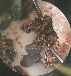
CONCLUSION
Rhinolithiasis is an uncommon etiology of unilateral nasal symptoms such as obstruction, pain, epistaxis, or purulent drainage. This is a slowly developing condition that is easily overlooked due to similarities in presentation with nasal neoplasms. If left unresolved, there is risk of local destructive change. Work-up of unilateral nasal mass, epistaxis, or purulent drainage without clear etiology should include CT, as biopsy is risky given the broad differential of unilateral mass. Surgical removal has a high success rate with low risk of complication.
The authors attest that their institution does not require patient consent for publication, The Institutional Review Board approval has been documented and filed for publication of this case report. Documentation on file.
Address for Correspondence: Daniel Mitchell, BS, The University of Vermont-Larner College of Medicine, 89 Beaumont Ave, Burlington, VT, 05405. Email: Daniel.j.mitchell@med.uvm.edu.
Conflicts of Interest: By the CPC-EM article submission agreement, all authors are required to disclose all affiliations, funding sources and financial or management relationships that could be perceived as potential sources of bias. The authors disclosed none.
Copyright: © 2023 Mitchell et al. This is an open access article distributed in accordance with the terms of the Creative Commons Attribution (CC BY 4.0) License. See: http://creativecommons.org/ licenses/by/4.0/
REFERENCES
1. Esmaili A and Acharya A. Clinical assessment, diagnosis and management of nasal obstruction. Aust Fam Physician 2017;46(7):499-503.
2. London NR Jr, Reh DD. Differential diagnosis of chronic rhinosinusitis with nasal polyps. Adv Otorhinolaryngol. 2016;79:1-12.
3. Fraser L and Kelly G. An evidence-based approach to the management of the adult with nasal obstruction. Clin Otolaryngol 2009;34(2):151-5.
4. Belli S, Yildirim M, Eroglu S, et al. Single-sided sinonasal mass: a
Volume 7, no. 2: May 2023 83 Clinical Practice and Cases in Emergency Medicine Mitchell et al. Iatrogenic Rhinolith
Image 3. Specimen with calcified exterior and synthetic rubbery core indicated by white arrow.
Iatrogenic Rhinolith Mitchell et al.
retrospective study. North Clin Istanb. 2018;5(2):139-43.
5. Brehmer D and Riemann R. The rhinolith: a possible differential diagnosis of a unilateral nasal obstruction. Case Rep Med 2010;2010:845671.
6. Appleton SS, Kimbrough RE, Engstrom HI. Rhinolithiasis: a review. Oral Surg Oral Med Oral Pathol. 1988;65(6):693-8.
7. Hadi U, Ghossaini S, Zaytoun G. Rhinolithiasis: a forgotten entity. Otolaryngol Head Neck Surg. 2002;126(1):48-51.
8. Bayram B, Deniz K, Polat Ozsoy O, et al. A simple surgical approach for management of the rhinoliths: case report. J Oral Maxillofac Surg. 2011;69(5):1403-7.
9. Seyhun N, Toprak E, Kaya KS, et al. Rhinolithiasis, a rare entity: analysis of 31 cases and literature review. North Clin Istanb 2020;8(2):172-7.
10. Pacheco PC and Busquets JM. A 44-year-old rhinolith: a case report and review of the literature. P R Health Sci J. 2015;34(2):105-7.
11. Akkoca Ö, Tüzüner A, Demirci Ş, et al. Patient characteristics and frequent localizations of rhinoliths. Turk Arch Otorhinolaryngol 2016;54(4):154-7.
12. Orhan K, Kocyigit D, Kisnisci R, et al. Rhinolithiasis: an uncommon entity of the nasal cavity. Oral Surg Oral Med Oral Pathol Oral Radiol Endod. 2006;101(2):e28-32.
13. Keck T, Liener K, Sträter J, et al. Rhinolith of the nasal septum. Int J Pediatr Otorhinolaryngol. 2000;53(3):225-8.
14. Flood TR. Rhinolith: an unusual cause of palatal perforation. Br J Oral Maxillofac Surg. 1988;26(6):486-90.
15. Marr JT. Stone in the maxillary sinus. Radiology. 1955;64(5):718-20.
Clinical Practice and Cases in Emergency Medicine 84 Volume 7, no. 2: May 2023
Case Report of Malignant Hyperthermia in the Emergency Department
Mitchell McMurray, MD
Austin Sowers, MD
Raymond Orthober, MD Martin Huecker, MD
Section Editor: Austin Smith, MD
Submission history: Submitted July 6, 2022; Revision received February 2, 2022; Accepted February 27, 2023
Electronically published May 26, 2023
Full text available through open access at http://escholarship.org/uc/uciem_cpcem
DOI: 10.5811/cpcem.1402
Introduction: Malignant hyperthermia (MH) is a rare but deadly condition that may be encountered in the emergency department (ED). This report highlights a case of a patient who initially presented for acute agitation with hypertension and tachycardia and provides explanation for how to manage MH.
Case Report: A 44-year-old male presented to the ED with altered mental status, eventually requiring intubation with etomidate and succinylcholine. Despite being afebrile initially, the patient developed a rectal temperature of 105.3° Fahrenheit (F) with significantly elevated arterial carbon dioxide levels after intubation. The treating team initiated cooling measures and dantrolene, leading to a positive outcome.
Conclusion: Clinicians should strive for expeditious recognition of MH and treatment with an updated institutional protocol. [Clin Pract Cases Emerg Med. 2023;7(2):85–88]
Keywords: case report; malignant hyperthermia.
INTRODUCTION
Malignant hyperthermia (MH) describes a condition of a hypermetabolic response to anesthetic gases (eg, halothane and sevoflurane) or the depolarizing muscle relaxant succinylcholine 1 Although rare clinically (estimated incidence of 1 in 10,000 to 1 in 250,000 anesthesia procedures), the genetic abnormality may occur as frequently as one in 3,000 individuals.1 The earliest signs are tachycardia, rise in end-tidal carbon dioxide (EtCO2), and muscle rigidity with hyperthermia usually occurring later.1 Left untreated, MH leads to cellular hypoxia (metabolic acidosis), rhabdomyolysis (hyperkalemia), myoglobinuria (with acute renal failure), cardiac arrythmias, and usually death.1
Patients susceptible to MH have a defective calcium channel (the ryanodine receptor) in the sarcoplasmic reticulum membrane.2 The mutation in this receptor (traced to the ryanodine receptor 1 gene) results in uncontrolled intracellular calcium release with exposure to triggering agents.2 Rapid adenosine triphosphate depletion occurs, and the muscle
membrane degrades.2 Inheritance is an autosomal dominant pattern and genetic testing using muscle biopsy confirms the diagnosis.2 Of note, MH is significantly more common in pediatric patients (about 1 in 100,000 adults and 1 in 30,000 in children).2 As a result, succinylcholine is sparingly used for rapid sequence intubation (RSI) in pediatrics.2
Most people remain unaware of their susceptibility to MH until the disease occurs. Although rarely used, both the caffeine-halothane contracture test (CHCT) and in vitro contracture test (IVCT) can reliably diagnose MH.3,4 Both measure the contracture response of freshly biopsied muscle to different levels of caffeine and halothane.4 Improved recognition and testing capabilities have decreased mortality from 70% 30 years ago to less than 5% today.1,5,6
CASE REPORT
A 44-year-old male with a past medical history of depression and chronic back pain presented to the emergency
Volume 7, no. 2: May 2023 85 Clinical Practice and Cases in Emergency Medicine Case Report
University of Louisville School of Medicine, Department of Emergency Medicine, Louisville, Kentucky
Like
A Fever Dream: A Case Report of Malignant Hyperthermia in the Emergency Department McMurray
department (ED) with altered mental status. According to his wife, he had been in normal health early that morning. Later in the morning he began to have an abnormal gait “like Frankenstein,” but retained normal speech and mentation. His wife returned home to find the patient with his extremities convulsing, diaphoretic, and eyes open but unresponsive. The patient had reportedly consumed alcohol the evening before but did not use recreational drugs. Emergency medical services administered naloxone 2 milligrams (mg) intravenous (IV), lorazepam 2 mg IV, and one liter of normal saline IV without improvement in mental status. The ground crew called a flight ambulance who transported the patient to our Level I trauma center.
Upon presentation to the ED, the patient had tachycardia in the 130s but was normotensive, afebrile, and had normal oxygenation saturation on room air. On physical exam, he was extremely diaphoretic and tremulous, but he moved all extremities spontaneously. His eyes (pupils 5 millimeters [mm] and reactive) opened spontaneously, he withdrew from painful stimuli, and he made incomprehensible sounds (Glasgow Coma Score 10). The patient had clear breath sounds bilaterally, no abdominal distention or tenderness, and no obvious signs of trauma. The initial chest radiograph (CXR) showed no acute findings; point-of-care lab testing was significant only for mild lactic acidosis, 1.8 millimoles per liter (mmol/L) (reference range: 0.5-2.2 mmol/L). The treatment team ordered two 2-mg doses of midazolam in an attempt to control agitation, but the patient remained combative. Given the patient’s declining mental status and need for emergent clinical workup, the treatment team made the decision to perform RSI.
Intubation was performed successfully using etomidate and succinylcholine followed by propofol infusion for sedation. After CXR confirmed endotracheal tube placement, the patient was transported for computed tomography (CT). Non-contrast head CT showed no acute findings and initial laboratory work was significant for acute kidney injury with creatinine of 2.31 mg per deciliter (mg/dL) (reference range: 0.7-1.3 mg/dL) and a leukocytosis of 27,400 thousand per microliter (uL) (reference range: 4,000-11,000 thousand per uL) with 84% neutrophils (55-70%). Urine and serum toxicology studies, troponin level, and coronavirus disease 2019 polymerase chain reaction (PCR) were all negative.
The team provided further fluid resuscitation, empiric antibiotic coverage for meningitis, and prepared for a lumbar puncture. Prior to the procedure, the patient developed rigid muscles diffusely and felt warm to the touch. Rectal temperature was 105.3° F, a significant increase from his triage temperature of 98.9° F approximately 90 minutes prior. His arterial carbon dioxide (pCO2) also increased to 65.3 mm of mercury (Hg) from 46.6 mm Hg (reference range: 35-45 mm Hg) immediately post-intubation despite no changes in ventilator settings. With concern for MH, the team began
CPC-EM Capsule
What do we already know about this clinical entity?
Malignant hyperthermia (MH) is a rare, life-threatening condition that occurs during anesthesia procedures involving succinylcholine or anesthetic gases.
What makes this presentation of disease reportable?
We diagnosed and treated a case of MH in the ED, where this condition is not typically described .
What is the major learning point?
Emergency departments should have an institutional protocol in place and access to dantrolene for management of MH.
How might this improve emergency medicine practice?
Succinylcholine is a common paralytic used in the ED. We hope this case encourages clinicians to be prepared to recognize and treat MH.
active external cooling measures, initiated our MH protocol, and consulted the anesthesia service for dantrolene administration. Fifty minutes after dantrolene administration, the patient’s rectal temperature decreased to 101.3° F. The patient’s rigidity resolved and pCO2 decreased to 31.2 mm Hg two hours after dantrolene. The patient was admitted to the intensive care unit (ICU) under the anesthesia service.
The patient received dantrolene for 24 hours while inpatient. Neurology service consultation led to an electroencephalogram, which showed no seizure activity. The ICU team added acyclovir to cover for possible herpes simplex virus (HSV) infection and performed a lumbar puncture. Cerebrospinal fluid analysis was consistent with viral meningitis. HSV-1 and -2 PCR testing were negative, and the team discontinued antimicrobials on hospital day three. After full diagnostic workup, the final diagnosis made by the ICU team for his original presentation of altered mental status was determined to be viral meningitis. The patient passed spontaneous breathing trials, began following commands, and was subsequently extubated. He was discharged on hospital day four in good condition with planning for a CHCT through
Clinical Practice and Cases in Emergency Medicine 86 Volume 7, no. 2: May 2023
al.
et
Like A Fever Dream: A Case Report of Malignant Hyperthermia in the Emergency Department
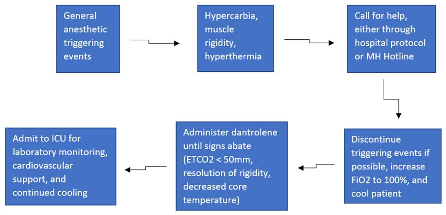
the University of Minnesota. The patient was lost to follow up; so, it is unclear whether he received confirmatory testing.
DISCUSSION
Malignant hyperthermia is a well described and rare condition that emergency physicians (EP) may encounter, although exact incidence of ED presentations is not described in the literature. While EPs will likely not directly use anesthetic gases, MH has a well-known association with succinylcholine, which is commonly used by EPs for RSI.7 A database analysis and systematic review of over six million perioperative cases determined that use of succinylcholine without volatile anesthetics triggered 24 MH cases.7 Another study examined the risk of MH based on whether or not a patient had received succinylcholine.8 They determined that for all cases the relative risk was 19.6 for those with compared to without succinylcholine.8 It is clear that any clinician using anesthetic gases or depolarizing paralytic agents should understand the presentation and management of MH. Although there is no known screening test for MH, it would be prudent for EPs to gather information on family history of MH or prior anesthesia complications before intubation, if possible, given the high mortality.9 If a patient with a known personal or family history of MH is being treated in an ED, clinicians and staff should be notified immediately so that use of succinylcholine is avoided.
Our patient initially presented for acute agitation with hypertension and tachycardia, a common presentation in the ED setting. This presentation yielded a broad differential diagnosis list: substance-induced (i.e., methamphetamine, cocaine, phencyclidine), serotonin syndrome, neuroleptic malignant syndrome; encephalitis; and malignant catatonia, among others. After RSI, the patient had rapid increases in temperature, pCO2, and muscle rigidity, leading to suspicion for MH.
Diagnosis of MH is made by a combination of clinical findings and laboratory testing.4 Signs and symptoms suggestive of MH are unexplained increased EtCO2 concentration, muscular rigidity, muscle breakdown, combined metabolic and respiratory acidosis, hyperthermia, cardiac arrhythmia, etc.4 An international panel of 11 experts on MH created a clinical grading scale to estimate the likelihood of an MH event.4 The score assigns point totals to different physiologic markers and also takes into account family history.4 A diagnosis of MH is likely when scores are greater than 20 and is almost certain if greater than 50.4 Our patient’s score exceeded 50 when using this scale.
The current gold standard for diagnosing MH is the CHCT or IVCT, which is mainly used in Europe.4 The IVCT has a sensitivity of 99.0% and a specificity of 93.6%, while CHCT demonstrates a sensitivity of 97% and a specificity of 78%.4 It is unclear in the literature how often suspected patients receive confirmatory testing, but it is estimated to be low given the rarity of MH and the limited diagnostic centers that perform the testing.
There are currently only four available centers for CHCT testing in North America listed on the Malignant Hyperthermia Association of the United States (MHAUS) website.10 Overall treatment goals of MH include discontinuation of trigger agents, active cooling measures, hyperventilation, and administration of dantrolene.1 A postsynaptic muscle relaxant, dantrolene acts as an antagonist to muscle ryanodine receptors.2 When indicated, dantrolene is given as a bolus initial dose (2.5 mg per kilogram [kg]) followed by maintenance infusion (1 mg/kg) for 24-48 hours with careful observation for reappearance of symptoms.2 The figure demonstrates a possible algorithm for management of MH.
The MHAUS recommends initiating dantrolene infusion less than 10 minutes after diagnosis.10 Clinicians must act quickly to mobilize the product because most patients require multiple vials mixed with sterile water prior to administration.10 In our case, the patient received dantrolene in a timely fashion with significant improvement in symptoms and a positive outcome. Dantrolene availability varies due to cost, storage requirements, and difficulty in administration.10
The best way to be prepared for a case of MH in the ED is having a detailed protocol in place. Our EPs relied on the institutional protocol throughout the treatment course. At a minimum, the MHAUS recommends facilities that stock and administer any triggering medication to have dantrolene available in case MH occurs.11 Many facilities have an MH cart that includes dantrolene and interventions to treat the effects of the hypermetabolic condition such as sodium bicarbonate, insulin, calcium, amiodarone, etc.10 Lack of rapid identification and treatment of the disease process leads to patient death due to severe hyperthermia and acidosis.2 Patients will require admission to the ICU, as mortality is up to 5% even with proper treatment.2
Throughout management of the case, the treatment team used the MH hotline (1-800-644-9737). This resource affords
Figure.
Volume 7, no. 2: May 2023 87 Clinical Practice and Cases in Emergency Medicine
McMurray et al.
Diagnosing and treating suspected malignant hyperthermia in the emergency department MH, malignant hyperthermia; ICU, intensive care unit; ETCO2, end-tidal carbon dioxide; Fi02, fraction of inspired oxygen.
Like A Fever Dream: A Case Report of Malignant Hyperthermia in the Emergency Department McMurray et al. clinicians with continuous access to experienced staff that gives medical oversight and could mean the difference between life and death for patients.
CONCLUSION
Although more commonly described in the anesthesia literature, malignant hyperthermia can occur in the ED and must be diagnosed and managed without delay for favorable outcomes. Patients should be screened when feasible for prior anesthesia complications, and all patients should be closely monitored following succinylcholine administration. It is strongly recommended that EDs have an institutional protocol and clinicians have access to dantrolene for management of MH.
The authors attest that their institution does not require Institutional Review Board approval. Patient consent has been obtained and filed for the publication of this case report. Documentation on file.
REFERENCES
1. Rosenberg H, Pollock N, Schiemann A, et al. Malignant hyperthermia: a review. Orphanet J Rare Dis. 2015;10:93.
2. Watt S. and McAllister RK. Malignant hyperthermia. 2022 Available at: https://www.ncbi.nlm.nih.gov/books/NBK430828/. Accessed July 29, 2022.
3. Rosenberg H, Antognini JF, Muldoon S. Testing for malignant hyperthermia. Anesthesiology. 2002;96(1):232-7.
4. Yang L, Tautz T, Zhang S, et al. The current status of malignant hyperthermia. J Biomed Res. 2019;34(2):75-85.
5. Brandom BW, Larach MG, Chen MS, et al. Complications associated with the administration of dantrolene 1987 to 2006: a report from the North American Malignant Hyperthermia Registry of the Malignant Hyperthermia Association of the United States. Anesth Analg 2011;112(5):1115-23.
6. Denborough M. Malignant hyperthermia. Lancet 1998;352(9134):1131-6.
7. Larach MG, Klumpner TT, Brandom BW, et al. Succinylcholine use and dantrolene availability for malignant hyperthermia treatment. Anesthesiology. 2019;130(1):41-54.
Address for Correspondence: Mitchell McMurray, MD, University of Louisville School of Medicine, Department of Emergency Medicine, 550 South Jackson Street, Louisville, KY 40202. Email: mitchell. mcmurray@louisville.edu
Conflicts of Interest: By the CPC-EM article submission agreement, all authors are required to disclose all affiliations, funding sources and financial or management relationships that could be perceived as potential sources of bias. The authors disclosed none.
Copyright: © 2023 McMurray et al. This is an open access article distributed in accordance with the terms of the Creative Commons Attribution (CC BY 4.0) License. See: http://creativecommons.org/ licenses/by/4.0/
8. Dexter F, Epstein RH, Wachtel RE, et al. Estimate of the relative risk of succinylcholine for triggering malignant hyperthermia. Anesth Analg. 2013;116(1):118-22.
9. Krivosic-Horber R and Adnet P. Tests de dépistage de la sensibilité à l’hyperthermie maligne [Screening tests for malignant hyperthermia susceptibility]. Ann Fr Anesth Reanim. 1989;8(5):444-56.
10. Healthcare Professionals. Malignant Hyperthermia Association of the United States. 2023. Available at: http://www.mhaus.org/. Accessed January 28, 2022.
11. Litman RS, Smith VI, Larach MG, et al. Consensus statement of the Malignant Hyperthermia Association of the United States on unresolved clinical questions concerning the management of patients with malignant hyperthermia. Anesth Analg. 2019;128(4):652-6.
Clinical Practice and Cases in Emergency Medicine 88 Volume 7, no. 2: May 2023
A Case Report of a LVAD Driveline Infection Diagnosed by Point-of-care Ultrasound
Nicholas Bielawa, MS, PA-C
Allison Cohen, MD
Milan Patel, DO
Brendon Stankard, PA-C
Mathew J. Nelson, DO
Section Editor: Manish Amin, DO
Submission history: Submitted September 9, 2022; Revision received March 3, 2023; Accepted March 9, 2023
Electronically published May 26, 2023
Full text available through open access at http://escholarship.org/uc/uciem_cpcem
DOI: 10.5811/cpcem.1403
Introduction: As the prevalence of patients with left ventricular assist devices (LVAD) presenting to the emergency department (ED) increases, clinicians must be aware of LVAD-associated infections.
Case Report: A well-appearing, 41-year-old male with history of heart failure status post prior-LVAD placement presented to the ED for swelling of his chest. What appeared initially as a superficial infection was further assessed with point-of-care ultrasound and found to represent a chest wall abscess involving the driveline, ultimately resulting in sternal osteomyelitis and bacteremia.
Conclusion: Point-of-care ultrasound should be considered an important tool in the initial assessment of potential LVAD-associated infection. [Clin Pract Cases Emerg Med. 2023;7(2):89–92]
Keywords: left ventricular assist device; driveline infection; abscess; point-of-care ultrasound; case report.
INTRODUCTION
Continuous-flow left ventricular assist devices (LVAD) have improved the survival of patients with end-stage systolic heart failure.1 Their complications include but are not limited to bleeding, device thrombosis, stroke, and infections.2,3 Of these complications, infections are less prevalent; however, they are the second most common cause of morbidity and mortality in patients who survive the initial six months after continuous-flow placement and are a leading cause of hospital admission in this patient population.2
According to the International Society for Heart and Lung Transplantation, LVAD infections are broken up into LVADspecific infections, LVAD-related infections, and non-LVAD infections.2 Infections specific to LVADs include pump and/or cannula infections, pump pocket infections, and driveline infections (DLI), which are further broken down into superficial and deep infections.2 Related infections include infective endocarditis, bacteremia, and sternal wound/surgical site infections.2 The Interagency Registry for Mechanically
Assisted Circulatory Support found a 19% prevalence in DLIs 12 months post-device implantation.4,5
It is difficult to discern the extent of the infection with only radiographic imaging. Computed tomography (CT) images are limited by artifact from the device. Point-of-care ultrasound (POCUS) can detect pockets of concern but cannot effectively inform whether deeper structures are involved.4 As a result, surgical exploration is often needed.4
CASE REPORT
A 41-year-old Hispanic male with a history of American College of Cardiology/American Heart Association Stage D chronic systolic heart failure secondary to prior anabolic steroid use, status post HeartMate 3 LVAD placement three years prior to presentation, on warfarin, presented to the emergency department (ED) due to concern for midsternal chest wall swelling that developed spontaneously two days prior. The patient denied any trauma to the chest, fever, chills, or rash. The patient reported a small amount of pain to the
Volume 7, no. 2: May 2023 89 Clinical Practice and Cases in Emergency Medicine Case Report
North Shore University Hospital, Manhasset, Department of Emergency Medicine, New York
Just Below the Surface: A Driveline Infection Diagnosed by POCUS
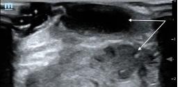
area with palpation only, and no active drainage. Initial vital signs were significant for a mean arterial pressure of 62 millimeters of mercury, heart rate of 78 beats per minute, respiratory rate of 20 breaths per minute, oxygen saturation of 98% on room air, and oral temperature of 37.2 degrees Celsius (98.9° Fahrenheit). Physical examination revealed a 2 centimeter (cm) x 2 cm, elevated, mildly tender, midsternal collection without overlying erythema or crepitus. Laboratory results found a white blood cell count of 8.94 thousand per microliter (K/uL) (reference range: 3.8-10.5 K/uL), hemoglobin of 7.6 grams per deciliter (g/dL) (13-17 g/dL), platelet count of 279 (K/uL) (150-400 K/uL), and an international normalized ratio of 2.53 (0.88-1.16). Serum electrolytes were within normal limits.
A point-of-care ultrasound was performed to further evaluate the fluid collection. The POCUS revealed a complex, mixed echogenic, collection that extended deep to chest wall and appeared contiguous with the driveline wire (Image 1).
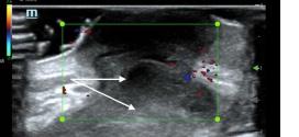
Swirling of complex material was evident with compression. Use of color flow Doppler demonstrated an avascular heterogeneous structure, concerning for a deep space infection with driveline involvement (Image 2).
After POCUS was performed, a CT of the chest with intravenous contrast was ordered, and blood cultures were drawn due to concern for acute infection. The patient was given 2000 milligrams (mg) of cefepime and 1000 mg of vancomycin for antibiotic coverage. Computed tomography of the chest revealed a fluid-attenuating midline chest-wall lesion extending to the anterior sternal border measuring 2 cm x 1.9 cm x 3.4 cm with mild peripheral enhancement suggestive of a chest wall abscess.
While in the ED the patient developed a fever of 38.3° Celsius and became tachycardic. He was admitted to the cardiothoracic surgery service for definitive management. On
CPC-EM Capsule
What do we already know about this clinical entity?
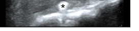
Left ventricular assist device (LVAD) infections are a significant cause of morbidity and mortality; computed tomography diagnosis can be limited by artifact.
What makes this presentation of disease reportable?
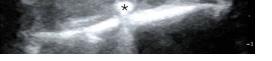
To date, there have been no reports on the use of ultrasound to diagnose LVAD driveline infections.
What is the major learning point?
Point-of-care ultrasound should be considered an important tool in the initial assessment of potential LVAD driveline infections.
How might this improve emergency medicine practice?
Point-of-care ultrasound can allow for rapid detection of LVAD driveline infections, leading to earlier identification and management.
admission, the fluid collection opened spontaneously and drained purulent material. A culture was sent and grew Staphylococcus epidermidis, and the first set of blood cultures grew Streptococcus mitis/oralis. The patient went to the
Clinical Practice and Cases in Emergency Medicine 90 Volume 7, no. 2: May 2023
et al.
Bielawa
Image 1. A point-of-care ultrasound view of the anterior chest wall, showing a complex fluid collection with irregular borders and mixed echogenicity (arrows) and surrounding inflammatory changes extending deep to the hyperechoic driveline (star).
Image 2. A point-of-care ultrasound of the anterior chest wall with color flow Doppler demonstrating a heterogeneous structure (arrows), extending to the driveline (star) without significant internal color flow.
Bielawa et al.
Just Below the Surface: A Driveline Infection Diagnosed by POCUS
operating room (OR) for irrigation and debridement of the sternal wound, removal of sternal wires except the inferiormost wire, and application of a wound vacuum-assisted closure device. Intraoperative transesophageal echocardiogram did not reveal valvular vegetations, and a wound culture obtained during debridement grew rare Propionibacterium acnes. The patient returned to the OR later in his hospital stay for further wound exploration and debridement. He was discharged on hospital day 17 on long-term antibiotic therapy with ceftriaxone 2000 mg via a peripherally inserted central catheter line.
DISCUSSION
In this case, POCUS was essential for making the diagnosis of a deep space infection. What appeared on physical exam to be only a superficial pustule, was discovered on POCUS to be a deep soft tissue infection involving the driveline. Point-of-care ultrasound is a safe, quick, and accurate way for emergency clinicians to differentiate between cellulitis and abscess.6 Previous studies have shown the diagnostic utility of POCUS for skin infections by enhancing the clinician’s ability to distinguish an abscess from simple cellulitis, which can be difficult to evaluate on physical exam.7,8 Sonographic findings of an abscess include heterogeneity, often with irregular borders, that is largely anechoic or hypoechoic containing mixed echogenic foci, and displaying posterior acoustic enhancement.9 In an abscess with mixed echogenicity, gentle pressure can produce a “swirl” sign, or “ultrasonic fluctuance,” to help confirm the presence of purulent material.9 Additionally, the use of color flow Doppler is also helpful in evaluating suspected abscess by excluding the presence of vascular flow and demonstrating peripheral hyperemia.9
In this patient, a complex-appearing fluid collection was visualized extending deep into the chest wall and communicating with the driveline wires concerning for a driveline infection. The results of this POCUS exam allowed for rapid diagnosis and early antibiotic administration. Although CT is typically preferred to assess for deep LVAD infections, the sensitivity and specificity of CT for the detection of these LVAD infections are not well-defined given the lack of a gold standard test for comparison.3,10 On CT imaging, driveline infections may appear as rim-enhancing fluid collections containing gas pockets or soft-tissue stranding adjacent to the device components.11 However, this imaging modality may be limited by the interpreter’s familiarity with LVAD anatomy and pathology and significant artifact from the LVAD hardware.10
Point-of-care ultrasound allows for the rapid and accurate evaluation of skin and soft tissue infections at the bedside, and although there have been no reports to date on the use of POCUS to detect LVAD infections, prior research has shown that ultrasound is more sensitive than CT for diagnosing certain soft tissue abscesses and provides more details
regarding the contents of the abscess cavity. 6-8,12 Other specialty imaging studies including leukocyte-labeled scintigraphy and positron emission tomography in combination with CT imaging may be more sensitive and specific for LVAD infection; however, cost, availability, and practicality in the ED limit the use of these modalities.10
This patient, who presented with superficial swelling of the chest wall, suffered from several infectious complications associated with his LVAD, including a DLI with associated chest wall abscess, sternal osteomyelitis, and LVAD-associated bloodstream infection (BSI). Driveline infections are the most frequent LVAD infection overall and occur most often within the first year of LVAD implantation10,13,14 Superficial DLIs spare the muscle and fascial layers while deep DLIs affect deeper structures, as was the case in this patient. Superficial DLIs have an unclear effect, while deep DLIs increase mortality in this group.13 The management of this patient involved a prolonged course of intravenous antibiotics and surgical debridement, which is often required in the treatment of deep LVAD infections.4,10,13 Clinicians should have a low threshold for evaluating and treating for possible LVAD infection in the ED, as the majority of these patients may not present with typical systemic inflammatory response syndrome in response to serious infections such as BSIs.3,13
CONCLUSION
Overall, the rate of LVAD implantation has increased greatly in recent years, and as the prevalence of LVADs increases, so too will the prevalence of LVAD patients in the ED.3,15 Although advances in LVAD technology have reduced the rate of complications, infections such as deep DLIs and BSIs still account for significant morbidity and mortality in this group.10,13-15 It is imperative that emergency clinicians be aware of infectious complications associated with LVADs and how to efficiently and effectively manage patients with these devices.
This case presents an LVAD recipient with a DLI associated with a chest wall abscess, sternal osteomyelitis, and bloodstream infection. Point-of-care ultrasound provides a fast, portable, and accurate method of detecting soft tissue infections that may be missed on physical exam, and in this case detection of a fluid collection on ultrasound ultimately led to the diagnosis of serious infectious complications in an LVAD recipient.6-8,12 Point-of-care ultrasound was essential in making this initial diagnosis and should be considered an important tool in the initial assessment of potential LVAD driveline infection. Further research is necessary to determine the accuracy of POCUS for the diagnosis of certain LVADrelated infections such as driveline infections compared to CT.
The authors attest that their institution does not require Institutional Review Board approval. Patient consent has been obtained and filed for the publication of this case report. Documentation on file.
Volume 7, no. 2: May 2023 91 Clinical Practice and Cases in Emergency Medicine
Just Below the Surface: A Driveline Infection Diagnosed by POCUS
Address for Correspondence : Nicholas Bielawa, MS, PA-C, North Shore University Hospital, Department of Emergency Medicine, 300 Community Dr, Manhasset, NY 11030. Email: nbielwa1@northwell.edu.
Conflicts of Interest: By the CPC-EM article submission agreement, all authors are required to disclose all affiliations, funding sources and financial or management relationships that could be perceived as potential sources of bias. The authors disclosed none.
Copyright: © 2023 Bielawa et al. This is an open access article distributed in accordance with the terms of the Creative Commons Attribution (CC BY 4.0) License. See: http://creativecommons.org/ licenses/by/4.0/
6. Gottlieb M, Avila J, Chottiner M, et al. Point-of-care ultrasonography for the diagnosis of skin and soft tissue abscesses: a systematic review and meta-analysis. Ann Emerg Med . 2020;76(1):67-77.
7. Squire BT, Fox JC, Anderson C. ABSCESS: Applied Bedside Sonography for Convenient Evaluation of Superficial Soft Tissue Infections. Acad Emerg Med. 2005;12(7):601-6.
8. Tayal VS, Hasan N, Norton HJ, et al. The effect of soft-tissue ultrasound on the management of cellulitis in the emergency department. Acad Emerg Med. 2006;13(4):384-8.
9. Dewitz A. (2014). Musculoskeletal, soft tissue, and miscellaneous application. In: Ma and Mateer’s Emergency Ultrasound (503-568). New York, New York: McGraw Hill.
REFERENCES
1. Slaughter MS, Rogers JG, Milano CA, et al. Advanced heart failure treated with continuous-flow left ventricular assist device. N Engl J Med. 2009;361(23):2241-51.
2. Trachtenberg BH, Cordero-Reyes A, Elias B, et al. A review of infections in patients with left ventricular assist devices: prevention, diagnosis and management. Methodist Debakey Cardiovasc J 2015;11(1):28-32.
3. Long B, Robertson J, Koyfman A, et al. Left ventricular assist devices and their complications: a review for emergency clinicians. Am J Emerg Med. 2019;37(8):1562-70.
4. Leuck AM. Left ventricular assist device driveline infections: recent advances and future goals. J Thorac Dis. 2015;7(12):2151-7.
5. Holman WL. Interagency Registry for Mechanically Assisted Circulatory Support (INTERMACS): What have we learned and what will we learn? Circulation. 2012;126(11):1401-6.
10. Zinoviev R, Lippincott CK, Keller SC, et al. In full flow: left ventricular assist device infections in the modern era. Open Forum Infect Dis 2020;7(5):ofaa124.
11. Shroff GS, Ocazionez D, Akkanti B, et al. CT imaging of complications associated with continuous-flow left ventricular assist devices (LVADs). Semin Ultrasound CT MR. 2017;38(6):616-28.
12. Gaspari R, Dayno M, Briones J, et al. Comparison of computerized tomography and ultrasound for diagnosing soft tissue abscesses. Crit Ultrasound J. 2012;4(1):5.
13. Aburjania N, Hay CM, Sohail MR. Continuous-flow left ventricular assist device systems infections: current outcomes and management strategies. Ann Cardiothorac Surg. 2021;10(2):233-9.
14. O’Horo JC, Abu Saleh OM, Stulak JM, et al. Left ventricular assist device infections: a systematic review. ASAIO J. 2018;64(3):287-94.
15. Molina EJ, Shah P, Kiernan MS, et al. The Society of Thoracic Surgeons Intermacs 2020 Annual Report. Ann Thorac Surg 2021;111(3):778-92.
Clinical Practice and Cases in Emergency Medicine 92 Volume 7, no. 2: May 2023
Bielawa et al.
Droperidol in the Management of Phantom Limb Pain: Case Report
Madison C. Winstead*
Kimberly J. Wells, MD†
Gavin T. Howington, PharmD‡§
University of Kentucky College of Pharmacy, Lexington, Kentucky
University of Kentucky HealthCare, Department of Emergency Medicine, Lexington, Kentucky
University of Kentucky College of Pharmacy, Department of Pharmacy Practice and Science, Lexington, Kentucky
University of Kentucky HealthCare, Department of Pharmacy Services, Lexington, Kentucky
Section Editor: Steven Walsh, MD
Submission history: Submitted October 17, 2022; Revision received January 5, 2023; Accepted February 2, 2023
Electronically published May 26, 2023
Full text available through open access at http://escholarship.org/uc/uciem_cpcem
DOI: 10.5811/cpcem.1405
Introduction: Phantom limb pain (PLP) is a poorly understood phenomenon experienced by amputees. The pain is typically classified as neuropathic, and there is no established first-line therapy. Droperidol is an antipsychotic with a wide array of pharmacologic activity including gammaaminobutyric acid-A channel modulation, μ opioid receptor potentiation, dopamine-2-receptor blockade, and alpha-2-receptor agonism. Due to this broad therapeutic activity, droperidol is used for many off-label indications.
Case Report: Our patient was a 25-year-old male with a history of lower limb amputation who presented for evaluation and management of an acute exacerbation of PLP. On arrival, the patient was in 10/10 pain (numeric pain rating scale) described as cramping and burning. He had been previously successfully managed with subdissociative ketamine. However, during a recent exacerbation he experienced an emergence reaction to ketamine. Literature guiding pharmacotherapy in the management of PLP is sparse and of low quality. Based on the prior emergence reaction to subdissociative ketamine we explored other pharmacotherapy options. Droperidol has a wide array of pharmacologic activity and is used off label for the management of some pain syndromes. Therefore, we administered an intravenous dose of droperidol 5 milligrams. Approximately 15 minutes after receiving droperidol the patient’s pain was visibly improved, and 30 minutes later he rated his pain at 3/10.
Conclusion: The success in treating this patient provides encouragement for future research and bolsters confidence that droperidol could be another tool in the management of complex pain syndromes. [Clin Pract Cases Emerg Med. 2023;7(2):93–96]
Keywords: phantom limb pain; droperidol; case report; neuropathic pain; psychogenic.
INTRODUCTION
Phantom limb pain (PLP) is a phenomenon experienced by patients with both upper and lower limb amputations and is estimated to occur in 50-80% of amputees.1 These patients experience pain in a region of the body that is no longer present. Phantom limb pain is categorized as a neuropathic
pain syndrome due to the description patients provide: tingling; throbbing; and pins and needles.2 Therefore, pharmacologic management is often extrapolated from other neuropathic pain syndromes.1 Many theoretical mechanisms have been postulated to explain PLP; one such theory is that PLP is psychogenic in nature as stress, anxiety, and depression
Volume 7, no. 2: May 2023 93 Clinical Practice and Cases in Emergency Medicine Case Report
* † ‡ §
are proposed to exacerbate PLP.3 Yet there are no established first-line pharmacologic treatments for this phenomenon. Based on current literature, a multimodal approach has the most success.3
Droperidol is a first-generation antipsychotic belonging to the butyrophenone class. Droperidol has similar dopaminergic blockade (D2-receptor antagonism) as haloperidol. However, droperidol also functions as an alpha-adrenergic blocker, alpha-2-receptor agonist, 5-hydroxytryptamine-3-receptor antagonist, histamine-1-receptor antagonist, gammaaminobutyric acid (GABA)-A receptor modulator, sodium channel blocker, and μ opiate receptor potentiator.4 Droperidol has a US Food and Drug Administration (FDA)-approved indication for the management of postoperative nausea and vomiting.5 However, it is used off label for many indications including acute undifferentiated agitation, migraine, vertigo, acute on chronic abdominal pain, and refractory nausea and vomiting.6 Droperidol pharmacokinetics are as follows: onset of action is 3-10 minutes; peak effect is around 30 minutes; duration of action is 2-4 hours; absorption is rapid when administered intramuscularly; and half-life is approximately 1.7 hours for children and two hours for adults.6 Doses less than 10 milligrams (mg) typically are associated with few adverse effects.7
In 2001 the FDA mandated the inclusion of a boxed warning to droperidol concerning the risk of cardiovascular complications primarily associated with QT-prolonging effects and association with torsades de pointes.8 Utilization of droperidol then significantly decreased in the US, and many hospitals removed it from their formulary. Since that time a thorough review of the reports leading to the boxed warning has demonstrated many duplicate cases and harm associated more specifically with excessively high doses of droperidol (50 mg or greater).8 Over the past two decades new research has been published supporting the safety and efficacy of droperidol for many indications, and pharmaceutical manufacturer American Regent, Inc (Shirley, NY) has begun producing droperidol once again. Thus, there has been a resurgence in its use.
To our knowledge, no previous literature has described the use of droperidol for management of PLP. Phantom limb pain is a particularly challenging disorder to treat; therefore, we felt compelled to share our experience. We hope that the successful outcome in our patient might provide the impetus for further research into the use of droperidol for the management of PLP.
CASE REPORT
A 25-year-old male presented to the emergency department (ED) for evaluation due to PLP. The patient was noted in the room to be visibly uncomfortable and in excruciating pain (numeric pain rating scale: 10/10.) He described his pain as cramping and burning in nature. This patient had a prior history of PLP consistent with his presentation as corroborated by a significant other at bedside and prior documented ED visits. Past medical history included
CPC-EM Capsule
What do we already know about this clinical entity?
Phantom limb pain (PLP) is poorly understood and difficul to treat. Pain is typically classified as neuropathic, and there is no established first-line therapy.
What makes this presentation of disease reportable?
We successfully managed this acute exacerbation of PLP with droperidol. Medical literature has not previously reported this use of droperidol.
What is the major learning point?
Complex pain syndromes are difficult to manage. Leveraging what is known about the pharmacology of uncommonly used medications can lead to treatment success.
How might this improve emergency medicine practice?
Adding droperidol to the ED armamentarium of therapies for patients experiencing acute exacerbations of PLP can improve pain control.
above the knee amputation secondary to a high-speed motorcycle crash occurring approximately 1.5 years prior. The patient typically managed his chronic pain at home with gabapentin but would occasionally run out of his medication, provoking an exacerbation of PLP symptoms.
The patient was evaluated for an acute episode of PLP the day prior in a different ED within the same health system. At that visit, he was noted to be afebrile and hemodynamically stable. Vitals during the initial ED visit were as follows: blood pressure 104/57 millimeters of mercury (mm Hg), heart rate 118 beats per minute, temperature 36.9°Celsius (C), respiratory rate 22 breaths per minute, and his oxygen saturation was 94% on room air. The patient reported a 10 /10 on a numeric pain rating scale (worst pain ever). The patient affirmed that ketamine had worked in the past for breakthrough symptoms of PLP. Therefore, subdissociative ketamine (0.3 mg per kilogram) was ordered. Approximately four minutes after administering subdissociative ketamine, the patient experienced confusion, disorientation, and hallucinations, seemingly consistent with emergence phenomenon.10 This represented the first occurrence of
Clinical Practice and Cases in Emergency Medicine 94 Volume 7, no. 2: May 2023
Winstead et al.
Does Droperidol Work for Everything?
emergence phenomenon for the patient, and it spontaneously resolved with no intervention provided. Additional painrelieving medication provided during this hospital stay included intravenous (IV hydromorphone. Upon reassessment, the patient affirmed that his pain had resolved, and he felt comfortable going home.
Approximately 24 hours later, the patient presented to our ED for another acute exacerbation of PLP. He was again noted to be in visible discomfort and reported 10/10 pain. His vitals upon arrival were as follows: blood pressure 127/78 mm Hg; heart rate 118 beats per minute; temperature 36.7°C; respiratory rate 16 breaths per minute, and his oxygen saturation was 97% on room air. The ED pharmacist was consulted for analgesic recommendations due to the patient’s recent emergence reaction to ketamine. The pharmacist either subdissociative ketamine with a plan to treat emergence if it occurred or droperidol. The patient was presented with each option but was hesitant to receive subdissociative ketamine based on his prior emergence reaction. Droperidol was suggested due to its wide array of pharmacologic activity, safety profile, and the fact that most pain has some degree of psychogenic component.
Pharmacy recommended a dose of droperidol 5 mg administered via IV. Approximately 15 minutes after receiving droperidol, the patient’s pain had visibly improved, and he reported a 5/10 on the numeric pain rating scale. Thirty minutes later his pain continued to improve, noting a numeric pain rating scale score of 3/10. An hour later, the patient was in no acute distress, his pain was relieved, and he was ready to be discharged home from the ED. The patient was notably satisfied with the efficacy, safety, and onset of pain relief. Additionally, he has not presented again to our healthcare system for management of an acute exacerbation of PLP.
However, it is difficult to appreciate whether this was due to long-term effects of droperidol or other factors as this case represents a single success.
DISCUSSION
The medical literature has not previously reported the use of droperidol for PLP. We opted to use this therapy because PLP is considered neuropathic in nature. Droperidol antagonizes dopamine, serotonin, and histamine receptors, inhibits sodium channels, and potentiates GABA-A receptors. This novel case makes an argument for the use of droperidol for PLP when other medications have been trialed with failures or adverse reactions.
A Cochrane review published in 2011 describes the presentation, pathophysiology, and management of PLP. Included studies were both randomized or quasi-randomized in design and evaluated a variety of pharmacologic agents vs placebo, differing classes of pharmacologic agents, or no therapy. The review incorporates many studies detailing pharmacologic and nonpharmacologic management of PLP and discusses current uncertainty in the optimal treatment modality.9 Pharmacologic therapies currently described for the management of PLP include botulinum toxin A, opioids, N-methyl-D-aspartate (NMDA) receptor antagonists (ketamine, memantine, dextromethorphan), anticonvulsants, antidepressants, calcitonin, and local anesthetics. These medications can be used to help improve pain, function, mood, sleep, and quality of life. A summary of agents from the Cochrane review along with their proposed mechanism of action and evidence for usage is described below (Table).
In our patient’s case, opioids and a NMDA receptor antagonist had been previously trialed in the management of PLP. However, due to a lack of response and the development of an adverse event, alternative pharmacologic therapies were
Table. Evidence summary for management of phantom limb pain from 2016 Cochrane review.9
Drug/drug class
Botulinum Toxin A8
Opioids8
Proposed mechanism
Prevents release of acetylcholine therefore blocking activation of the neuromuscular junction
Decrease cortical reorganization, block presynaptic nerve terminals and postsynaptic neurons involved in pain transmission
NMDA receptor antagonists8 Blocks NMDA receptors on dorsal horn potentially decreasing pain manifestations
Anticonvulsants8
Antidepressants8
Binds presynaptically to modulate release of excitatory neurotransmitters
Inhibit presynaptic reuptake of serotonin and norepinephrine
Evidence
Did not lower pain intensity assessed monthly for six months
Oral and intravenous forms of morphine significantly reduced intensity of pain
Equivocal results
Contradictory results
Amitriptyline has been considered first-line for neuropathic pain, but a recent meta‐analysis demonstrated a lack of good‐quality studies and benefit; possibly only useful in select patients
Calcitonin8
Direct central action to inhibit neuronal firing in response to peripheral stimulation
Local anesthetics8 Decrease spontaneous hyperactivity
NMDA, N-methyl-D-aspartate.
Contrasting results
Variable results
Volume 7, no. 2: May 2023 95 Clinical Practice and Cases in Emergency Medicine Winstead
al. Does
Work for Everything?
et
Droperidol
Does Droperidol Work for Everything? Winstead et
explored. Droperidol demonstrates its therapeutic effects by modulating serotonin, dopamine, histamine, and alpha-2 receptors. Therefore, it is not unreasonable to associate potential effectiveness of droperidol with improvement in PLP. For our patient, ketamine had worked in the past, but due to the development of emergence he requested another agent. Mechanisms of droperidol that are helpful in the analgesic effect are muscarinic antagonism, sodium channel blockade, and opiate receptor potentiation.4 This combination of psychogenic effects and analgesic effects make it a promising pharmacologic therapy in the management of PLP.
CONCLUSION
We present a case that illustrates a potential unique indication for droperidol. Phantom limb pain can be difficult to manage, and the literature guiding pharmacologic therapy is sparse and of low quality. Droperidol was selected based on its broad array of pharmacologic activity and the underlying pathophysiology of PLP. The success in treating PLP in this patient gives encouragement for future research and bolsters confidence that droperidol could be another tool in the management of complex pain syndromes.
The authors attest that their institution does not require Institutional Review Board approval. Patient consent has been obtained for publication of this case report. Documentation on file.
REFERENCES
1. Richardson C and Kulkarni J. A review of the management of phantom limb pain: challenges and solutions. J Pain Res 2017;10:1861-70.
2. Kaur A and Guan Y. Phantom limb pain: a literature review. Chin J Traumatol. 2018;21(6):366-8.
3. Subedi B and Grossberg GT. Phantom limb pain: mechanisms and
Address for Correspondence: Gavin T. Howington, PharmD, Department of Pharmacy Services, University of Kentucky Healthcare, 3100 Glengarth Parkway, Lexington, KY 40509. Email: gavin.howington@uky.edu.
Conflicts of Interest: By the CPC-EM article submission agreement, all authors are required to disclose all affiliations, funding sources and financial or management relationships that could be perceived as potential sources of bias. The authors disclosed none.
Copyright: © 2023 Winstead et al. This is an open access article distributed in accordance with the terms of the Creative Commons Attribution (CC BY 4.0) License. See: http://creativecommons.org/ licenses/by/4.0/
treatment approaches. Pain Res Treat. 2011;2011:864605.
4. Mattson A, Friend K, Brown CS, et al. Reintegrating droperidol into emergency medicine practice. Am J Health Syst Pharm 2020;77(22):1838-45.
5. Inapsine (droperidol) injection [package insert]. Decatur, IL. Taylor Pharmaceuticals; 2001.
6. Droperidol. Lexi-Drugs. Lexicomp. Wolters Kluwer Health, Inc. Riverwoods, IL. Available at: http://online.lexi.com. Accessed August 6, 2022.
7. Spevak C, Hamsher C, Brown CQ, et al. The clinical significance of QT interval prolongation in anesthesia and pain management: What you should and should not worry about Pain Med. 2012;13(8):1072-80.
8. Cisewski D, Long B, Gottlieb M. Emergency medicine updates: droperidol. Am J Emerg Med. 2022;53:180-4.
9. Alviar MJM, Hale T, Lim-Dungca M. Pharmacologic interventions for treating phantom limb pain. Cochrane Database of Syst Rev. 2016, Issue 10. Art. No.: CD006380.
10. Mihaljević S, Pavlović M, Reiner K, et al. Therapeutic mechanisms of ketamine. Psychiatr Danub. 2020;32(3-4):325-33.
Clinical Practice and Cases in Emergency Medicine 96 Volume 7, no. 2: May 2023
al.
Tension Pneumomediastinum and Coronary Artery Thrombosis Following a Motorcycle Accident: A Case Report
Samuel G. Rouleau, MD*
Martin A.C. Manoukian, MD*
Gordon X. Wong, MD†
David K. Barnes, MD*
University of California Davis Health, Department of Emergency Medicine, Sacramento, California
University of California Davis Health, Division of Cardiovascular Medicine, Department of Internal Medicine, Sacramento, California
Section Editor: Christopher San Miguel, MD
Submission history: Submitted October 24, 2022; Revision received March 3, 2023; Accepted March 8, 2023
Electronically published May 27, 2023
Full text available through open access at http://escholarship.org/uc/uciem_cpcem
DOI: 10.5811/cpcem.1410
Introduction: Tension pneumomediastinum and coronary artery thrombosis (CAT) secondary to blunt polytrauma are, rare yet have the potential for serious complication.
Case Report: A 40-year-old man presented to the emergency department following a motorcycle accident. He was found to have multiple orthopedic injuries, pneumothorax, and pneumomediastinum. An electrocardiogram showed myocardial infarction. He developed obstructive shock physiology that resolved with mediastinal percutaneous needle drainage. Subsequent coronary angiography revealed acute thrombosis of the left circumflex artery
Conclusion: This is a rare case of traumatic tension pneumomediastinum associated with coronary artery thrombosis requiring coronary stenting. Emergency physicians should be mindful of CAT in the setting of blunt chest injury. [Clin Pract Cases Emerg Med. 2023;7(2):97–100]
Keywords: blunt trauma; coronary artery thrombosis; pneumomediastinum; motorcycle accident; case report.
INTRODUCTION
Polytrauma following motorcycle accidents is associated with significant morbidity and mortality. In the United States, motorcycle accidents have a fatality rate 28 times higher than other motor vehicle collisions and represent 14% of all motor vehicle fatalities.1 Blunt chest trauma (BCT) is common after motorcycle accidents and can lead to life-threatening injuries including blunt cardiac injury (BCI), pulmonary injury, airway compromise, and damage to the great vessels.
Blunt cardiac injury is associated with approximately one fifth of all deaths caused by BCT in motor vehicle collisions.2 Traumatic pneumomediastinum is also associated with increased mortality in BCT.3 Although rare, coronary artery injury and coronary artery thrombosis (CAT) are life-threating complications of BCI. Blunt chest trauma causing CAT in the absence of coronary artery dissection has been sparsely reported in the literature.4 Herein, we report a unique case of a 40-year-old man who presented with pneumothorax, pneumomediastinum, and CAT following a motorcycle accident.
CASE REPORT
A 40-year-old man presented to the emergency department (ED) via emergency medical services following a high-speed motorcycle versus automobile collision. Due to respiratory distress and decreased breath sounds in the right lung field, needle decompression was performed on scene by paramedics. Upon arrival to the ED, the patient was hemodynamically stable with a heart rate of 89 beats per minute and blood pressure 120/74 millimeters of mercury (mm Hg). His mentation was intact. Physical exam revealed extensive right-sided chest wall ecchymosis and tenderness. Point-of-care ultrasound (POCUS) demonstrated reduced lung sliding on the right and no intraperitoneal free fluid; however, cardiac windows were suboptimal and nondiagnostic. Bedside chest radiograph showed multiple rightsided rib fractures with apical and lateral pneumothorax. Computed tomography (CT) demonstrated fractures of right ribs 3-11 with accompanying pneumothorax, sternomanubrial joint dislocation with retrosternal hematoma, and
Volume 7, no. 2: May 2023 97 Clinical Practice and Cases in Emergency Medicine Case Report
* †
pneumomediastinum (Image 1). Additionally, the patient was found to have a grade 1 liver laceration and first lumbar (L1) vertebral body fracture with 10 mm of retropulsion.

A 14-French pigtail catheter was placed into the right hemithorax without immediate complication. Twelve-lead electrocardiogram was remarkable for ST-segment elevation in leads aVR and aVL with diffuse ST depression in leads II, III, aVF, and V1-V6 concerning for BCI and prompting cardiology consultation (Image 2). An initial high-sensitivity troponin T returned at 225 nanograms/milliliter (ng/mL) (reference range: <19). During the cardiology team’s evaluation, the patient complained of worsening chest pain and difficulty breathing, becoming hypotensive with a blood pressure of 78/59 mm Hg and tachycardic with a heart rate of 119 bpm.
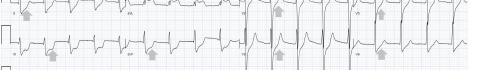

Repeat POCUS demonstrated bilateral lung sliding and no intra-abdominal free fluid; similarly, the chest tube and pleural drainage apparatus were functioning normally. Again, cardiac windows could not be obtained due to presumed air scattering. Because of significant pneumomediastinum seen on CT, obstructive shock physiology from the mediastinal free air was suspected. An emergent bedside percutaneous needle drainage in the left fifth intercostal space was performed, yielding 9 mL of blood with scant air bubbles. The patient’s blood pressure immediately improved, and cardiac windows were subsequently visualized on POCUS.
With high suspicion for acute myocardial infarction, the patient was taken for emergent coronary angiography. Transthoracic and transesophageal ultrasounds obtained in the cardiac catheterization lab did not demonstrate pericardial fluid or cardiac tamponade. Angiography demonstrated 95% thrombotic occlusion of the proximal left circumflex artery at the origin of the first obtuse marginal branch without evidence of coronary artery dissection (Image 3). Percutaneous coronary intervention (PCI) with aspiration thrombectomy and deployment of two drug-eluting stents resulted in complete restoration of coronary
CPC-EM Capsule
What do we already know about this clinical entity?
Coronary artery thrombosis (CAT) and tension pneumomediastinum are potential causes of morbidity and mortality in blunt chest trauma.
What makes this presentation of disease reportable?

A CAT in the absence of coronary dissection is rare and may be attributable to the vascular stasis caused by obstructive shock physiology.

What is the major learning point?
Clinicians should consider CAT in polytrauma patients with chest pain and concerning ECG findings, particularly when tension physiology occurs.
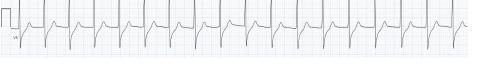
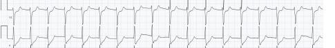
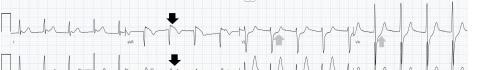
How might this improve emergency medicine practice?
Early involvement of multidisciplinary specialists and careful consideration of interventional strategies can improve patient outcomes in CAT associated with polytrauma.
blood flow. The patient was transferred to the surgical intensive care unit.
Aspirin and ticagrelor were initiated immediately after PCI. Norepinephrine and amiodarone were administered for cardiogenic shock and non-sustained ventricular tachycardia, respectively, both of which resolved within 48 hours. Following cardiac stabilization, the inpatient team addressed the patient’s spinal injury, although urgent operative intervention was
Clinical Practice and Cases in Emergency Medicine 98 Volume 7, no. 2: May 2023 Tension Pneumomediastinum and CAT Following a Motorcycle Accident Rouleau et al.
Image 1. Computed tomography of the chest with intravenous contrast demonstrating subcutaneous emphysema (black arrows), pneumomediastinum (red arrow), and hemopneumothorax (white arrows with black outline).
Image 2. Electrocardiogram with ST-segment elevation in leads aVR and aVL (black arrows), and diffuse ST depression in leads II, III, aVF, and V1-V6 (red arrows).
deferred because of hemodynamic instability and the bleeding risk associated with antiplatelet therapy. Prior to spinal fixation on hospital day three, ticagrelor was changed to tirofiban infusion, which was held prior to the first incision and resumed postoperatively. Ticagrelor was re-started following the removal of a spinal drain on postoperative day two. The patient remained stable for the remainder of hospitalization and was discharged on aspirin and ticagrelor on hospital day 14.
On a follow-up phone call with the patient almost six months after the injury, he reported that he was back to working full time and feeling fully recovered. He recalled the accident and arriving to the ED, although he did not recall undergoing tube thoracostomy placement or percutaneous needle drainage. He was advised to continue aspirin and ticagrelor for at least six months.
DISCUSSION
This patient’s ED course and management were complex due to the number and severity of his injuries. He developed obstructive shock physiology. We hypothesize that both blood and air in the mediastinum resulted in obstructive shock. In general, the differential diagnoses of obstructive shock in the setting of BCT include tension pneumothorax, pneumomediastinum, pneumopericardium, hemopericardium, and large retrosternal hematoma.5–9 The presence of retrosternal hematoma on CT and aspiration of blood during percutaneous needle drainage suggest that the retrosternal hematoma may have contributed to the obstructive shock physiology by exerting extrapericardial force on the heart. Retrosternal hematomas have been reported to accumulate over time and result in delayed decompensation hours after injury.10 Although the rate of fluid accumulation is typically understood to be a more important factor in causing obstructive shock physiology and has been well defined for cardiac tamponade, the volume and rate of accumulation necessary for a retrosternal hematoma to cause obstructive shock secondary to extra-pericardial compression is unknown.
In addition to the retrosternal hematoma, pneumomediastinum identified on CT and the presence of air
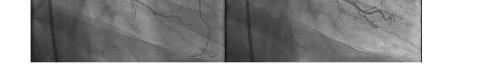


bubbles during percutaneous needle drainage suggest that the air in the mediastinum also contributed to the development of obstructive shock. Traumatic pneumomediastinum in BCT can be caused by several injuries, such as tracheoesophageal injury, alveolar injury, hollow viscus injury, or facial (sinus) injury.5 In this case, we suspect pneumomediastinum was likely secondary to extensive right-sided pneumothorax, pulmonary injury, and alveolar injury combined with the Macklin effect. The Macklin effect occurs when blunt trauma ruptures alveoli allowing air to dissect along bronchovascular sheaths resulting in mediastinal air.5 Although only a small amount of air was aspirated, needle insertion may have created a communication between the mediastinum and right hemithorax where a functional thoracostomy tube was already in place, thereby allowing for additional drainage of air. While the exact etiology of the obstructive shock physiology remains unknown, we hypothesize that the presence of blood (retrosternal hematoma) and air (pneumomediastinum) played significant roles. The bedside percutaneous needle drainage resolved the hemodynamic instability and allowed patient care to proceed.
Isolated CAT following BCT in the absence of concurrent coronary artery dissection is a rare complication that has been reported sparingly in the literature; therefore, its epidemiology is difficult to define.4 As with coronary artery dissection, there is predilection for the left anterior descending artery, which is hypothesized to be due to its anatomically vulnerable location immediately behind the sternum.11 The pathophysiology of thrombus formation is hypothesized to involve endothelial injury to the coronary artery wall caused by direct blunt trauma as well as indirect kinetic energy transmission that causes further damage or stasis of flow.4
In this instance, the presence of a CAT in the left circumflex artery may suggest an etiology other than direct trauma. It has been postulated that tension physiology from a pneumothorax can lead to reduced coronary artery blood flow thereby causing vascular stasis and thrombus formation.12 Extracardiac forces from pneumomediastinum and retrosternal hematoma may have compressed the left circumflex artery, leading to vascular congestion and thrombosis. The initial high-sensitivity troponin, drawn prior to the development of obstructive shock physiology, was 225 ng/mL and consistent with BCI. However, after development of obstructive shock and subsequent percutaneous needle drainage of retrosternal hematoma and pneumomediastinum, troponin increased to 1,161 ng/mL, eventually peaking at 4,012 ng/mL. While a delayed rise in troponin is expected after BCI, the significant rise in troponin after the development of obstructive shock support our hypothesis. Importantly, this patient had no risk factors for coronary artery disease, which also support our hypothesis.
Co-occurrence of an unstable spinal injury and liver injury complicated the decision to administer anti-platelet and anticoagulant medication in this case. Antiplatelet and anticoagulation therapy to treat CAT must be weighed against the risk of hemorrhage in a critically injured trauma patient.
Volume 7, no. 2: May 2023 99 Clinical Practice and Cases in Emergency Medicine Rouleau et al. Tension Pneumomediastinum and CAT Following a Motorcycle Accident
Image 3. Left: pre-stent right anterior oblique (RAO) view of proximal left circumflex artery sub-total occlusion (white arrow). Right: post-stent RAO view showing recanalized left circumflex artery (red arrow).
Tension Pneumomediastinum and CAT Following a Motorcycle Accident
Severity of concomitant injuries, compressibility of current or possible hemorrhage, underlying hematologic conditions, and current medication usage must all be considered. Contrary to aspirin, clopidogrel, and ticagrelor, whose antiplatelet effects may continue for days, heparin and tirofiban are favored in patients with CAT. Both agents are associated with low to medium risk of bleeding because of their relatively short half-life and titratability.13 While tranexamic acid administration has been shown to decrease mortality in multisystem trauma, it is also associated with an increased risk of thrombotic events and should be avoided in patients with known or suspected thrombosis.14 This patient received aspirin and ticagrelor after stent placement but was subsequently switched to tirofiban to allow titration. After surgical fixation of the L1 fracture on hospital day three and removal of spinal drain on hospital day five, ticagrelor was restarted, and the patient was discharged on dual antiplatelet therapy without any major bleeding events.
CONCLUSION
Coronary artery thrombosis and pneumomediastinum are uncommon complications of blunt cardiac injury, yet both injuries are associated with significant complications and present diagnostic challenges and treatment dilemmas, particularly in the setting of multisystem trauma. Emergency clinicians must be cognizant of the complications of blunt chest trauma and be prepared to identify and stabilize these patients while definitive care is pending.
The authors attest that their institution does not require Institutional Review Board approval. Patient consent has been obtained and filed for the publication of this case report. Documentation on file.
Address for Correspondence: Samuel Rouleau, MD, Department of Emergency Medicine, UC Davis Health,4150 V Street PSSB 2100, Sacramento, CA 95817. Email: sgrouleau@icloud.com
Conflicts of Interest: By the CPC-EM article submission agreement, all authors are required to disclose all affiliations, funding sources and financial or management relationships that could be perceived as potential sources of bias. The authors disclosed none.
Copyright: © 2023 Rouleau et al. This is an open access article distributed in accordance with the terms of the Creative Commons Attribution (CC BY 4.0) License. See: http://creativecommons.org/ licenses/by/4.0/
REFERENCES
1. Elsemesmani H, Bachir R, el Sayed MJ. Association between trauma center level and outcomes of adult patients with motorcycle crash–related injuries in the United States. J Emerg Med . 2020;59(4):499-507.
2. Marcolini EG and Keegan J. Blunt cardiac injury. Emerg Med Clin North Am . 2015;33(3):519-27.
3. Lee WS, Chong VE, Victorino GP. Computed tomographic findings and mortality in patients with pneumomediastinum from blunt trauma. JAMA Surg . 2015;150(8):757-62.
4. Sinha A, Sibel M, Thomas P, et al. Coronary thrombosis without dissection following blunt trauma. Case Rep Cardiol. 2016;2016:1-4.
5. Mansella G, Bingisser R, Nickel CH. Pneumomediastinum in blunt chest trauma: a case report and review of the literature. Case Rep Emerg Med . 2014;2014:685381.
6. Bhullar IS and Wagner S. Management of cardiac tamponade secondary to pneumomediastinum after blunt thoracic trauma. Am Surg . 2010;76(6):E50-1.
7. Beg MH, Reyazuddin, Ansari MM. Traumatic tension pneumomediastinum mimicking cardiac tamponade. Thorax 1988;43(7):576-7.
8. Rambaud G, Desachy A, François B, et al. Extrapericardial cardiac tamponade caused by traumatic retrosternal hematoma. J Cardiovasc Surg (Torino) . 2001;42(5):621-4.
9. Arabi RI, Aljudaibi A, Althumali AA, et al. Traumatic retrosternal hematoma leading to extra-pericardial cardiac tamponade-Case report. Int J Surg Case Rep . 2019;61:30-2.
10. Rodgers-Fischl PM, Makdisi G, Keshavamurthy S. Extrapericardial tamponade after blunt trauma. Ann Thorac Surg. 2021;111(1):e49-50.
11. Bjørnstad JL, Pillgram-Larsen J, Tønnessen T. Coronary artery dissection and acute myocardial infarction following blunt chest trauma. World J Emerg Surg 2009 4:1. 2009;4(1):1-4.
12. Arao K, Mase T, Nakai M, et al. Concomitant spontaneous tension pneumothorax and acute myocardial infarction. Intern Med 2019;58(8):1131.
13. Eikelboom JW, Hirsh J, Spencer FA, et al. Antiplatelet Drugs: Antithrombotic Therapy and Prevention of Thrombosis, 9th ed: American College of Chest Physicians Evidence-Based Clinical Practice Guidelines. Chest . 2012;141(2 Suppl):e89S.
14. CRASH-2 trial collaborators, Shakur H, Roberts I, et al. Effects of tranexamic acid on death, vascular occlusive events, and blood transfusion in trauma patients with significant haemorrhage (CRASH-2): a randomised, placebo-controlled trial. Lancet 2010;376(9734):23-32.
Clinical Practice and Cases in Emergency Medicine 100 Volume 7, no. 2: May 2023
et al.
Rouleau
Emergency-physician Performed, Ultrasound-guided Lateral
Femoral Cutaneous Nerve Block for Meralgia Paresthetica: A Report of Two Cases
Matthew M. Kongkatong, MD
Christopher D. Thom, MD, RDMS
Jakob Ottenhoff, DO
Section Editor: Rachel Lindor, MD, JD
University of Virginia Health System, Department of Emergency Medicine, Charlottesville, Virginia
Submission history: Submitted November 7, 2022; Revision received February 22, 2023; Accepted March 9, 2023
Electronically published May 27, 2023
Full text available through open access at http://escholarship.org/uc/uciem_cpcem
DOI: 10.5811/cpcem.1409
Introduction: Neuropathy of the lateral femoral cutaneous nerve, also known as meralgia paresthetica, causes pain and paresthesia to the anterolateral thigh. It commonly results from nerve irritation from extrinsic compression; however, it may occur spontaneously. Symptoms from this condition can be debilitating, and the pain may be ascribed to other conditions leading to delays in diagnosis. Peripheral nerve blockade can be useful both diagnostically and therapeutically for meralgia paresthetica.
Case Report: Two female patients in their sixties presented to the emergency department for chronic, atraumatic, left upper thigh pain. In both cases the patients had hyperalgesia and paresthesia to the anterolateral, upper thigh. The emergency physician performed an ultrasoundguided nerve block of the lateral femoral cutaneous nerve for each patient, which resulted in temporary, complete resolution of their pain.
Conclusion: Meralgia paresthetica is an uncommon but painful condition that can elude diagnosis. Physical exam findings such as allodynia and hyperalgesia of the anterolateral thigh in the absence of back pain is suggestive of the diagnosis. Utrasound-guided nerve blockade can be helpful to the emergency physician to confirm the diagnosis and provide non-opioid pain relief to the patient. [Clin Pract Cases Emerg Med. 2023;7(2):101–105]
Keywords: case series; meralgia paresthetica; regional anesthesia; lateral femoral cutaneous neve; ultrasound-guided nerve block.
INTRODUCTION
Meralgia paresthetica (MP) also referred to as lateral femoral cutaneous nerve (LFCN) neuropathy is characterized by painful paresthesia and numbness of the anterolateral thigh. The incidence of this disorder is 4.3 per 10,000 person per year and usually presents in the third or fourth decade of life but can occur at any age. Causes of MP are grouped into spontaneous and iatrogenic categories. In spontaneous cases, external compression from tight clothing or belts can irritate the nerve. Compression may also result from intra-abdominal or pelvic processes, such as obesity, pregnancy, and uterine tumors. Spontaneous neuropathy can be caused by metabolic abnormalities such as diabetes, hypothyroidism, and nutritional deficiencies.1 Iatrogenic MP may
result from inadvertent nerve transection from surgical incision, compression or traction from retractor placement, and extrinsic compression in the setting of body positioning for surgical exposure or restraint belt placement.1,2
Typically, patients will present with unilateral symptoms, but bilateral symptoms occur in 20% of patients.2 Patients often complain of uncomfortable tingling in the anterolateral thigh and can experience allodynia. Symptoms are often exacerbated by direct palpation of the area around the anterior superior iliac spine (ASIS) and by hip extension maneuvers.1,2 The presence of concurrent abdominal or urogenital complaints or abnormal examination findings like palpable masses should prompt the clinician to evaluate for other causes of the patient’s symptoms,
Volume 7, no. 2: May 2023 101 Clinical Practice and Cases in Emergency Medicine Case Report
Ultrasound-guided LFCN Block for Meralgia Paresthetica Kongkatong
which may involve abdominopelvic imaging such as ultrasound or computed tomography (CT).
Lumbar radiculopathy may mimic symptoms of MP, but in cases of isolated MP, back pain is absent. Muscle strength and deep tendon reflexes should be normal as the LCFN is a purely sensory nerve. Patients may have an antalgic gait.1,2 Symptoms and exam findings concerning for nerve root or spinal cord compression should be worked up with imaging, typically magnetic resonance imaging (MRI) or CT.
Meralgia paresthetica can cause significant patient distress and disability. Clinician unfamiliarity with this condition may lead to delay in diagnosis, leading to prolonged patient discomfort and unnecessary workup for other conditions.2 Diagnostic workup into the underlying cause depends on clinician suspicion for a secondary cause, such as compression from an abdominal mass.1,2 Nerve conduction studies may be performed as an outpatient but are usually not available to clinicians in the emergency department (ED). Diagnostic nerve blockade may be pursued in the ED or clinic. Rapid improvement or resolution of the patient’s symptoms after LFCN blockade can confirm the diagnosis as well as treat pain.1 Herein we present two cases where a patient presented to our ED with MP and received LFCN blocks.
CASE REPORT
Case 1
A 67-year-old female presented to the ED for left thigh pain worsening over the course of one month. The pain began after abdominal aortic aneurysm surgery three months prior. She had one ED presentation for severe pain as well as several clinic visits with workup including CT angiogram of the abdominal aorta with iliofemoral run-off and Doppler ultrasound for venous thrombosis without any surgical complications or venous thrombosis noted. Her vital signs on presentation were blood pressure (BP) of 183/88 millimeters of mercury (mm Hg), temperature 36.4°Celsius (C), heart rate 67 beats per minute (bpm), respiratory rate 20 breaths per minute, and oxygen saturation (SPO2) 98%. On physical examination, she had hyperalgesia and allodynia of the skin of her proximal, left anterolateral thigh. Tenderness and paresthesia to palpation inferior to the ASIS was present. She had normal strength to hip flexion, knee extension, and ankle dorsiflexion/plantarflexion and palpable ankle pulses.
Using a 6-15 megahertz linear array X-Porte (FUJIFILM Sonosite, Inc, Bothell, WA), we performed an ultrasoundguided LFCN block on the patient with seven milliliters (mL) of 0.25% bupivacaine after obtaining consent. On reevaluation 10 minutes later, the patient reported complete resolution of the pain. She was given the diagnosis of MP and recommended to see pain management. On chart review follow-up, the initial block gave her two days of symptom relief. She received another block with pain management that lasted two days. She was started on pregabalin with improvement of symptoms over the next several weeks.
CPC-EM Capsule
What do we already know about this clinical entity?
Meralgia paresthetica (MP) is an uncommon cause of upper thigh pain. While not lifethreatening, it can be distressing for patients and difficult to diagnose.
What makes this presentation of disease reportable?
Because it is uncommon, pain from MP may be ascribed to other conditions leading to negative workups and repeat patient presentations for pain.
What is the major learning point?
Ultrasound-guided lateral femoral cutaneous nerve blockade is a low-risk option that can aid physicians in correctly diagnosing MP and provide non-opioid pain control.
How might this improve emergency medicine practice?
Correct diagnosis of MP in the emergency department allows physicians to initiate specific therapy and plan appropriate outpatient follow-up for MP patients.
Case 2
A 60-year-old female presented to the ED for acute worsening of a five-month history of atraumatic left thigh pain. The patient had multiple ED, primary care, and specialty care visits for the pain without definitive diagnosis. She had been worked up previously with negative Doppler ultrasound for venous thrombosis and MRI of the lumbar spine showing only mild neuroforaminal stenosis. On presentation she had normal vital signs: temperature 36.7°C, BP 128/73 mm Hg, respiratory rate 18 breaths per minute, and SPO2 100% (besides mildly elevated heart rate of 99 bpm). On physical examination she had an antalgic gait, but a negative, straightleg raise test bilaterally and normal strength to hip flexion, knee extension, and ankle movement bilaterally. She had normal ankle pulses. Skin examination was notable for hyperesthesia and allodynia to the region of the left, anterolateral thigh without erythema, induration, or rashes. After consenting the patient, we performed an ultrasoundguided LFCN block with the X-Porte using six mL of 0.25% bupivacaine with four milligrams of preservative-free dexamethasone. After 10 minutes, the patient was pain-free and ambulated out of the ED with a normal gait. She was
Clinical Practice and Cases in Emergency Medicine 102 Volume 7, no. 2: May 2023
et al.
Kongkatong et al.
recommended to continue following up with her primary care physician; however, because they were outside our health system we could not perform chart follow-up.
DISCUSSION
The LFCN is a purely sensory nerve originating from the posterior divisions of L2 and L3 of the lumbar plexus. The nerve courses in the retroperitoneal space through the psoas muscle and over the iliacus muscle in the region of the inguinal ligament (Figure 1).3 The most common (87%) anatomical position of the LFCN is deep to the inguinal ligament medial to the ASIS and sartorius muscle. However, there are variants described where the nerve may course through or superficial to the inguinal ligament, superficial or lateral to the ASIS, and through the sartorius muscle. Typically (79%), the nerve exits from the fascia lata and then divides into an anterior and posterior branch.4


To perform the block, expose the affected thigh and inguinal area with the patient in a supine position. A high frequency linear transducer should be used to visualize the nerve and surrounding structures. The probe can initially be placed just inferior to the ASIS in a transverse orientation (Image 1). Scanning inferiorly, the nerve may be visualized as a round, hyperechoic, honeycombed structure deep to the subcutaneous tissue and fascia lata, positioned between the sartorius muscle medially and the tensor fascia lata muscle laterally (Image 2). As with other nerves, the LFCN may appear hypoechoic if the angle of insonation is oblique with respect to the nerve. This can be remedied by fanning the probe such that the nerve becomes hyperechoic on the screen.3,5 In cases of
Ultrasound-guided LFCN Block for Meralgia Paresthetica


entrapment, the nerve may be a larger diameter than the unaffected side or have a more hypoechoic appearance of the fascicles. The ultrasound transducer can also be used to provoke a Tinel sign (elicitation of symptoms with palpation of the nerve) similar to sonographic Murphy’s sign in cholecystitis.3

Once the nerve is identified, the patient should be prepped in an antiseptic fashion. A probe cover and sterile conduction medium should be used. A small gauge needle is then guided to the soft tissue surrounding this nerve, and local anesthetic is deposited after negative aspiration of blood (Image 2). An in-plane or out-of-plane approach may be used; however, we prefer the in-plane approach to visualize the whole course of the needle.5 We prefer using a longer acting local anesthetic such as bupivacaine to maximize duration of effect; however, shorter acting local anesthetics such as lidocaine have been described. Volumes used in the literature range from 1 mL-10 mL and adjunctive use of steroids such as methylprednisolone and triamcinolone have been described.6-10 The use of adjunctive steroids has been shown to extend the duration of nerve blocks.11


Meralgia paresthetica is an uncommon entity that can cause significant discomfort. Conservative treatment of MP consists of addressing any extrinsic causes of compression like tight clothing and weight loss and use of non-steroidal anti-inflammatory drugs (NSAID). Persistent neuropathic pain may be treated with tricyclic antidepressants or anticonvulsants such as gabapentin.2 Nerve blockade of the LFCN offers a low-risk, non-opioid treatment for this condition.1 Although ultrasound-guided LFCN blockade has been described in the physical medicine and rehabilitation literature as well as the anesthesia literature; however, it has not commonly been described in the ED context. In both cases, the patients had severe pain that was refractory to treatment with acetaminophen, NSAIDs, and oxycodone. Both patients experienced rapid improvement after nerve blockade without any complications. In the first case, our diagnosis led the patient to be started on specific therapy for MP as opposed to renewed opioid prescription.

Volume 7, no. 2: May 2023 103 Clinical Practice and Cases in Emergency Medicine
Figure 1. A) Anatomic drawing of the course of the lumbar plexus and lateral cutaneous femoral nerve (highlighted in green); B) dermatomal distribution of the lateral femoral cutaneous nerve (black arrow). Image obtained from https://commons.wikimedia. org/wiki/File:Gray824.png.
Image 1. Superficial landmarks of the anterior superior iliac spine (ASIS) (circle) and inguinal ligament (dotted line). The transducer is placed axially just inferior to the ASIS to begin looking for the lateral femoral cutaneous nerve.
Image 2. A) A labeled image of the lateral femoral cutaneous nerve (white arrow) lying deep to the fascia lata (dotted line) between the sartorius muscle (SM) and tensor fascia lata muscle (TFL). Also visualized is the illiopsoas muscle (IPM) medially and anterior inferior iliac spine (AIIS) in the far field; B) Image of a needle (white arrows) depositing local anesthetic (dotted area) around the lateral femoral cutaneous nerve (asterisk).
In both cases, the patients had undergone multiple comprehensive imaging studies and specialist evaluation without a diagnosis or improvement in symptoms. Performance of the LFCN block in the ED was diagnostic and therapeutic without any additional imaging studies or specialty consultation. In both cases our patients experienced great satisfaction with the result of the block. The first patient was able to be transitioned off opioid therapy for symptom control.
Limited studies of ultrasound-guided nerve blocks in MP patients show that some require only a single injection to improve symptoms. Tagliafico et al. showed symptom improvement in 16 out of 20 MP patients with a single injection. They also demonstrated a significant mean improvement of patient-reported quality of life.6 Another study performed by Klauser et al. showed that some patients may experience sustained pain control at 12
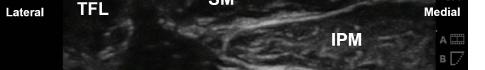
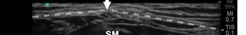
months after a single injection. However, most patients (15/20) required more than one block to be pain-free at 12 months.9 This demonstrates the importance of outpatient follow-up for MP that is diagnosed in the ED.
Ultrasound-guided nerve blocks have been shown to be efficacious for the treatment of musculoskeletal pain and neuropathic pain.12,13 With the progressive integration of ultrasound in emergency medicine practice, ultrasound-guided nerve blockade for procedural analgesia, fracture analgesia, and atraumatic conditions such as sciatica, has become more commonplace.14,15 The LFCN is a superficial, purely sensory nerve that is amenable to visualization on ultrasound. Additionally, the nerve is typically not directly adjacent to significant neurovascular structures, such as the femoral vasculature, making it a low-risk target amenable to even the novice ultrasound operator. Finally, only a small volume of anesthetic is needed to achieve the desired effect, decreasing the risk of local anesthetic toxicity. This makes it an attractive block for the emergency clinician to diagnose and treat MP. Our report adds to the body of literature demonstrating that ultrasound-guided peripheral nerve blockade can be effectively and safely performed in the ED.

CONCLUSION

Meralgia paresthetica, although uncommon, can be a painful and chronic condition that may go misdiagnosed. Ultrasound-guided peripheral nerve blockade performed in the ED can help the emergency physician differentiate MP from other entities, which can improve diagnostic confidence and avoid potentially unnecessary workups. Ultrasound-guided LFCN blockade also provides the emergency physician an opioid-sparing option for treatment of MP in ED patients.


ACKNOWLEDGMENTS

We would like to thank David G Moyer, MD, and the UVA School of Medicine gross anatomy staff.
The authors attest that their institution does not require Institutional Review Board approval. Patient consent has been obtained and filed for the publication of this case report. Documentation on file.
Address for Correspondence: Matthew Kongkatong, MD, University of Virginia Health System, Department of Emergency Medicine, 1215 Lee St, Charlottesville, VA 22908. Email: mk3wk@uvahealth.org
Conflicts of Interest: By the CPC-EM article submission agreement, all authors are required to disclose all affiliations, funding sources and financial or management relationships that could be perceived as potential sources of bias. The authors disclosed none.
Copyright: © 2023 Kongkatong et al. This is an open access article distributed in accordance with the terms of the Creative Commons Attribution (CC BY 4.0) License. See: http:// creativecommons.org/licenses/by/4.0/

Clinical Practice and Cases in Emergency Medicine 104 Volume 7, no. 2: May 2023
Ultrasound-guided LFCN Block for Meralgia Paresthetica Kongkatong et al.
A B
Kongkatong et al.
REFERENCES
1. Patijn J, Mekhail N, Hayek S, et al. Meralgia paresthetica. Pain Practice. 2011;11(3):302-8.
2. Grossman MG, Ducey SA, Nadler SS, et al. Meralgia paresthetica: diagnosis and treatment. J Am Acad Orthop Surg. 2001;9(5):336-44.
3. Becciolini M, Pivec C, Riegler G. Ultrasound of the lateral femoral cutaneous nerve: a review of the literature and pictorial essay. J Ultrasound Med. 2021;41(5):1273-84.
4. Tomaszewski KA, Popieluszko P, Henry BM, et al. The surgical anatomy of the lateral femoral cutaneous nerve in the inguinal region: a meta-analysis. Hernia. 2016;20(5):649-57.
5. Lateral femoral cutaneous nerve block. In: Hadzic’s Peripheral Nerve Blocks and Anatomy for Ultrasound-Guided Regional Anesthesia, 3rd Edition (265-269). New York, NY: McGraw Hill; 2022.
6. Tagliafico A, Serafini G, Lacelli F, et al. Ultrasound-guided treatment of meralgia paresthetica (lateral femoral cutaneous neuropathy). J Ultrasound Med. 2011;30(10):1341-6.
7. Kim JE, Lee SG, Kim EJ, et al. Ultrasound-guided lateral femoral cutaneous nerve block in meralgia paresthetica. Korean J Pain 2011;24(2):115-8.
8. Öztürk G MD. Conservative treatment versus ultrasound guided injection in the management of meralgia paresthetica: a randomized
Ultrasound-guided LFCN Block for Meralgia Paresthetica
controlled trial. Pain Physician. 2020;3;23(6;3):253-64.
9. Klauser AS, Abd Ellah MMH, Halpern EJ, et al. Meralgia paraesthetica: ultrasound-guided injection at multiple levels with 12-month follow-up. Eur Rad. 2015;26(3):764-70.
10. Hurdle MF, Weingarten TN, Crisostomo RA, et al. Ultrasound-guided blockade of the lateral femoral cutaneous nerve: technical description and review of 10 cases. Arch Phys Med and Rehabil 2007;88(10):1362-4.
11. Pehora C, Pearson AM, Kaushal A, et al. Dexamethasone as an adjuvant to peripheral nerve block. Cochrane Database Syst Rev 2017;2017(11).
12. Yürük D, Akkaya ÖT, Polat ÖE, et al. Ultrasound‐guided erector spinae plane block and trapezius muscle injection for myofascial pain syndrome. J Ultrasound Med. 2021;41(1):185-91.
13. Nwawka OK, Meyer R, Miller TT. Ultrasound-guided subgluteal sciatic nerve perineural injection: report on safety and efficacy at a single institution. J Ultrasound Med 2017;36(11):2319-24.
14. Goldsmith AJ, Liteplo A, Hayes BD, et al. Ultrasound-guided transgluteal sciatic nerve analgesia for refractory back pain in the ED. Am J Emerg Med 2020;38(9):1792-5.
15. Wilson C. Feeling blocked? Another pain management tool in the emergency department. Ann Emerg Med. 2018;72(2):120-6.
Volume 7, no. 2: May 2023 105 Clinical Practice and Cases in Emergency Medicine
Diagnosing Atypical Flutter in the Post-atrial Fibrillation Ablation Patient: A Case Report
Alexandra Nicole Fuher, MD*
Ryan Borne, MD†
John Cunningham, MD*‡
Section Editor: Lev Libet, MD
University of Colorado Anschutz Medical Campus, Department of Internal Medicine, Aurora, Colorado University of Colorado Health, Division of Cardiology, Colorado Springs, Colorado Denver Health and Hospital Authority, Division of Internal Medicine, Denver, Colorado
Submission history: Submitted July 23, 2022; Revision received March 30, 2023; Accepted March 31, 2023
Electronically published May 30, 2023
Full text available through open access at http://escholarship.org/uc/uciem_cpcem
DOI: 10.5811/cpcem.1413
Introduction: Late atrial arrhythmias after catheter ablation for atrial fibrillation occur in up to 30% of post-ablation patients and are increasingly encountered by emergency physicians. However, diagnosing the exact mechanism of the arrhythmia on the surface electrocardiogram (ECG) remains challenging due to atrial scarring leading to heterogeneous P-wave morphology.
Case Report: A 74-year-old male with a history of prior catheter ablation for atrial fibrillation presented with palpitations and subacute symptoms of heart failure. The patient’s ECG revealed narrow complex tachycardia with more P waves than QRS complexes. The differential diagnosis included typical flutter, atypical flutter, and focal atrial tachycardias with 2:1 conduction block. P waves were positive in V1 and across all precordial leads (absent precordial transition). This favors atypical flutter originating from the left atrium over typical cavotricuspid isthmus-dependent right atrial flutter. Transthoracic echocardiogram showed a reduced ejection fraction due to tachycardiamediated cardiomyopathy. The patient underwent a repeat electrophysiology study and ablation, which confirmed the presence of an atypical flutter circuit using the mitral annulus, known as perimitral flutter. Repeat catheter ablation resulted in maintenance of sinus rhythm. At follow-up, his ejection fraction recovered.
Conclusion: Recognizing ECG findings suggestive of atypical flutter impacts initial emergency department decisions and triage as atypical flutter post-atrial fibrillation ablation is frequently resistant to rate-controlling medications and often requires cardiology and/or electrophysiology consultation if available. [Clin Pract Cases Emerg Med. 2023;7(2):106–109]
Keywords: case report; atrial fibrillation; atrial flutter; typical, atypical.
INTRODUCTION
Catheter ablation with pulmonary vein isolation is increasingly used in the management of paroxysmal atrial fibrillation (AF). Post-ablation late atrial tachycardias are common, occurring in up to 30% of patients.1,2 As a result, emergency physicians and other acute care clinicians are encountering post-ablation arrhythmias more frequently. The electrocardiogram (ECG) interpretation in this population, however, is challenging. Analysis of P-wave morphology to
determine the mechanism of arrhythmia is limited by the altered atrial conduction, which results from atrial scarring. This case report demonstrates ECG findings that distinguish atypical flutter circuits originating from the left atrium (LA) from typical atrial flutter in patients who are post ablation.
CASE REPORT
A 74-year-old male with a history of symptomatic drug-refractory paroxysmal AF treated with a catheter ablation
Clinical Practice and Cases in Emergency Medicine 106 Volume 7, no. 2: May 2023 Case Report
* † ‡
with pulmonary vein isolation 14 years prior and a second catheter ablation two years prior, presented to the emergency department (ED) with three months of progressive shortness of breath and palpitations. Upon presentation, he was afebrile, his heart rate was 137 beats per minute, respiratory rate was 18 breaths per minute, blood pressure was 106/76 millimeters of mercury, and oxygen saturation was 90% on room air. Cardiovascular examination revealed tachycardia with normal heart sounds and no murmurs, rubs or gallops. Jugular venous pressure was elevated. Pulmonary exam was negative for rales, and there was no lower extremity edema.
The ECG (Image 1) revealed narrow complex tachycardia with more P waves than QRS complexes. The differential diagnosis included typical and atypical atrial flutter and focal atrial tachycardias (AT) with 2:1 conduction block. Notably, P waves are positive in V1 and across all precordial leads (absent precordial transition), favoring atypical flutter from the left atrium over typical cavotricuspid isthmus (CTI)dependent right atrial flutter. Positive flutter waves in the inferior leads indicate high to low atrial activation, most often caused by counterclockwise perimitral flutter and left atrial (LA) roof-dependent AT. Focal AT, due to enhanced automaticity from an ectopic focus, cannot be ruled out based on the ECG findings; however, reentrant arrhythmias are more common post-AF ablation.
A transthoracic echocardiogram (TTE) revealed a severely reduced ejection fraction of 20% without valvular disease or wall motion abnormalities. The patient was started on amiodarone and underwent direct current cardioversion. An electrophysiology study revealed perimitral isthmus flutter. Image 2 indicates how the pattern of right atrial activation correlates with the ECG flutter wave morphology on the patient’s initial ECG. Repeat catheter ablation successfully terminated the arrhythmia. One month later, a follow-up TTE
CPC-EM Capsule
What do we already know about this clinical entity?
Late atrial arrhythmias after atrial fibrillation ablation occur in up to 30% of post-ablation patients. However, the diagnosis remains challenging due to heterogeneous P wave morphology.
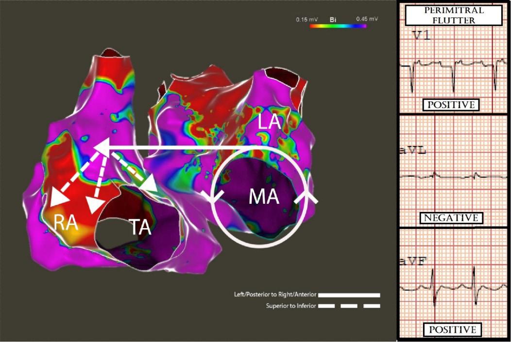
What makes this presentation of disease reportable?
This case highlights the electrocardiogram (ECG) findings suggestive of atypical flutter in a patient with prior catheter ablation.
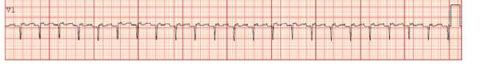
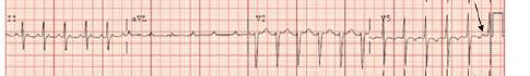
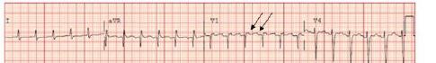
What is the major learning point?
The absence of precordial transition favors atypical flutter originating in the left atrium in the postablation patient, among other ECG findings.
How might this improve emergency medicine practice?
Atypical flutter is often resistant to control medications. Symptomatic patients presenting with atypical flutter warrant early consultation or referral to cardiac electrophysiology.
Image 1. Electrocardiogram on presentation. Two P waves occur for every QRS complex. Upright flutter waves in the precordial leads remain positive across all precordial leads (noted by black arrows). Upright inferior P waves in the inferior leads, annotated with blue arrows.
Image 2. The patient’s intracardiac voltage mapping is adjacent to the associated P-wave polarity of electrocardiogram leads V1, aVF, and aVL. Solid white lines with arrows indicate the counterclockwise perimitral annulus flutter circuit. Solid lines indicate left to right and posterior to anterior initial atrial activation toward the right atria resulting in a positive P wave in lead V1 and negative P wave in lead aVL. Dashed lines indicate superior to inferior RA depolarization resulting in positive inferior P waves. MA, mitral annulus; TA, tricuspid annulus; RA, right atrium; LA, left atrium.
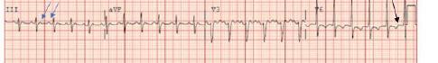
Volume 7, no. 2: May 2023 107 Clinical Practice and Cases in Emergency Medicine Fuher et al. Diagnosing Atypical
in
Patient: A Case Report
Flutter
the Post-atrial Fibrillation Ablation
Diagnosing Atypical Flutter in the Post-atrial Fibrillation Ablation Patient: A Case Report Fuher et
showed a recovered ejection fraction of 50%, indicating a likely tachycardia-mediated cardiomyopathy.
DISCUSSION
Delayed atrial arrhythmias post-AF ablation present a challenge as the degree of atrial scarring and location of prior radiofrequency ablation sites result in variable P-wave morphology. Most post-ablation arrhythmias are caused by either macroreentry or localized microreentrant atrial tachycardias (circuits <3 centimeters in diameter), and true focal AT due to an ectopic focus is less common.3 The most common macroreentrant circuits post-ablation include typical atrial flutter, perimitral flutter, and left atrial-roof dependent reentry.3 Reentry results from either gaps in ablation lines or ablation lesions that act as obstacles resulting in tunneled conduction through one or more isthmuses within the left atrium, thereby allowing reentry.4 Despite limitations, the surface ECG is essential when initially determining the type of arrhythmia post catheter ablation. The ECG can frequently differentiate typical from atypical atrial flutter originating from the left atrium. This is important as it impacts clinical management as the outcomes of repeat ablation differ between typical and atypical flutter.3,5
Typical atrial flutter is defined as a macroreentrant circuit (most often counterclockwise) around the tricuspid annulus, using the CTI.5 This results in negative flutter waves in the inferior leads and initial positive flutter waves in the precordial leads that transition to negative by V6 (Image 3).3 The classic “sawtooth” pattern with negative flutter waves in the inferior leads may be absent in over half of patients with CTI-dependent flutter post-ablation. However, precordial transition is 98% specific for typical counterclockwise flutter with a high negative predictive value of 95%.3
Atypical atrial flutter refers to macroreentrant tachycardias that are not CTI-dependent. Atypical flutter circuits can originate in the left or right atrium, usually around an atrial scar in patients with structural heart disease.5 Multiple circuits may be present in the same patient. Studies suggest that P-wave morphology in post-PVI arrhythmias primarily results from right atrial activation, likely due to an electrically inert LA scar with loss of electrical forces toward the posterior LA wall.6 Acknowledging the relationship between the left and right atria is also posterior to anterior explains the positive P waves in V1 in LA flutter (Image 1). Negative P waves in any precordial lead suggests a right atrial circuit over a left atrial circuit, with 83% and 100% sensitivity and specificity, respectively.1,4 Therefore, the absence of precordial transition or negative precordial flutter waves in Image 1 suggests the left atrial origin of the arrhythmia.
However, an electrophysiology study is needed to map the exact reentrant circuit. Flutter wave polarity in the inferior leads does not separate typical from atypical flutter but rather indicates that the macroreentrant circuit initially activated the superior right atrium with subsequent superior to inferior right
Image 3. A) Electrocardiogram demonstrating typical cavotricuspid-isthmus dependent flutter in a patient with a prior ablation. Blue arrows indicate negatively directed inferior flutter waves. B) Demonstrates precordial transition from positive flutter waves to negative within the solid, vertical black lines. Typically, the negative flutter waves in the left sided leads proceeds the positive flutter wave in V1.3
atrial activation.6 P-wave polarity in leads I and aVL are unreliable when distinguishing post-ablation macroreentrant tachycardias, likely due to the degree of left atrial scarring.3 Surface ECG cannot reliably separate LA roof-dependent reentry from PMFL, highlighting the need for electrophysiology referral.
A notable feature in Image 1 is the lack of a typical undulating flutter wave present in Image 3. A discrete isoelectric interval greater than 80 milliseconds in all leads favors focal AT over macroreentrant rhythms. However, nearly a quarter of post-ablation macroreentrant arrhythmias will have discrete isoelectric intervals in all leads.3 Atrial scarring may result in decreased flutter-wave voltages leading to ECG findings more characteristic of AT. Thus, an electrophysiology study with intracardiac mapping is needed for definitive diagnosis when managing post-ablation arrhythmias.3,5
Distinguishing LA atypical flutter from an arrhythmia originating in the right atrium impacts clinical management as atypical flutter is often resistant to rate-controlling and antiarrhythmic medications and frequently requires repeat catheter ablation.3,7 Therefore, earlier consultation with cardiology is often required to manage these patients. Ablation of a left atrial atypical flutter post-pulmonary vein isolation has a lower acute success rate and higher major complication rate than a typical flutter ablation due to the increased complexity of the procedure and the need for septal puncture to perform electroanatomic mapping and ablation within the left atrium.5
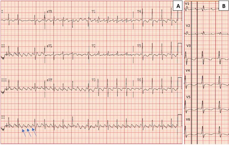
CONCLUSION
Late post-ablation arrhythmias are increasingly common as the number of catheter ablation procedures performed for AF increases. The presence of upright P waves across all precordial
Clinical Practice and Cases in Emergency Medicine 108 Volume 7, no. 2: May 2023
al.
Patient consent has been obtained and filed for the publication of this case report.
Address for Correspondence: Alexandra N Fuher, MD, University of Colorado Anschutz Medical Campus, Division of Internal Medicine, 12631 E 17th Ave, Mail Stop B177, Aurora, CO 80045. Email: alexandra.fuher@cuanschutz.edu.
Conflicts of Interest: By the CPC-EM article submission agreement, all authors are required to disclose all affiliations, funding sources and financial or management relationships that could be perceived as potential sources of bias. The authors disclosed none.
Copyright: © 2023 Fuher et al. This is an open access article distributed in accordance with the terms of the Creative Commons Attribution (CC BY 4.0) License. See: http://creativecommons.org/ licenses/by/4.0/
REFERENCES
1. Chang SL, Tsao HM, Lin YJ, et al. Differentiating macroreentrant from focal atrial tachycardias occurred after circumferential pulmonary vein isolation. J Cardiovasc Electrophysiol. 2011;22(7):748-55.
2. Dixit S. Characterization of left atrial Tachyarrhythmias in patients following atrial fibrillation ablation: correlation of surface ECG with intracardiac mapping. J Atr Fibrillation. 2008;1(1).
Fuher et al. Diagnosing Atypical Flutter in the Post-atrial Fibrillation Ablation Patient: A Case Report leads favors atypical flutter from the left atrium over typical CTI dependent right atrial flutter. Once recognized, atypical flutter, including perimitral flutter, is often resistant to rate and rhythm controlling medications. Patients with atrial fibrillation or typical flutter who are asymptomatic once rate controlled or who spontaneously convert in the emergency department are often appropriate for outpatient follow-up with primary care, with cardiology follow-up based on symptoms and clinical course. Symptomatic patients presenting to the ED with atypical flutter warrant early consultation or referral to cardiac electrophysiology for definitive diagnosis and management.
3. Pascale P, Roten L, Shah AJ, et al. Useful electrocardiographic features to help identify the mechanism of atrial tachycardia occurring after persistent atrial fibrillation ablation. JACC Clin Electrophysiol 2018;4(1):33-45.
4. Issa Z, Miller JM, Zipes DP. (2019). Clinical Arrhythmology and Electrophysiology: A Companion to Braunwald’s Heart Disease (3rd Ed.). Philadelphia, PA: Elsevier.
5. Page RL, Joglar JA, Caldwell MA, et al. 2015 ACC/AHA/HRS guideline for the management of adult patients with supraventricular tachycardia: a report of the American College of Cardiology/American Heart Association Task Force on Clinical Practice Guidelines and the Heart Rhythm Society [published correction appears in J Am Coll Cardiol. 2016 Dec 27;68(25):2922-3]. J Am Coll Cardiol. 2016;67(13):e27-e115.
6. Hu W, Zhou D, Hua B, et al. Flutter wave morphology of peri-mitral atrial flutters is mainly determined by right atrial activation. Circ Arrhythm Electrophysiol. 2020;13(9):e008446.
7. Aldaas OM, Lupercio F, Lin AY, et al. Ablation of mitral annular flutter ablation utilizing a left atrial anterior line versus a lateral mitral isthmus line: a systematic review and meta-analysis. J Interv Card Electrophysiol. 2022;63(1):87-95.
Volume 7, no. 2: May 2023 109 Clinical Practice and Cases in Emergency Medicine
Implanted Penile Prosthetic Visualized During Focused Assessment with Sonography for Trauma Examination: A Case Report
Kevin Chambers, DO* Geoffrey Comp, DO*†
Section Editor: Austin Smith, MD
Creighton University School of Medicine, Department of Emergency Medicine, Phoenix, Arizona
University of Arizona College of Medicine-Phoenix, Phoenix, Arizona
Submission history: Submitted December 27, 2022; Revision received April 6, 2023; Accepted April 7, 2023
Electronically published May 27, 2023
Full text available through open access at http://escholarship.org/uc/uciem_cpcem
DOI: 10.5811/cpcem.1407
Introduction: This is a case report of an implanted penile prosthetic visualized during focused assessment with sonography for trauma (FAST) examination. The case represents a unique finding near the lateral bladder that may confound assessment of intraperitoneal fluid collections during initial assessment of trauma patients.
Case Report: A 61-year-old Black male was brought to the emergency department from a nursing facility for evaluation after sustaining a ground-level fall. A FAST exam demonstrated an abnormal fluid collection anterior and lateral to the bladder, later identified as an implanted penile prosthetic.
Conclusion: Focused assessment with sonography for trauma examinations are often performed on unidentified patients in a time-sensitive manner. Understanding of potential false-positive results is crucial to appropriate use of the tool. This report demonstrates a novel false-positive result that may be difficult to differentiate from a true intraperitoneal bleed. [Clin Pract Cases Emerg Med. 2023;7(2):110–112.]
Keywords: case report; implanted penile prosthetic; point-of-care ultrasound; focused assessment with sonography for trauma (FAST).
INTRODUCTION
The focused assessment with sonography for trauma (FAST) examination is a widely used point-of-care ultrasound tool to evaluate for intraperitoneal bleeding in trauma patients. It is frequently used as part of the initial assessment of traumatic injuries and may determine whether a patient has need for additional imaging such as computed tomography (CT) or surgical intervention such as an exploratory laparotomy.1 The sensitivity and specificity of the FAST exam have been widely studied and are reportedly as high as 96% and 98%, respectively.2,3 The FAST exam uses a series of standardized sonographic windows that identify regions where intraperitoneal fluid tends to accumulate due to being dependent areas. The lateral aspects of the bladder are one region where such fluids can be visualized. Numerous false-
positive FAST results have been described in the literature including ascites, gastric contents, and perinephric fat pads.4 In this case report, we describe a potential false-positive FAST exam due to an implanted penile prosthetic in a patient presenting after a fall.
CASE REPORT
A 61-year-old Black male was brought to the emergency department from a nursing facility for evaluation after sustaining a ground-level fall. The patient, who ambulated with a walker, had a past medical history of hypertension, diabetes, mild dementia, and cerebrovascular accident (CVA) with residual right-sided spastic hemiplegia. He had a witnessed fall while attempting to transfer from the bed to his walker, falling forward into the
Clinical Practice and Cases in Emergency Medicine 110 Volume 7, no. 2: May 2023 Case Report
* †
walker and onto the ground. No loss of consciousness or use of anticoagulation were reported.
Upon arrival, the patient was noted to be alert and oriented to person, place, and time; however, he did have some challenge relaying recent historical details. The patient’s initial vital signs were notable for temperature of 36.4˚ Celsius, heart rate of 109 beats per minute, blood pressure of 124/78 millimeters of mercury, respiratory rate of 18 breaths per minute, and oxygen saturation of 96% on room air. He was protecting his airway, with clear and equal breath sounds bilaterally. Heart sounds were non-muffled, and his abdomen was soft. Pelvis was stable to compression, and the patient had spastic motor function of all extremities consistent with his known CVA.
Secondary survey revealed a superficial laceration to the right forehead and scattered abrasions over the anterior chest and abdomen. Due to evidence of abdominal trauma, a FAST exam was performed that revealed no free fluid in the right upper quadrant, left upper quadrant, or in subxiphoid views. However, the suprapubic view demonstrated an abnormal anechoic fluid collection anterior and lateral to the bladder (Image 1). The collection was visualized in transverse and sagittal views.
CPC-EM Capsule
What do we already know about this clinical entity?
Focused Assessment with Sonography for Trauma (FAST) examinations utilize a series of standardized sonographic windows that identify regions where intraperitoneal fluid tends to accumulate in dependent areas.
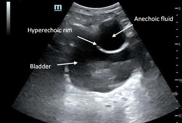
What makes this presentation of disease reportable?
This report demonstrates novel false positive images that may be difficult to differentiate from a true intraperitoneal bleed.
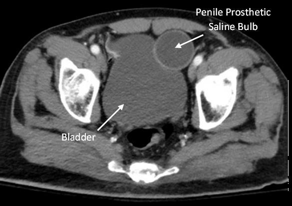
What is the major learning point?
Awareness of implanted penile prosthetic design and function is important to avoid false positive FAST examinations.
How might this improve emergency medicine practice?
FAST examinations are performed in a time sensitive manner and understanding of potential false positive results is crucial to appropriate use of the tool.
Vital signs remained stable, and the patient’s abdomen remained soft and non-tender. He was not able to provide further clarification of the abnormal finding during initial questioning secondary to presumed dementia. During further evaluation of his abnormal ultrasound findings, genitourinary surgical scars were noted to the groin and scrotum. Upon chart review, we found a CT of the abdomen and pelvis with intravenous contrast that had previously identified the findings as a prosthetic penile implant (Image 2). Ultimately, the patient was cleared by trauma service following negative imaging
of the head. No further evaluation of potential intraperitoneal bleeding was deemed necessary. The laceration to his forehead was repaired at bedside, and the patient was transported back to the nursing facility at his neurologic baseline.
Volume 7, no. 2: May 2023 111 Clinical Practice and Cases in Emergency Medicine Chambers et al. Implanted Penile Prosthetic Visualized During FAST Examination
Image 1. Point-of -care ultrasound image of anechoic fluid collection with hyperechoic rim lateral to bladder in transverse view.
Image 2. Computed tomography image of penile prosthetic saline bulb reservoir in transverse pelvic view.
Implanted Penile Prosthetic Visualized During FAST Examination Chambers et
DISCUSSION
In this presented case, the hyperechoic lucency with anechoic center is a reservoir for an implanted penile prosthetic (Image 3). Penile prosthetics are performed for a variety of reasons; they most commonly occur following prostatic surgical procedures to maintain sexual function and often incorporate discrete surgical scars for cosmetic purposes.5 There are approximately 20,000 such devices
tools alone should never replace a thorough history and physical exam.
The case we report here occurred in a patient who was hemodynamically stable with an overall low suspicion for intraperitoneal injury. Therefore, additional chart review allowed for identification of the abnormal ultrasonographic finding. However, in the unstable patient this finding may confound the diagnosis or resuscitation attempts or lead to potentially dangerous invasive measures.
The authors attest that their institution requires neither Institutional Review Board approval, nor patient consent for publication of this case report. Documentation on file.
Address for Correspondence: Kevin D. Chambers, DO, Creighton University School of Medicine, Department of Emergency Medicine, 2601 E. Roosevelt St., Phoenix, AZ, 85008. Email: kdc44846@creighton.edu.
Conflicts of Interest: By the CPC-EM article submission agreement, all authors are required to disclose all affiliations, funding sources and financial or management relationships that could be perceived as potential sources of bias. The authors disclosed none.
Copyright: © 2023 Chambers et al. This is an open access article distributed in accordance with the terms of the Creative Commons Attribution (CC BY 4.0) License. See: http://creativecommons.org/ licenses/by/4.0/
implanted annually.6 While many types of devices exist, this particular device encompassed cylindrical shafts placed within the corpus cavernosum, a pump mechanism, and a separate saline balloon reservoir implanted surgically in the inferior abdomen beneath the transversalis fascia.
The saline balloon can appear similar to a Foley catheter balloon but will be located outside the bladder wall. The reservoir is filled with saline and can change shape via the pump diverting pressure to the cylinder system.7 Therefore, the reservoir may not always appear as well circumscribed and could confound the FAST exam or even be interpreted as a false positive. Awareness of the implant design and function is important to avoid falsepositive FAST exams. Typically, intraperitoneal fluid collects in the most dependent portions of the abdomen making isolated findings near the lateral bladder unusual.
CONCLUSION
The case represents a unique finding near the lateral bladder that may confound assessment of intraperitoneal fluid collections. Focused assessment with sonography exams are often performed on unidentified patients in a time-sensitive manner; an understanding of potential false-positive results is crucial to appropriate use of the tool. Obtaining additional history from available sources and performing focused physical assessments can help explain potential false-positive FAST exams and avoid unnecessary interventions. Imaging
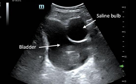
REFERENCES
1. Brenchley J, Walker A, Sloan JP, et al. Evaluation of focussed assessment with sonography in trauma (FAST) by UK emergency physicians. Emerg Med J. 2006;23(6):446-8.
2. Lee C, Balk D, Schafer J, et al. Accuracy of focused assessment with sonography for trauma (FAST) in disaster settings: a meta-analysis and systematic review. Disaster Med Public Health Prep. 2019;13(56):1059-64.
3. Christian NT, Burlew CC, Moore EE, et al. The focused abdominal sonography for trauma examination can reliably identify patients with significant intra-abdominal hemorrhage in life-threatening pelvic fractures. J Trauma Acute Care Surg. 2018;84(6):924-8.
4. Sierzenski PR, Schofer JM, Bauman MJ, et al. The double-line sign: a false positive finding on the focused assessment with sonography for trauma (FAST) examination. J Emerg Med. 2011;40(2):188-9.
5. Anderson PC, Jain S, Summerton DJ, et al. Surgical atlas. Insertion of an inflatable penile prosthesis. BJU Int. 2007;99(2):467-82.
6. Grewal S, Vetter J, Brandes SB, et al. A population-based analysis of contemporary rates of reoperation for penile prosthesis procedures. Urology. 2014;84(1):112-6.
7. Al-Enezi A, Al-Khadhari S, Al-Shaiji TF. Three-piece inflatable penile prosthesis: surgical techniques and pitfalls. J Surg Tech Case Rep. 2011;3(2):76-83.
Clinical Practice and Cases in Emergency Medicine 112 Volume 7, no. 2: May 2023
al.
Image 3. Point-of-care ultrasound image of saline reservoir in pelvic sagittal view.
Benign Episodic Mydriasis as a Cause of Isolated Anisocoria
Alyssa Seibold, MD*
Jonathan Barnett, MD†
Lawrence Stack, MD*
Charles Lei, MD*
Section Editor: Austin Smith, MD
Vanderbilt University Medical Center, Department of Emergency Medicine, Nashville, Tennessee
Vanderbilt University Medical Center, Department of Ophthalmology and Visual Sciences, Nashville, Tennessee
Submission history: Submitted October 7, 2022; Revision received December 21, 2022; Accepted January 20, 2023
Electronically published April 18, 2023
Full text available through open access at http://escholarship.org/uc/uciem_cpcem
DOI: 10.5811/cpcem.1316
Case Presentation: A 22-year-old female presented to the emergency department with a dilated right pupil and mild blurry vision. Physical examination revealed a dilated, sluggishly reactive right pupil, without other ophthalmic or neurologic abnormalities. Neuroimaging was normal. The patient was diagnosed with unilateral benign episodic mydriasis (BEM).
Discussion: BEM is a rare cause of acute anisocoria with an underlying pathophysiology that is not well understood. The condition has a female predominance and is associated with a personal or family history of migraine headaches. It is a harmless entity that resolves without intervention and results in no known permanent damage to the eye or visual system. Benign episodic mydriasis is a diagnosis of exclusion that may be considered only after the life- and eyesight-threatening causes of anisocoria. [Clin Pract Cases Emerg Med. 2023;7(2):113–114]
Keywords: benign episodic mydriasis; anisocoria; migraine headache.
CASE PRESENTATION
A 22-year-old female with no known medical history presented to the emergency department (ED) with a dilated right pupil and mild blurry vision that she first noticed after waking up in the morning. She reported no eye pain or redness, double vision, or headache. She had no history of trauma to the eye, prior ocular disease, or known exposure to medications or toxins. Her vital signs were normal, including a blood pressure of 115/59 millimeters (mm) of mercury. Her visual acuity was 20/20 in both eyes. Her right pupil was round, 7 mm in diameter (Image), and sluggishly reactive to light, while her left pupil was 4 mm and briskly reactive. She had no afferent pupillary defect. She had intact extraocular movements, full visual fields, and normal intraocular pressures in both eyes.
She had no ptosis or other neurologic deficits. Computed tomography angiography of the head and neck was normal. She was evaluated in the ED by an ophthalmologist who felt her presentation was most consistent with unilateral benign episodic mydriasis (BEM). The mydriasis resolved prior to her follow-up ophthalmology appointment three weeks later, and she had no recurrences.
DISCUSSION
The causes of anisocoria (greater than 1 mm difference in pupillary diameter) range from benign conditions to life- or eyesight-threatening emergencies. In patients presenting to the ED with acute anisocoria, it is imperative to first consider the most serious etiologies, including cerebral aneurysm, stroke, intracranial hemorrhage, meningitis, intracranial or ocular mass, acute angle-closure glaucoma, open globe, optic neuritis, and ocular infections. Benign causes of acute anisocoria include BEM, traumatic mydriasis, post-surgical changes, ocular migraine, and medication or chemical exposure (eg, anticholinergics, sympathomimetics).
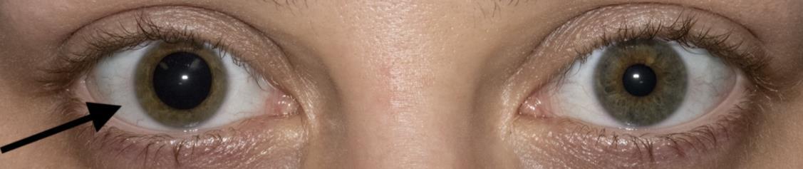
Volume 7, no. 2: May 2023 113 Clinical Practice and Cases in Emergency Medicine Images in Emergency Medicine
* †
Image. The patient’s right pupil (arrow) was dilated and sluggishly reactive to light, compared to her normal left pupil.
The first step in the evaluation of isolated anisocoria is to determine which pupil is abnormal.1 If the larger pupil is found to be abnormal and a third nerve palsy has been excluded, pilocarpine may be used to determine the cause of unilateral mydriasis. Pupillary constriction in response to dilute (0.1%) pilocarpine indicates Adie tonic pupil. In the absence of a response to dilute pilocarpine, concentrated (1%) pilocarpine may be administered; pupillary constriction indicates BEM, whereas the lack of a response suggests pharmacologic mydriasis.
Benign episodic mydriasis is a rare cause of acute anisocoria and a diagnosis of exclusion. While typically unilateral, the affected eye may alternate in recurrent episodes and there may be bilateral involvement during a single episode.2 Patients with BEM may have isolated anisocoria or experience a wide variety of concomitant symptoms, such as blurry vision, photophobia, orbital pain, nausea, eye redness, double vision, or headache.3
The underlying pathophysiology of BEM is not well understood. Several studies have suggested that BEM may be caused by hyperactivity of the sympathetic nervous system (which causes pupillary dilation) or hypoactivity of the parasympathetic nervous system (which causes pupillary constriction).4 It occurs predominantly in females and has been connected to a personal or family history of migraine headaches, particularly in patients with recurrent episodes, but the significance of these associations is not clear.2,3
An episode of BEM may resolve within minutes to hours or persist for weeks to months. 3 Aside from the cosmetic inconvenience and discomfort of migraineassociated symptoms (if present), BEM is a harmless entity, and there is no known irreversible damage to the eye or visual system. 2
The authors attest that their institution does not require Institutional Review Board approval. Patient consent has been obtained and filed for the publication of this case report. Documentation on file.
REFERENCES
1. Garcia KL, Breazzano MP, Bond JB 3rd, et al. A 3-year-old with sudden-onset anisocoria after playing outside. Pediatr Emerg Care 2018;34(6):e111-4.
2. Martín-Santana I, González-Hernández A, Tandón-Cárdenes L, et al. Benign episodic mydriasis. Experience in a specialist neuro-ophthalmology clinic of a tertiary hospital. Neurologia
CPC-EM Capsule
What do we already know about this clinical entity?
The causes of anisocoria range in seriousness from benign conditions to life- or eyesight-threatening emergencies.
What is the major impact of the image(s)?
The image illustrates a case of benign episodic mydriasis, a rare cause of acute anisocoria that has not been well described in the emergency medicine literature.
How might this improve emergency medicine practice?
Emergency physicians should have a stepwise approach to the evaluation of isolated anisocoria and consider benign episodic mydriasis as a diagnosis of exclusion.
Address for Correspondence: Charles Lei, MD, Vanderbilt University Medical Center, Department of Emergency Medicine, 1313 21st Avenue South, Nashville, TN 37332. Email: Charles.lei@vumc.org.
Conflicts of Interest: By the CPC-EM article submission agreement, all authors are required to disclose all affiliations, funding sources and financial or management relationships that could be perceived as potential sources of bias. The authors disclosed none.
Copyright: © 2023 Seibold et al. This is an open access article distributed in accordance with the terms of the Creative Commons Attribution (CC BY 4.0) License. See: http://creativecommons.org/ licenses/by/4.0/
2015;30(5):290-4.
3. Caty JT, Kogan M, Reidy JJ, et al. A case of recurrent benign episodic unilateral mydriasis and decreased visual acuity and review of the literature. N Am J Med Sci. 2015;8(3):139-42.
4. Skeik N and Jabr FI. Migraine with benign episodic unilateral mydriasis. Int J Gen Med. 2011;4:501-3.
Clinical Practice and Cases in Emergency Medicine 114 Volume 7, no. 2: May 2023 Benign Episodic
Seibold et al.
Mydriasis as a Cause of Isolated Anisocoria
Point-of-care Ultrasound Identification of Hepatic Abscess in the Emergency Department
Michael Blomquist, MS*
Taz Brinkerhoff, MD†
David Weech, DO†
Heesun Choi, DO†‡
Section Editor: Shadi Lahham, MD
Rocky Vista University of Osteopathic Medicine, Parker, Colorado Kingman Regional Medical Center, Department of Emergency Medicine, Kingman, Arizona University of California, Irvine, Department of Emergency Medicine, Orange, California
Submission history: Submitted June 15, 2022; Revision received August 19, 2022; Accepted December 6, 2022
Electronically published April 18, 2023
Full text available through open access at http://escholarship.org/uc/uciem_cpcem
DOI: 10.5811/cpcem.1299
Case Presentation: A 92-year-old female with past medical history of hypertension presented to the emergency department with pain in her right shoulder, right flank, and right upper quadrant of her abdomen. Point-of-care ultrasound (POCUS) and computed tomography imaging showed concerns for multiple large hepatic abscesses. Percutaneous drainage removed 240 millileters of purulent fluid that identified Fusobacterium nucleatum, a rare cause of pyogenic liver abscess.
Discussion: Emergency physicians should keep hepatic abscess on their differential for right upper quadrant abdominal pain and can use POCUS for expeditious diagnosis. [Clin Pract Cases Emerg Med. 2023;7(2):115–117]
Keywords: Point-of-care ultrasound; hepatic abscess; pyogenic liver abscess.
CASE PRESENTATION
A 92-year-old female with past medical history of hypertension presented to the emergency department with one week onset of pain in her right shoulder, right flank, and right upper quadrant of her abdomen. The patient’s initial presentation was consistent with a cholestatic pattern of gallbladder disease; however, further evaluation via point-of-care ultrasound (POCUS) of hepatobiliary structures revealed multiple large abscesses within the liver parenchyma (Image 1 & Video).
Computed tomography (CT) was obtained prior to labs due to concerning physical exam, which revealed large septated fluid collection in the right hepatic lobe most consistent with a large hepatic abscess and an additional fluid collection in the left hepatic lobe also consistent with hepatic abscess (Image 2).
DISCUSSION
The initial POCUS findings were indicative for large hepatic abscess and significantly raised clinical concern for sepsis. In this case, POCUS was performed prior to lab work as was CT, which facilitated an expedited diagnosis of a severe, life-threatening disease. The patient was started on empiric antibiotic therapy
and underwent percutaneous, ultrasound-guided drainage and drain placement that removed 240 of purulent fluid. During her
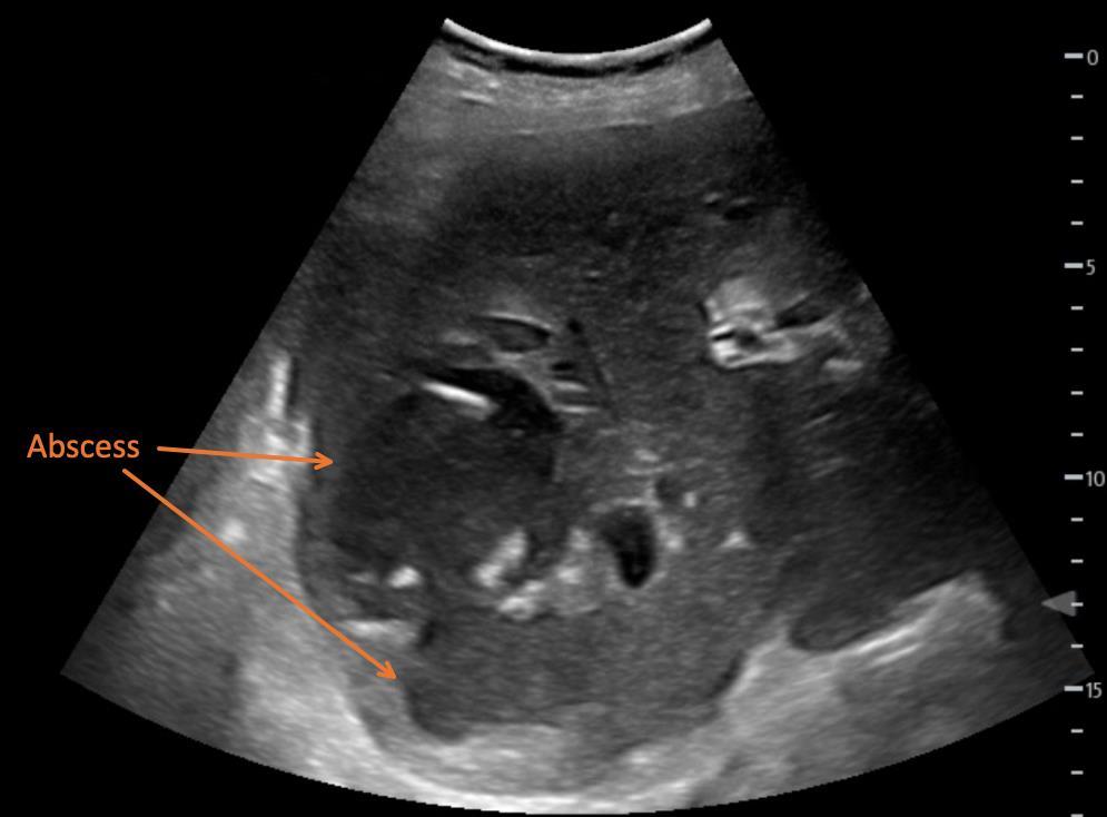
Volume 7, no. 2: May 2023 115 Clinical Practice and Cases in Emergency Medicine Images in Emergency Medicine
* † ‡
Image 1. Soft tissue point-of-care ultrasound showing multiple, hypoechoic, loculated fluid collections within the parenchyma of the right lobe of the liver, indicative of hepatic abscesses.
admission she developed multisystem organ failure and was ultimately transitioned to hospice care.
Pyogenic liver abscesses are relatively uncommon, with an annual estimated incidence of 3.6 cases per 100,000 people in the United States, and have an estimated mortality rate of 5.6%.1 In North America, the most common cause of pyogenic liver abscess is direct spread from biliary infection; however, almost any bacterial infection of the gastrointestinal tract can undergo hematogenous spread to the liver via the portal circulation.2 The most common pathogens consistently identified from abscess cultures are Escherichia coli, Klebsiella species, and Streptococcus species.1 Interestingly, cultures from the abscess fluid drained from this patient identified Fusobacterium nucleatum, a rare cause of pyogenic liver abscess that is thought to be related to periodontal disease and sigmoid diverticulitis.3
Patients with hepatic abscess most commonly present with fevers and abdominal pain; however, symptoms can include a broad range of complaints such as nausea, vomiting, and weight loss. Jaundice may be the first and
CPC-EM Capsule
What do we already know about this clinical entity?
Pyogenic liver abscess is rare yet its estimated mortality rate is 5.6%. The clinical presentation of pyogenic liver abscess is nonspecific which can cause delay of care. Performing POCUS early during the initial evaluation can provide narrow differentials. It is relatively easy to Identify large fluid collections in the liver as shown in the images.
What is the major impact of the image(s)?
Patient’s presentation was concerning for gallbladder pathology. However, POCUS finding indicated large septal fluid collection in the right hepatic lobe. This critical POCUS finding allowed physicians to deliver prompt treatment plans for the patient without a delay.
How might this improve emergency medicine practice?
Performing POCUS can provide faster, specific and accurate assessment and treatment plans on an elderly patient with undifferentiated abdomen pain. This can decreased financial burden to the patients, duration of ED stay, and ED wait time. treatment plans. Also providing specific labs and imaging studies can decreased financial burden to the patients, total hours of ED stay, and ED wait time.
only clinical manifestation of the disease.4 The diagnosis of pyogenic liver abscess relies heavily on prompt imaging. Computed tomography is somewhat more sensitive for liver abscesses than ultrasound (approximately 95% vs 85%).5 If ultrasound was performed initially but did not demonstrate any abnormalities, CT should be performed if concern for a possible liver abscess or additional underlying pathology remains high.
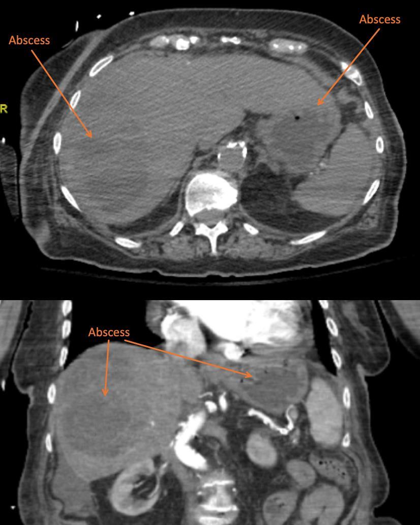
Clinical Practice and Cases in Emergency Medicine 116 Volume 7, no. 2: May 2023 POCUS Identification of Hepatic Abscess Blomquist et al.
Video. Soft tissue point-of-care ultrasound showing multiple, hypoechoic, loculated fluid collections within the parenchyma of the right lobe of the liver, indicative of hepatic abscesses.
Image 2. Computed tomography imaging with transverse (top) and coronal (bottom) plane views of the upper abdomen showing large septated fluid collection in the right hepatic lobe, most consistent with a large hepatic abscess, and additional fluid collection in the left hepatic lobe also consistent with hepatic abscess.
The authors attest that their institution does not require Institutional Review Board approval. Patient consent has been obtained and filed for the publication of this case report. Documentation on file.
POCUS Identification of Hepatic Abscess
Address for Correspondence: Heesun Choi, DO, Kingman Regional Medical Center, Department of Emergency Medicine, 3269 Stockton Hill Rd., Kingman, AZ 86409. Email: heesunchoido@gmail.com.
REFERENCES
1. Meddings L, Myers RP, Hubbard J, et al. A population-based study of pyogenic liver abscesses in the United States: incidence, mortality, and temporal trends. Am J Gastroenterol. 2010;105(1):117-24.
2. Altemeier WA, Culbertson WR, Fullen WD, et al. Intra-abdominal abscesses. Am J Surg. 1973;125(1):70-9.
3. Wijarnpreecha K, Yuklyaeva N, Sornprom S, et al. Fusobacterium nucleatum: Atypical organism of pyogenic liver abscess might be related to sigmoid diverticulitis. N Am J Med Sci. 2016;8(4):197-9.
4. Mohsen AH, Green ST, Read RC, et al. Liver abscess in adults: ten years experience in a UK centre. QJM. 2002;95(12):797-802.
Conflicts of Interest: By the CPC-EM article submission agreement, all authors are required to disclose all affiliations, funding sources and financial or management relationships that could be perceived as potential sources of bias. The authors disclosed none.
Copyright: © 2023 Blomquist et al. This is an open access article distributed in accordance with the terms of the Creative Commons Attribution (CC BY 4.0) License. See: http://creativecommons.org/ licenses/by/4.0/
5. Lin AC, Yeh DY, Hsu YH, et al. Diagnosis of pyogenic liver abscess by abdominal ultrasonography in the emergency department. Emerg Med J. 2009;26(4):273-5.
Volume 7, no. 2: May 2023 117 Clinical Practice and Cases in Emergency Medicine
Blomquist et al.
An Inexpensive Biomechanical Model to Help Teach and Learn Newer Mandible Reduction Techniques

Shantal Tummings, BS*
Sierra Garrett, BS*

Ja’Neil G. Humphrey, BS*

Jasmine Haddad, BS*

Jon Roper, MD*†
Deena I. Bengiamin, MD*†
Timothy P. Young, MD*†
Section Editor: Kenneth Whitlow, MD

Loma Linda University School of Medicine, Laboratory for Innovations in Medical Education, Loma Linda, California
Loma Linda University Medical Center and Children’s Hospital, Department of Emergency Medicine, Loma Linda, California
Submission history: Submitted September 28, 2021; Revision received March 19, 2022; Accepted August 23, 2022
Electronically published May 30, 2023
Full text available through open access at http://escholarship.org/uc/uciem_cpcem
DOI: 10.5811/cpcem.1416
[Clin Pract Cases Emerg Med. 2023;7(2):118–120]
Dear Editor:
While preparing for a mandible reduction teaching session, we were happy to find Lum and Poh’s description of the successful use of the wrist pivot method for a refractory dislocation.1 Like the authors, we have encountered difficulty with the conventional method of mandible dislocation reduction. In this technique, the clinician places both thumbs in the patient’s mouth along the lower molars and applies downward, backward force. Simultaneous reduction of both sides of a bilateral dislocation requires considerable force on the part of the clinician, which is difficult for the patient to tolerate. Even when successful, the conventional approach places the proceduralist’s thumbs at risk of injury from masseter spasm; they must be protected by gauze wraps or a bite block.
In the last two decades, two techniques have been proposed as alternatives to the conventional approach: the wrist pivot and extraoral methods.2,3 The authors used the wrist pivot method, which uses different hand positioning and is better tolerated than the conventional method but still requires the clinician’s fingers in the patient’s mouth.4 The extraoral technique allows the proceduralist to avoid placing fingers in the patient’s mouth, but it is more difficult for physicians to perform.4 However, these techniques are difficult to understand and learn from static images.
While the authors found success with the wrist pivot method after eight doctors attempted to use the conventional method with and without sedation, Image 1 of the authors’ article shows hand positioning that is different from the original description of the method (https://bit.ly/3Tf5RRO).2 This is not surprising, considering that they employed the technique following written instructions only. They also illustrate and discuss the extraoral method. Like the authors’ experience with the wrist pivot method,
we initially misread the original description of this technique and taught and used a slightly different method. In our experience, hands-on practice with a trainer is the best way to learn complicated techniques in the way that authors intended them to be performed. This can be aided by video demonstrations, which can help clarify hand positioning and force application. However, we could not locate any commercial mandible reduction trainers. To remedy this, we created an inexpensive, biomechanical model. We bought a plastic human skull model (www.amzn.com/ B07R9NYPCN), removed the temporomandibular joint (TMJ) capsules, and drilled 1/8” holes at the center of the TMJ and 7/64” holes through the top of the condyles (Image 1). We
Clinical Practice and Cases in Emergency Medicine 118 Volume 7, no. 2: May 2023 Letter To The Editor
* †
Image 1. We drilled a hole in the center of the articular fossa (arrow) and one through the condyle, and then secured the condyle with elastic string.
Tummings et al. Comments on “Refractory Temporomandibular Joint Dislocation: Reduction Using the Wrist Pivot Method” looped elastic string (www.amzn.com/B088CQR4PL) through the TMJ and condyles and secured it with toggle stoppers (www.amzn.com/B08M7Z2XY1). Resistance bands (www. amzn.com/B01AVDVHTI) simulate masseter muscles, looped around each zygomatic arch, tied at the base of the mandible, and held in place by a drywall screw on each side. Since the skull model that we bought had a removable cranium cap, we used a nut and fender washer to hold it in place. The total cost for parts was $67. A video explaining how the trainer works can be viewed here: youtu.be/OLPu0vwmTpo.
During our practice session, we had an assistant hold the trainer on a gurney with the head of the bed elevated (Image 2). This is the same positioning approach we use for real patients. For the conventional method, we were able to demonstrate and practice a sequential approach, in which each side is reduced in succession. This approach may be successful when a bilateral simultaneous approach fails.5 While practicing the extraoral technique, it became clear that the application of force in opposite directions on either side of the mandible is key (Image 3). This causes a rotational motion like the one employed in a
conventional sequential reduction. The motion of the wrist pivot method disengages the condyle from the skull base and allows it to clear the articular eminence located anterior to the mandibular fossa. We were able to directly visualize this with the trainer. The trainer’s utility is best conveyed by moving images; so we made a video that can be viewed (Video).
We agree with the authors’ statement that familiarity with multiple techniques is important for refractory mandible dislocations. We add that learning proper technique using trainers increases chances of success.
This project was a product of the Laboratory for Innovations in Medical Education, which is supported by a grant from the Kiwanis Cal-Nev-Ha Children’s Fund.

The authors attest that their institution requires neither Institutional Review Board approval nor patient consent for publication of this case report. Documentation on file.
Address for Correspondence: Timothy Young, MD, Loma Linda University Medical Center and Children’s Hospital, Department of Emergency Medicine, 11234 Anderson Street, Room A890A, Loma Linda, Ca. 92354. Email: tpyoung@llu.edu.


Conflicts of Interest: By the CPC-EM article submission agreement, all authors are required to disclose all affiliations, funding sources and financial or management relationships that could be perceived as potential sources of bias. The authors disclosed none.
Copyright: © 2023 Tummings et al. This is an open access article distributed in accordance with the terms of the Creative Commons Attribution (CC BY 4.0) License. See: http://creativecommons.org/ licenses/by/4.0/
REFERENCES
1. Lum VWM and Poh J. Refractory temporomandibular joint dislocation: reduction using the wrist pivot method. Clin Pract Cases Emerg Med . 2017 Oct 18;1(4):380-3.


2. Lowery LE, Beeson MS, Lum KK. The wrist pivot method, a novel technique for temporomandibular joint reduction. J Emerg Med 2004;27(2):167-70.
3. Chen YC, Chen CT, Lin CH, et al. A safe and effective way for reduction of temporomandibular joint dislocation. Ann Plast Surg 2007;58(1):105-8.

4. Ardehali MM, Tari N, Bastaninejad SH, et al. Comparison of different approaches to the reduction of anterior temporomandibular joint dislocation: a randomized clinical trial. Int J Oral Maxillofac Surg . 2016;45(8):1009-14.
5. Mellick LB. Pediatric jaw dislocation reduction. Available at: https://www.youtube.com/watch?v=-Zm7ev8LUAA Accessed March 16, 2022.
Volume 7, no. 2: May 2023 119 Clinical Practice and Cases in Emergency Medicine
Image 2. The completed mandible reduction trainer.
Image 3. The extraoral reduction technique. On one side, the thumb pushes on the coronoid process while the other hand grasps the angle of the mandible and pulls. The process is repeated on the opposite side for bilateral dislocations.
Video. Trainer explanation of mandible reduction.
Response to “An Inexpensive Biomechanical Model to Help Teach and Learn Newer Mandible Reduction Techniques”
Vincent W.M. Lum, MBBS, MCEM* Juliana Poh, MBBS, MRCSEd†
Changi General Hospital, Department of Accident & Emergency, Singapore, Singapore
Singapore General Hospital, Department of Emergency Medicine, Bukit Merah, Singapore
Dear Editors,
We thank Tummings et al. for their interest in our article and congratulate them on the use of their innovative mandible reduction trainer. We agree that video demonstrations and
hands-on practice are superior to text and static pictures for learning mandibular reduction techniques. Slight variations in techniques may indeed occur with different clinicians as their experience with using these techniques grows.
Clinical Practice and Cases in Emergency Medicine 120 Volume 7, no. 2: May 2023 Letter to the Editor
* †

editor@westjem.org


E volv ED: Redefining EM
CALIFORNIA ACEP’S ANNUAL CONFERENCE 2023


Education is targeted to Medical Students and Residents, but all are welcome to attend.

Friday, September 8, 2023
The Sheraton Pasadena
