Clinical Practice and Cases in Emergency Medicine


In Collaboration with the Western Journal of Emergency Medicine


Case Series 272 Ultrasound-Guided Posterior Tibial Nerve Block for Frostbite of the Plantar Surfaces: A Case Series Burl T, Latshaw P, Dreyfuss A 276 Diaphragmatic Excursion as a Novel Objective Measure of Serratus Anterior Plane Block Efficacy: A Case Series Lentz B, Kharasch S, Goldsmith AJ, Brown J, Duggan NM, Nagdev A
Case Reports 280 Point-of-Care Ultrasound Diagnosis of Tetralogy of Fallot Causing Cyanosis: A Case Report Addepalli A, Guillen M, Dreyfuss A, Mantuani D, Nagdev A, Martin DA 284 Recurrent Infantile Hypertrophic Pyloric Stenosis in the Emergency Department: A Case Report Kosoko, AA Tobar DC 288 Legionnaires’ Disease Causing Severe Rhabdomyolysis and Acute Renal Failure: A Case Report Branstetter A, Wyler B 292 Acute Isolated Thenar Compartment Syndrome in a Patient with Evans Syndrome: A Case Report Jiang E, Kim KH, Babigian A 296 Tibial Spine Fracture in an Adolescent Male After Minor Injury: A Case Report Nunez A, Sleight S, Khan Z, Blasko B, Kim TY 298 Life-Threatening Complications Associated with Bladder Decompression: A Case Report Knapp BJ, Apgar L, Pennell K
Volume 6, Number 4, November 2022 Open Access at www.cpcem.org ISSN: 2474-252X A Peer-Reviewed, International Professional Journal
Clinical Practice and Cases in Emergency Medicine VOLUME 6 ISSUE 4 November 2022 PAGES 272-335
Contents continued on page iii

www.acoep.org
ACOEP
stands with all
emergency physicians and providers on the front line. We thank you for your tireless work and effort.
Clinical Practice and Cases in Emergency Medicine
Indexed in PubMed and full text in PubMed Central
Rick A. McPheeters, DO, Editor-in-Chief
Kern Medical/UCLA- Bakersfield, California
University
Mark I. Langdorf, MD, MHPE, Senior Associate Editor
University of California, Irvine School of Medicine- Irvine, California
Shahram Lotfipour, MD, MPH, Senior Associate Editor
University of California, Irvine School of Medicine- Irvine, California

Shadi Lahham, MD, MS, Associate Editor
Kaiser Permanente- Orange County, California
Manish Amin, DO, Associate Editor
Kern Medical/UCLA- Bakersfield, California
John Ashurst,
Kingman
Anna McFarlin, MD, Decision Editor
Louisiana State University Health Science Center- New Orleans, Louisiana
Amin A. Kazzi, MD, MAAEM
The American University of Beirut, Beirut, Lebanon
Anwar Al-Awadhi, MD
Mubarak Al-Kabeer Hospital, Jabriya, Kuwait
Arif A. Cevik, MD
United Arab Emirates University College of Medicine and Health Sciences, Al Ain, United Arab Emirates
Abhinandan A.Desai, MD
University of Bombay Grant Medical College, Bombay, India
Bandr Mzahim, MD
King Fahad Medical City, Riyadh, Saudi Arabia
Barry E. Brenner, MD, MPH
Case Western Reserve University
Brent King, MD, MMM
University of Texas, Houston
Daniel J. Dire, MD
University of Texas Health Sciences Center San Antonio
David F.M. Brown, MD
Massachusetts General Hospital/Harvard Medical School
Amal Khalil, MBA
Edward Michelson, MD Texas Tech University
Edward Panacek, MD, MPH University of South Alabama
Editorial Board
Christopher Sampson, MD, Decision Editor
University of Missouri- Columbia, Missouri
Joel Moll, MD, Decision Editor
Virginia Commonwealth University School of Medicine- Richmond, Virginia
Steven Walsh, MD, Decision Editor
Einstein Medical Center Philadelphia-Philadelphia, Pennsylvania
Melanie Heniff, MD, JD, Decision
Editor
University of Indiana School of Medicine- Indianapolis, Indiana
Austin Smith, MD, Decision Editor
Vanderbilt University Medical Center-Nashville, Tennessee
Rachel A. Lindor, MD, JD, Decision Editor
Mayo Clinic College of Medicine and Science
Jacqueline K. Le, MD, Decision Editor
Desert Regional Medical Center
Christopher San Miguel, MD, Decision Editor
Ohio State Univesity Wexner Medical Center
Robert Suter, DO, MHA
UT Southwestern Medical Center
Erik D. Barton, MD, MBA Icahn School of Medicine, Mount Sinai, New York
Francesco Dellacorte, MD
Azienda Ospedaliera Universitaria “Maggiore della Carità,” Novara, Italy
Francis Counselman, MD Eastern Virginia Medical School
Gayle Galleta, MD
Sørlandet Sykehus HF, Akershus Universitetssykehus, Lorenskog, Norway
Hjalti Björnsson, MD Icelandic Society of Emergency Medicine
Jacob (Kobi) Peleg, PhD, MPH Tel-Aviv University, Tel-Aviv, Israel
Jonathan Olshaker, MD Boston University
Katsuhiro Kanemaru, MD University of Miyazaki Hospital, Miyazaki, Japan
Advisory Board
UC Irvine Health School of Medicine
Elena Lopez-Gusman, JD
California ACEP
American College of Emergency Physicians
Adam Levy, BS
American College of Osteopathic Emergency Physicians
John B. Christensen, MD
California Chapter Division of AAEM
Randy Young, MD
California ACEP
American College of Emergency Physicians
Mark I. Langdorf, MD, MHPE
UC Irvine Health School of Medicine
Jorge Fernandez, MD
California ACEP


American College of Emergency Physicians University of California, San Diego

Peter A. Bell, DO, MBA
American College of Osteopathic Emergency Physicians
Baptist Health Science University
Robert Suter, DO, MHA
American College of Osteopathic Emergency Physicians
UT Southwestern Medical Center
Shahram Lotfipour, MD, MPH
UC Irvine Health School of Medicine
Brian Potts, MD, MBA
California Chapter Division of AAEM
Alta Bates Summit-Berkeley Campus
Khrongwong Musikatavorn, MD King Chulalongkorn Memorial Hospital, Chulalongkorn University, Bangkok, Thailand
Leslie Zun, MD, MBA Chicago Medical School
Linda S. Murphy, MLIS University of California, Irvine School of Medicine Librarian
Nadeem Qureshi, MD St. Louis University, USA Emirates Society of Emergency Medicine, United Arab Emirates
Niels K. Rathlev, MD Tufts University School of Medicine
Pablo Aguilera Fuenzalida, MD Pontificia Universidad Catolica de Chile, Región Metropolitana, Chile
Peter A. Bell, DO, MBA Baptist Health Science University
Peter Sokolove, MD University of California, San Francisco
Robert M. Rodriguez, MD University of California, San Francisco
Robert W. Derlet, MD University of California, Davis
Rosidah Ibrahim, MD Hospital Serdang, Selangor, Malaysia
Samuel J. Stratton, MD, MPH Orange County, CA, EMS Agency
Scott Rudkin, MD, MBA University of California, Irvine
Scott Zeller, MD University of California, Riverside
Steven Gabaeff, MD Clinical Forensic Medicine
Steven H. Lim, MD
Changi General Hospital, Simei, Singapore
Terry Mulligan, DO, MPH, FIFEM
ACEP Ambassador to the Netherlands Society of Emergency Physicians
Vijay Gautam, MBBS
University of London, London, England
Wirachin Hoonpongsimanont, MD, MSBATS
Siriraj Hospital, Mahidol University, Bangkok, Thailand
Editorial Staff
Isabelle Nepomuceno, BS Executive Editorial Director
Anuki Edirimuni, BS and Visha Bajaria, BS WestJEM Editorial Director
Zaynab Ketana, BS
CPC-EM Editorial Director Associate Marketing Director
Stephanie Burmeister, MLIS WestJEM Staff Liaison
June Casey, BA Copy Editor
Cassandra Saucedo, BS Executive Publishing Director
Jordan Lam, BS WestJEM Publishing Director
Rubina Rafi, BS CPC-EM Publishing Director
Avni Agarwal, BS
CPC-EM Associate Publishing Director Associate Marketing Director
Anthony Hoang, BS WestJEM Associate Publishing Director
Available in MEDLINE, PubMed, PubMed Central, Google Scholar, eScholarship, DOAJ, and OASPA.
Editorial and Publishing Office: WestJEM/Depatment of Emergency Medicine, UC Irvine Health, 333 City Blvd, West, Rt 128-01, Orange, CA 92866, USA Office: 1-714-456-6389; Email: Editor@westjem.org
Volume 6, no. 4: November 2022 i Clinical Practice and Cases in Emergency Medicine
Official Journal of the California Chapter of the American College of Emergency Physicians, the America College of Osteopathic Emergency Physicians, and the California Chapter of the American Academy of Emergency Medicine
DO, Decision Editor/ ACOEP Guest Editor
Regional Health Network, Arizona
R. Gentry Wilkerson, MD, Deputy Editor
of Maryland School of Medicine
Clinical Practice and Cases in Emergency Medicine
Indexed in PubMed and full text in PubMed Central
This open access publication would not be possible without the generous and continual financial support of our society sponsors, department and chapter subscribers.
Professional Society Sponsors
American College of Osteopathic Emergency Physicians California ACEP
Academic Department of Emergency Medicine Subscribers
Albany Medical College Albany, NY
American University of Beirut Beirut, Lebanon
Arrowhead Regional Medical Center Colton, CA
Augusta University Augusta GA
Baystate Medical Center Springfield, MA Beaumont Hospital Royal Oak, MI
Beth Israel Deaconess Medical Center Boston, MA
Boston Medical Center Boston, MA
Brigham and Women’s Hospital Boston, MA
Brown University Providence, RI
Carl R. Darnall Army Medical Center Fort Hood, TX
Conemaugh Memorial Medical Center Johnstown, PA
Desert Regional Medical Center Palm Springs, CA
Doctors Hospital/Ohio Health Columbus, OH
Eastern Virginia Medical School Norfolk, VA
Einstein Healthcare Network Philadelphia, PA
Emory University Atlanta, GA
Genesys Regional Medical Center Grand Blanc, Michigan
Hartford Hospital Hartford, CT
Hennepin County Medical Center Minneapolis, MN
Henry Ford Hospital Detroit, MI
State Chapter Subscribers
Arizona Chapter Division of the American Academy of Emergency Medicine
California Chapter Division of the American Academy of Emergency Medicine Florida Chapter Division of the American Academy of Emergency Medicine
International Society Partners
Emergency Medicine Association of Turkey
Lebanese Academy of Emergency Medicine
INTEGRIS Health Oklahoma City, OK
Kaweah Delta Health Care District Visalia, CA
Kennedy University Hospitals Turnersville, NJ
Kern Medical Bakersfield, CA
Lakeland HealthCare St. Joseph, MI
Lehigh Valley Hospital and Health Network Allentown, PA
Loma Linda University Medical Center Loma Linda, CA
Louisiana State University Health Sciences Center New Orleans, LA
Madigan Army Medical Center Tacoma, WA
Maimonides Medical Center Brooklyn, NY
Maricopa Medical Center Phoenix, AZ
Massachusetts General Hospital Boston, MA
Mayo Clinic College of Medicine Rochester, MN
Mt. Sinai Medical Center Miami Beach, FL
North Shore University Hospital Manhasset, NY
Northwestern Medical Group Chicago, IL
Ohio State University Medical Center Columbus, OH
Ohio Valley Medical Center Wheeling, WV
Oregon Health and Science University Portland, OR
Penn State Milton S. Hershey Medical Center Hershey, PA
Presence Resurrection Medical Center Chicago, IL
California Chapter Division of AmericanAcademy of Emergency Medicine
Robert Wood Johnson University Hospital New Brunswick, NJ
Rush University Medical Center Chicago, IL
Southern Illinois University Carbondale, IL
St. Luke’s University Health Network Bethlehem, PA
Stanford/Kaiser Emergency Medicine Residency Program Stanford, CA
Staten Island University Hospital Staten Island, NY
SUNY Upstate Medical University Syracuse, NY
Temple University Philadelphia, PA
Texas Tech University Health Sciences Center El Paso, TX
University of Alabama, Birmingham Birmingham, AL
University of Arkansas for Medical Sciences Little Rock, AR
University of California, Davis Medical Center Sacramento, CA
University of California Irvine Orange, CA
University of California, Los Angeles Los Angeles, CA
University of California, San Diego La Jolla, CA
University of California, San Francisco San Francisco, CA
UCSF Fresno Center Fresno, CA
University of Chicago, Chicago, IL
University of Colorado, Denver Denver, CO
University of Florida Gainesville, FL
Great Lakes Chapter Division of the AmericanAcademyofEmergencyMedicine
Tennessee Chapter Division of the AmericanAcademyofEmergencyMedicine
Norwegian
MediterraneanAcademyofEmergencyMedicine
University of Florida, Jacksonville Jacksonville, FL
University of Illinois at Chicago Chicago, IL
University of Illinois College of Medicine Peoria, IL
University of Iowa Iowa City, IA
University of Louisville Louisville, KY
University of Maryland Baltimore, MD
University of Michigan Ann Arbor, MI
University of Missouri, Columbia Columbia, MO
University of Nebraska Medical Center Omaha, NE
University of South Alabama Mobile, AL
University of Southern California/Keck School of Medicine Los Angeles, CA
University of Tennessee, Memphis Memphis, TN
University of Texas, Houston Houston, TX
University of Texas Health San Antonio, TX
University of Warwick Library Coventry, United Kingdom University of Washington Seattle, WA
University of Wisconsin Hospitals and Clinics Madison, WI
Wake Forest University Winston-Salem, NC
Wright State University Dayton, OH
Uniformed Services Chapter Division of the American Academy of Emergency Medicine
Virginia Chapter Division of the American Academy of Emergency Medicine
Sociedad Chileno Medicina Urgencia ThaiAssociationforEmergencyMedicine
To become a WestJEM departmental sponsor, waive article processing fee, receive print and copies for all faculty and electronic for faculty/residents, and free CME and faculty/fellow position advertisement space, please go to http://westjem.com/subscribe or contact:
Alissa Fiorentino
WestJEM Staff Liaison
Phone: 1-800-884-2236 Email: sales@westjem.org
Clinical Practice and Cases in Emergency Medicine ii Volume 6, no. 4: November 2022
Society for Emergency Medicine Sociedad Argentina de Emergencias
Clinical Practice and Cases in Emergency Medicine
Indexed in PubMed and full text in PubMed Central
JOURNAL FOCUS
Clinical Practice and Cases in Emergency Medicine (CPC-EM) is a MEDLINE-indexed internationally recognized journal affiliated with the Western Journal of Emergency Medicine (WestJEM). It offers the latest in patient care case reports, images in the field of emergency medicine and state of the art clinicopathological and medicolegal cases. CPC-EM is fully open-access, peer reviewed, well indexed and available anywhere with an internet connection. CPC-EM encourages submissions from junior authors, established faculty, and residents of established and developing emergency medicine programs throughout the world.
Table of Contents continued
302 Acute Brachial Artery Occlusion on Point-of-Care Ultrasound in the Emergency Department: A Case Report
AY Mughal, P Kishi
305 Cells Gone Wild: A Case Report on Missed Acute Leukemia and Subsequent Disseminated Intravascular Coagulation in the Emergency Department
O Okorji, R Kern, S Klein, B Jordan, K Kaur
310 Consideration of Acute Porphyria in an Emergency Department Patient: A Case Report and Discussion of Common Pitfalls
A Rios, L Kehrberg, HE Davis
314 Ultrasound-Guided Erector Spinae Plane Block in Emergency Department for Abdominal Malignancy Pain: A Case Report H Ashworth, N Sanders, D Mantuani, A Nagdev
318 A Case Report of a Lebanon Viper (Montivipera bornmuelleri) Envenomation in a Child F Tabbara, SS Abdul Nabi, R Sadek, Z Kazzi, T El Zahran
323 Pancreatitis, with a Normal Serum Lipase, a Rare Post-Esophagogastroduodenoscopy Complication: A Case Report
M Sturlis, K McGrane
Images in Emergency Medicine
327 19-Year-Old with Sudden Onset Left Testicular Pain E Small, N Ashenburg, K Schertzer
330 Spontaneous Tension Hemothorax in a Patient with Asbestosis T Suzuki, T Takada
333 Absent Pulmonary Artery Presenting as High-Altitude Pulmonary Edema D Dllon, AT Smith
Policies for peer review, author instructions, conflicts of interest and human and animal subjects protections can be found online at www.cpcem.org.
Clinical Practice and Cases in Emergency
Volume 6, no. 4: November
iii
2022
Medicine
Revenue Cycle & Practice Management Solutions.
Brault offers scalable services to support your emergency medicine practice.
Whether you’re looking for a full-service RCM partner, a support team to manage your business functions, or a group of experts to advise and help grow your practice—Brault has everything you need under one umbrella.


Schedule a Free Review today at www.Brault.us


Call for Reviewers! Please send your CV and letter of interest to editor@westjem.org

SPRING SEMINAR 2023 April 1-5, 2023 Hilton Phoenix Tapatio Cliffs Resort Phoenix, AZ www.acoep.org #ACOEP23 SAVE THE DATE


Education Fellowship at Eisenhower Medical Center, Rancho Mirage, CA ABOUT THE PROGRAM SAEM-approved Education Fellowship Opportunities to learn in both Graduate and Undergraduate Medical Education Offer “Training to Teach in Medicine” certificate program from Harvard Medical School One- or two-year fellowship Competitive salary with full-benefits from Eisenhower Health ABOUT EISENHOWER MEDICAL CENTER Rated among the region’s Best Hospitals by U.S. News & World Report More than 85,000 visits per year Advanced Primary Stroke Center, STEMI Center, Accredited Geriatric Emergency Department and Level Four Trauma Center State-of-art medical center 50 private patient rooms Best EMR: Epic Three-year Emergency Medicine residency program LIVING IN THE DESERT Affordable cost of living Variety of activities: hiking, shopping, dining, golfing, etc. Within two hours from many big cities (L.A. and San Diego) CONTACT Wirachin Hoonpongsimanont, MD, MS Cell: 862-216-0466 Email: wirachin@gmail.com website: gme.eisenhowerhealth.org 39000 Bob Hope Drive, Rancho Mirage, CA 92270 EisenhowerHealth.org LIVE. WORK. PLAY. PROSPER.
Case Series
Ultrasound-Guided Posterior Tibial Nerve Block for Frostbite of the Plantar Surfaces: A Case Series
Taylor Burl, MD*
Parker Latshaw, MD*
Andrea Dreyfuss, MD†
Section Editor: Shadi Lahham, MD
Submission History: Submitted March 09, 2022; Revision received July 08, 2022; Accepted July 15, 2022
Electronically published October 24, 2022
Full text available through open access at http://escholarship.org/uc/uciem_cpcem DOI: 10.5811/cpcem.2022.7.56727
Introduction: Frostbite is a painful condition that requires rapid identification and wound care to optimize outcomes. The posterior tibial nerve (PTN) block, however, has yet to be described in the literature for pain control of frostbite injuries on the plantar surfaces
Case Series: In this case series we discuss three patients who presented with bilateral frostbite on the plantar surfaces. Ultrasound-guided PTN blocks were performed on these patients and pain control was achieved in under 10 minutes, facilitating burn care. No patient experienced adverse effects. All patients had been scheduled for future debridement that was either not performed or performed using intravenous (IV) medications due to pain control issues.
Conclusion: The ultrasound-guided PTN block facilitated proper wound debridement that was previously intolerable with oral and IV pain medications. This case series highlights the efficacy, safety, and accessibility of this block for frostbite pain control in the emergency department. Additionally, it emphasizes the potential role of ultrasound-guided PTN blocks as part of a multi-modal pain control strategy in other clinical settings. [Clin Pract Cases Emerg Med. 2022;6(4):272–275.]
Keywords: frostbite; ultrasound-guided nerve block; posterior tibial nerve block; case series.
INTRODUCTION
Frostbite injuries exist on a spectrum but have one thing in common: they are painful and debilitating. Peripheral nerve blocks for frostbite have been described mostly in the context of military medicine, where simple and effective pain control is needed in a prehospital setting.1,2 The PTN block is effective for pain control in distal foot amputations, surgeries, foot fractures, and foreign body removal.3-8 In the civilian setting, the emergency department (ED) is often the first point of care for patients with frostbite, where there is a need for safe, effective, and timely management of frostbite injuries to prevent longterm consequences such as chronic pain, necrosis, and amputation.9 This case series presents ED patients with frostbite on bilateral plantar surfaces who received posterior tibial nerve (PTN) blocks to facilitate debridement and wound care.
Originating from the sciatic nerve, the PTN provides both motor and sensory input to the plantar aspect of the foot.10
Previous studies have demonstrated the accessibility and effectiveness of PTN blocks for calcaneal fractures and foreign body removal in pediatric patients.3-6 Nerve blocks at the stellate ganglion, lumbar epidural space, and at the distal nerve of the wrist have been shown to provide substantial pain relief prior to frostbite treatment.1,2,11 To our knowledge there is no documented case of the PTN block used for frostbite management, despite its ease of application and historically high success rates for pain control.7,8
CASE SERIES
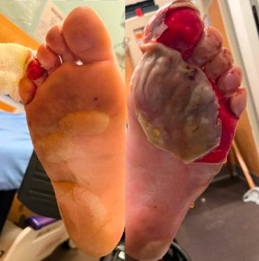
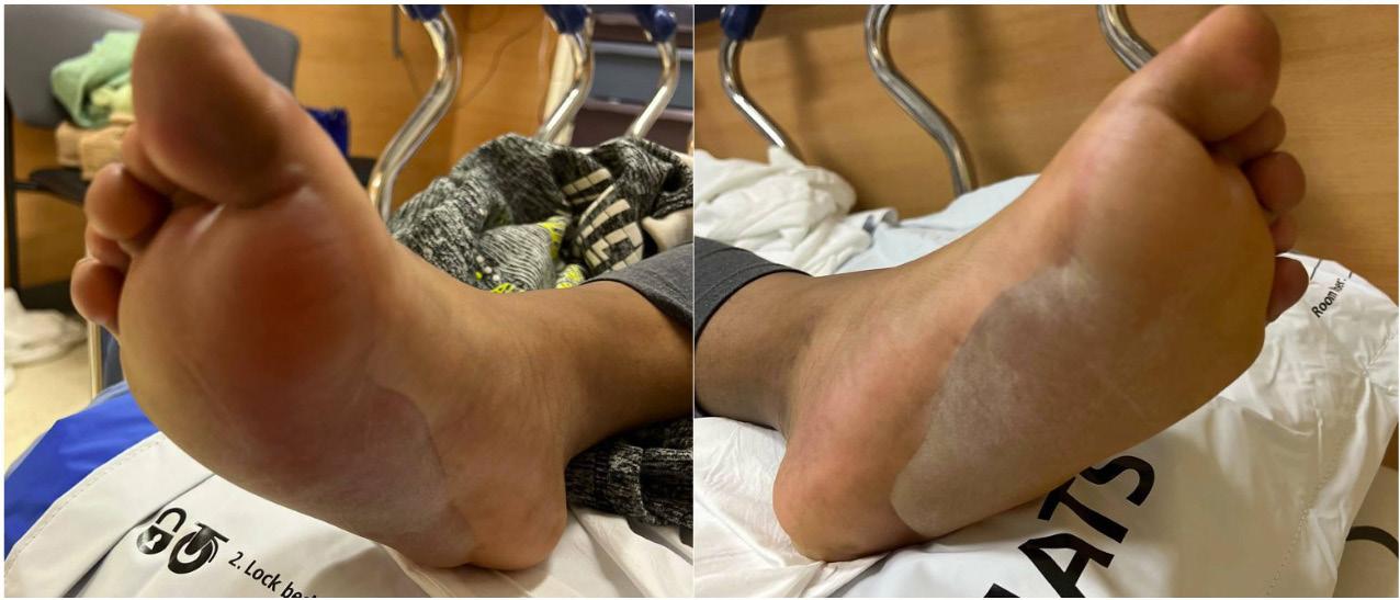
All three of our patients presented with bilateral plantar surface frostbite injuries that occurred during the winter months. Patient 1 was a 17-year-old male with 4% total body surface area (TBSA) partial-thickness burns that occurred approximately 24 hours prior (Image 1). Patient 2 was an 18-year-old female with 5% TBSA partial-thickness burns
Clinical
Medicine 272 Volume 6, no. 4: November 2022
Practice and Cases in Emergency
Hennepin County Medical Center, Department of Emergency Medicine, Minneapolis, Minnesota University of Minnesota Medical School, Minneapolis, Minnesota
* †
from a more recent exposure (Image 2). Patient 3 was a homeless male with 4% TBSA partial-thickness burns who frequently walked outside barefoot. The patients reported their pain from 7/10 to 10/10 prior to pain medication, with only modest improvement after receiving oral analgesia. The specific oral analgesia given and subsequent pain scores are delineated in the Table.
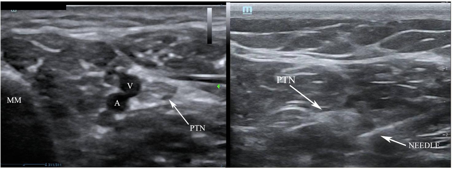
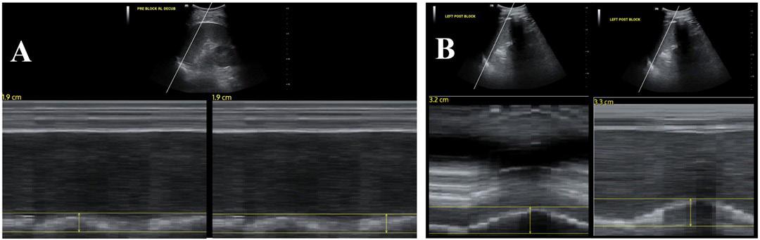
Ultrasound-guided PTN blocks were performed using the same technique for each patient (Image 3), blocking the left PTN for patient 1 and bilateral PTNs for patients 2 and 3. Using a linear transducer, the PTN was identified adjacent to the medial malleolus, keeping the posterior tibial artery and vein in view. The needle was advanced using an in-plane approach and normal saline was introduced to confirm location at the nerve and away from surrounding structures. Following conformation, five milliliters of local anesthetic was injected and observed to surround the nerve. After 10 minutes the patients were re-evaluated, and each patient reported significant improvement in their pain, all scoring 0/10 on the pain scale. The table highlights the type of local anesthetic and post-block pain scores for each patient. Successful blister debridement was performed in the ED for patients 1 and 3. Patient 2 had her burn care performed on the burn surgery service floor immediately following PTN block in the ED. There were no reports of local or systemic toxicity from the anesthetic.
Patient 1 was discharged home after debridement in the ED, while patients 2 and 3 were admitted for further
CPC-EM Capsule
What do we already know about this clinical entity?
Frostbite injuries are debilitating, painful and require rapid identification and appropriate pain control and wound care to optimize outcomes.
What makes this presentation of disease reportable?
Data is limited on the use of the posterior tibial nerve (PTN) block to effectively provide pain control for frostbite injuries of the plantar surfaces.
What is the major learning point?
The PTN block can be used to provide pain control for patients with plantar surface frostbite in the emergency department.
How might this improve emergency medicine practice?
Emergency physicians can utilize the PTN block for plantar surface frostbites to improve pain control and wound management.
management. None of the patients underwent repeat PTN blocks for wound care while outpatient or inpatient. Patient 1 was seen in the burn clinic three days after his ED visit. In that visit, the tissue of his left foot was pink and moist, and the team planned to debride a serous blister on his right foot. He was given oral (PO) oxycodone but was unable to tolerate the procedure due to pain. Patient 2 had multiple repeat debridements on the burn surgery service where IV opiate medications were utilized for pain management. She described these debriedments as “unbearable” and “so painful.” On hospital day #5, the patient required procedural sedation with
Volume 6, no. 4: November 2022 273 Clinical Practice and Cases in Emergency Medicine
Burl et al.
Ultrasound-guided Posterior Tibial Nerve Block for Frostbite of the Plantar Surfaces
Image 1. Bilateral partial-thickness frostbite on patient 1 with a broken serous blister on the left.
Image 2. Bilateral partial-thickness frostbite present on patient 2. Large serous blisters are present.
Table. Patient pain scores after oral analgesia and after posterior tibial nerve block.
Patient % TBSA burn PO medications Pain score after PO medications Local anesthetic used for PTN block Pain score after PTN block
1 4%
Acetaminophen, ibuprofen, oxycodone, olanzapine 6/10 – 10/10 5 mL 0.25% bupivacaine 0/10
2 5% Acetaminophen, ibuprofen, oxycodone 7/10 5 mL 0.5% ropivacaine 0/10
3 4% Ibuprofen 10/10 5 mL 0.25% bupivacaine 0/10
TBSA, total body surface area; PO, oral medication; PTN, posterior tibial nerve; mL, milliliter.
propofol for proper dressing changes and additional debridement. Once she was able to tolerate dressing changes without intravenous pain medications, she was deemed safe for discharge. Patient 3 required a 10-day hospital admission for co-management of frostbite pain and alcohol withdrawal.
DISCUSSION
This case series demonstrates the severity of frostbite pain and the challenge it creates to receiving appropriate wound care. Ultrasound-guided PTN blocks bypass this challenge and achieve effective analgesia in the ED, allowing for optimal wound debridement as shown in these patients’ experiences.
There are multiple strengths and limitations in our approach. The most clinically relevant benefit of the PTN block is the analgesia it provides. Pain is a significant barrier to treating frostbite injuries, originating from the burn itself as well as from rewarming and debridement.2,9 All patients reported pain scores of 7/10 to 10/10 with PO medications only. Following the PTN block, patients reported drastic improvement in their pain levels and tolerated debridement without additional medications. This outcome aligns with prior studies that have shown pain control success rates of 95-100% when using the PTN block for foot surgeries.7,8 Later attempts at repeat debridement were either unsuccessful or performed under procedural sedation for patient one and patient two, highlighting the superiority of the PTN block analgesia.
The addition of ultrasound guidance further improved the PTN block. Peripheral nerve blocks performed with ultrasound provide better pain control, require fewer additional pain medications, and have fewer complications as compared to landmark guidance.4,12,13 A study by Kakhi et al revealed shorter time to onset and longer duration of analgesia with ultrasound guidance.13 Shah et al demonstrated the superior accuracy of ultrasound for targeting the posterior tibial nerve and avoiding surrounding structures.14 This accuracy translates into increased block success and fewer incidents of intravascular injection and systemic toxicity.4,13,14 Each of our patients reported significant pain relief that was achieved in less than 10 minutes, and no patient experienced adverse effects.
Our experience using the PTN block for these patients demonstrates the accessibility and relevance of this block for future patient encounters. The PTN block is a well described and thoroughly examined nerve block that is accessible to clinicians of different training levels and experience.7,8,15 In the series we report, it was performed by first- and third-year emergency medicine residents under the guidance of an ultrasound-trained faculty member. Emergency departments in colder climates frequently see frostbite as a chief complaint, and this case series can help guide the use of PTN blocks for pain control in such patients.
Despite the accessibility of the PTN block, clinician comfort with nerve blocks and the availability of ultrasoundtrained faculty could limit its use. The ultrasound-guided block is also more time intensive when compared to PO or IV pain medications.4 Lastly, our case series only focuses on the experiences of three patients with second-degree frostbite of bilateral plantar surfaces. Patients with other levels and locations of frostbite injury may have different outcomes.
CONCLUSION
This case series demonstrates that the ultrasound-guided PTN block provides superior pain relief, has low risk of systemic toxicity, allows for necessary wound care, and is accessible to clinicians of varying training levels. Ultrasoundguided PTN blocks have the potential to play a major role within a multi-modal pain control strategy for frostbite management. This role is unequivocally applicable within the
Clinical Practice and Cases in Emergency Medicine 274 Volume 6, no. 4: November 2022
Burl et al.
Ultrasound-guided Posterior Tibial Nerve Block for Frostbite of the Plantar Surfaces
Image 3. Posterior tibial nerve (PTN) block. Left demonstrates anatomical landmarks of the medial malleolus (MM), vein (V) and artery (A). Right, the needle with anesthetic spread around the PTN coming from a posterior approach.
Burl et al.
Ultrasound-guided Posterior Tibial Nerve Block for Frostbite of the Plantar Surfaces
emergency department. The ease and accessibility of the block also lends its use to other clinical contexts, including outpatient wound clinics and inpatient burn units. These potential applications of the PTN block warrant further research on its use for frostbite management in different clinical scenarios.
The authors attest that their institution does not require Institutional Review Board approval. Patient consent has been obtained and filed for the publication of this case report. Documentation on file.
4. Redborg KE, Antonakakis JG, Beach ML, et al. Ultrasound improves the success rate of a tibial nerve block at the ankle. Reg Anesth Pain Med. 2009;34(3):256-60.
5. Moake MM, Presley BC, Barnes RM. Ultrasound-guided posterior tibial nerve block for plantar foot foreign body removal. Pediatr Emer Care. 2020;36(5):262-5.
6. Binder ZW, Murphy KM, Constantine E. Ultrasound guided posterior tibial nerve block to facilitate foreign body removal in a school-aged child. Glob Pediatr Health. 2020;7:1-5.
7. Wassef MR. Posterior tibial nerve block. A new approach using the bony landmark of the sustentaculum tali. Anaesthesia 1991;46(10):841-4.
Address for Correspondence: Andrea Dreyfuss, MD, Hennepin County Medical Center, Department of Emergency Medicine, 701 Park Ave, Minneapolis, MN 55404. Email: adreyfus@umn.edu.
Conflicts of Interest: By the CPC-EM article submission agreement, all authors are required to disclose all affiliations, funding sources and financial or management relationships that could be perceived as potential sources of bias. The authors disclosed none.
Copyright: © 2022 Burl et al. This is an open access article distributed in accordance with the terms of the Creative Commons Attribution (CC BY 4.0) License. See: http://creativecommons.org/ licenses/by/4.0/
REFERENCES
1. Zaeem K, Janjua S, Arain I. Stellate ganglion block for the immediate treatment of frostbite of upper limb. Pak Armed Forces Med J. 2008;58(1):41-4.
2. Taylor MS. Lumbar epidural sympathectomy for frostbite injuries of the feet. Mil Med. 1999:164(8):566-7.
3. Clattenburg E, Herring A, Hahn C, et al. ED ultrasound-guided posterior tibial nerve blocks for calcaneal fracture analgesia. Am J Emerg Med. 2016;34(6):1183.e1-3.
8. Rudkin GE, Rudkin AK, Dracopoulos GC. Ankle block success rate: a prospective analysis of 1,000 patients. Can J Anesth. 2005;52(2):209-10.
9. Zafren K and Danzi DF. Frostbite and nonfreezing cold injuries. In: Walls R, Hockberger, R, Gausche-Hill M, eds. Rosen’s Emergency Medicine: Concepts and Clinical Practice. Vol 2, 9th Ed. Elsevier; 2018:1735-1742.e1.
10. Granger CJ and Cohen-Levy WB. Anatomy, bony pelvis and lower limb, posterior tibial nerve. 2022. Available at: https://www.ncbi.nlm. nih.gov/books/NBK546623/. Accessed February 12, 2022.
11. Pasquier M, Ruffinen GZ, Brugger H, et al. Pre-hospital wrist block for digital frostbite injuries. High Alt Med Biol. 2012;13(1):65-6.
12. Lewis SR, Price A, Walker KJ, et al. Ultrasound guidance for upper and lower limb blocks. Cochrane Database Syst Rev. 2015;2015(9).
13. Kakhki B, Ebrahimi M, Foroughian M, et al. The success rate of posterior tibial nerve block in the ankle with and without ultrasound guidance: A clinical trial study for pain management in emergency departments. J Emerg Pract Trauma. 2021;7(1):12-6.
14. Shah A, Morris S, Alexander B, et al. Landmark technique vs ultrasound-guided approach for posterior tibial nerve block in cadaver models. Indian J Orthop. 2020;54(1):38-42.
15. Benimeli-Fenolla M, Montiel-Company JM, Almerich-Silla JM, et al. Tibial nerve block: supramalleolar or retromalleolar approach? A randomized trial in 110 participants. Int J Environ Res Public Health 2020;17(11):3860.
Volume 6, no. 4: November 2022 275 Clinical Practice
Emergency Medicine
and Cases in
Diaphragmatic Excursion as a Novel Objective Measure of Serratus Anterior Plane Block Efficacy: A Case Series
Brian Lentz, MD, MS*
Sigmund Kharasch, MD†
Andrew J. Goldsmith MD, MBA‡
Joseph Brown, MD§
Nicole M. Duggan, MD‡
Arun Nagdev, MD*
* † ‡ §
Highland Hospital-Alameda Health System, Department of Emergency Medicine, Oakland, California
Massachusetts General Hospital, Department of Emergency Medicine, Boston, Massachusetts
Brigham and Women’s Hospital, Department of Emergency Medicine, Boston, Massachusetts
University of Colorado, Department of Emergency Medicine, Aurora, Colorado
Section Editors: Christopher Sampson, MD
Submission history: Submitted May 19, 2022; Revision received July 12, 2022; Accepted July 21, 2022 Electronically published November 4, 2022
Full text available through open access at http://escholarship.org/uc/uciem_cpcem
DOI: 10.5811/cpcem.2022.7.57457
Introduction: Pain scales are often used in peripheral nerve block studies but are problematic due to their subjective nature. Ultrasound-measured diaphragmatic excursion is an easily learned technique that could provide a much-needed objective measure of pain control over time with serial measurements.
Case Series: We describe three cases where diaphragmatic excursion was used as an objective measure of decreased pain and improved respiratory function after serratus anterior plane block in emergency department patients with anterior or lateral rib fractures.
Conclusion: Diaphragmatic excursion may be an ideal alternative to pain scores to evaluate serratus anterior plane block efficacy. More data will be needed to determine whether this technique can be applied to other ultrasound-guided nerve blocks. [Clin Pract Cases Emerg Med. 2022;6(4):276–279.]
Keywords: ultrasound; diaphragmatic excursion; nerve block; pain scale; case report.
INTRODUCTION
Peripheral nerve blocks are an important component of multimodal analgesia for thoracic pain.1 The serratus anterior plane block (SAPB) involves placing local anesthetic into the fascial plane between the serratus anterior and latissimus dorsi muscles, or between serratus anterior and an underlying rib, using real-time ultrasound guidance.2 Serratus anterior plane block can be used in a variety of settings including after surgery involving the chest wall or in the emergency care setting for anterior and/or lateral rib fractures. Rib fractures occur in 9-10% of all trauma patients. Controlling chest wall pain in these patients is crucial as inadequately treated pain is associated with increased risk of chest wall splinting leading to hypoventilation, atelectasis and, eventually, pneumonia.3,4 Diaphragmatic excursion has been
proposed as a surrogate objective method of respiratory status in this patient subpopulation.5
Point-of-care ultrasound (POCUS) evaluation of diaphragmatic excursion can provide the quantification of diaphragmatic function over time through serial evaluation, and it has high sensitivity and specificity compared to chest radiography.5-7 Further, diaphragmatic dysfunction can be caused by a variety of interventions and diseases including mechanical ventilation, cardiac and abdominal surgery, phrenic nerve injury, neuromuscular disorders, lung hyperinflation, and multi-organ dysfunction in critical illness.8 Several reviews describing POCUS uses and techniques to evaluate the diaphragm have been published.9-11
Specifically, diaphragmatic excursion may provide a quantification method of respiratory status after intervention. As
Clinical Practice and
in Emergency Medicine 276 Volume 6, no. 4: November 2022
Cases
Case Series
visual pain scores are a subjective perception of an individual’s pain, objectively comparing this measurement has been a challenge in peripheral nerve block studies.12 The excursion of the dome of the diaphragm can be used to guide clinicians on the degree of respiratory compromise in specific pulmonary pathologies.13,14 As splinting secondary to rib fractures is a known phenomenon, diaphragmatic ultrasound may provide an objective outcome of successful nerve blocks for rib fractures. We propose diaphragmatic excursion as a new objective outcome of block efficacy in thoracic nerve blocks. Here we describe three cases where diaphragmatic excursion was used as an objective measure of SAPB efficacy in emergency department (ED) patients with anterior or lateral rib fractures.
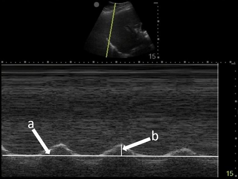
To measure diaphragmatic excursion the patient is first placed in a supine position. A curvilinear probe is placed in the midaxillary line and oriented cephalad to optimally visualize the inferior aspect of the lungs, diaphragm, and upper abdomen (ie, spleen or liver). Diaphragmatic excursion was quantified on M-mode imaging, with the M-mode cursor directed through the diaphragm. The amplitude of diaphragmatic excursion was measured from the baseline to the point of maximal excursion on the vertical axis (Image 1).
Nagdev et al provide a complete description of SAPB.15 In summary, the patient is placed in a lateral decubitus or supine position. A high-frequency linear transducer is placed in the midaxillary line at the level of the nipple to locate the serratus anterior muscle. A blunt-tip block needle is then used to inject anesthetic into the plane between the serratus anterior muscle and latissimus dorsi muscles. To perform the block, the needle is visualized using an in-plane approach until the tip is located just above the serratus anterior muscle. Once the correct position is confirmed, a large volume of anesthetic is injected into the fascial layer. As with all blocks, intralipid should be
CPC-EM Capsule
What do we already know about this clinical entity? Pain scales are often used to measure the efficacy of peripheral nerve blocks but are problematic due to their subjective nature.
What makes this presentation of disease reportable? This is the first description of diaphragmatic excursion as an objective measure of appropriate pain control in acute rib fractures in the emergency care setting.
What is the major learning point? Diaphragmatic excursion is a promising novel tool to quantify improved pain and respiratory function after serratus anterior nerve block and possibly other blocks.
How might this improve emergency medicine practice?
This technique could be utilized in future studies of nerve block efficacy as well as clinically to guide appropriate pain control.
readily available in the event of local-anesthetic systemic toxicity syndrome.
CASE SERIES
Case 1
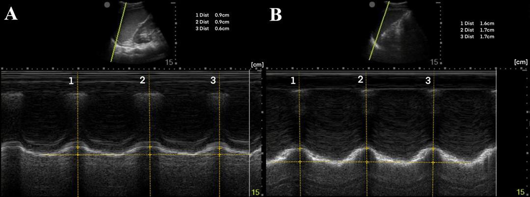
A 44-year-old man presented after a motorcycle collision and was found to have right-sided fractures of ribs 2-5 and 12 on computed tomography (CT). The patient continued to report severe pain after 100 micrograms (mcg) of intravenous (IV) fentanyl. M-mode of the right diaphragm was performed prior to SAPB and showed 5 millimeters (mm) of diaphragmatic excursion and a respiratory rate of 24 breaths per minute (BPM) (Image 2A). A right ultrasound-guided SAPB was performed with 20 milliliters (mL) of 1% ropivacaine. On re-evaluation approximately 60 minutes later, M-mode of the right diaphragm showed a respiratory rate of 16 BPM and 14 mm of diaphragmatic excursion (increase of 64%) (Image 2B). Increase in diaphragmatic excursion was calculated as the change in diaphragmatic excursion (14 mm minus 5 mm) divided by the post-block diaphragmatic excursion (14 mm).
Case 2
A 35-year-old man presented after an assault and was found to have a left lateral sixth rib fracture on CT. The patient received 1000 milligrams (mg) of IV acetaminophen but continued to
Volume 6, no. 4: November 2022 277 Clinical Practice
Cases in Emergency Medicine
and
Lentz et al. Diaphragmatic Excursion as a Novel Objective Measure of SAPB
Image 1. Diaphragmatic excursion is calculated by first determining a baseline (line a) and then measuring the distance of maximal vertical excursion (distance b).
report severe pain. M-mode of the left diaphragm was performed prior to SAPB and showed 8 mm of diaphragmatic excursion and a respiratory rate of 20 BPM (Image 3A). A left ultrasoundguided SAPB was performed with 20 mL of 1% ropivacaine. On re-evaluation approximately 60 minutes later, M-mode of the left diaphragm showed 17 mm of diaphragmatic excursion (increase of 53%) and a respiratory rate of 16 BPM (Image 3B).
Case 3
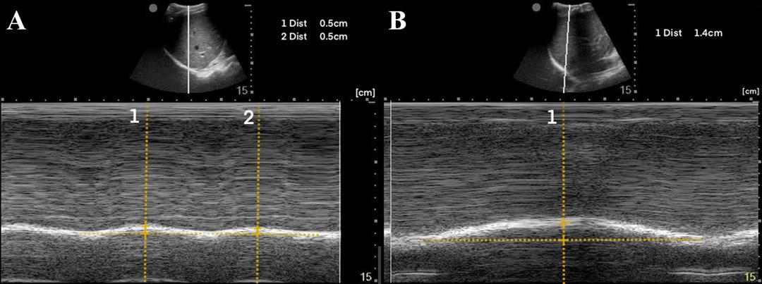
A 50-year-old man presented with left-sided chest wall pain after a fall four days prior when intoxicated and was found to have fractures of ribs 6-9 on CT. The patient initially rated his pain 10/10 and was given 100 mcg of IV fentanyl. Pain continued to be 10/10 and a SAPB was performed for pain control. A left ultrasound-guided SAPB was performed with 20 mL of 0.5% bupivacaine combined with 10 mg of dexamethasone. The patient’s pain 60 minutes after the block was 2/10. A diaphragmatic POCUS was performed both before and 60 minutes after the SAPB block. The initial respiratory rate was 20 BPM with 19 mm of diaphragmatic excursion (Image 4A). After 60 minutes from the SAPB, the patient’s respiratory rate was 14 BPM with a diaphragmatic excursion of 32 mm (increase of 41%) (Image 4B).
All blocks were performed by fellowship-trained ultrasound faculty. The vital signs of all the patients were stable; specifically, no patient had hypotension or hypoxia
prior to the block being performed. No additional pain medications were given prior to the second diaphragmatic excursion exam.
DISCUSSION
To our knowledge this is the first description of using diaphragmatic excursion as a measurement of appropriate pain control in acute rib fractures. In our cases, after SAPB the respiratory rate decreased while M-mode measured diaphragmatic excursion increased. This initial data may suggest a new objective measure of improved pain control for acute rib fractures as well as decreased respiratory splinting. Ultrasound-measured diaphragmatic excursion may provide a much-needed objective measure of respiratory improvement in acute rib fracture, since subjective pain scores are typically the only measures used during clinical care.12
Diaphragmatic excursion may also be used as a signal for overall pain. Pain can be evident in some patient populations by an increase in respiratory rate and more shallow breaths for all acute painful conditions. Diaphragmatic excursion may directly demonstrate improved pain control and respiratory function following ultrasoundguided peripheral nerve blocks, and not just for thoracic trauma as shown in these three cases. Although subjective pain scales have a role in measuring block effectiveness, they are an indirect measure of respiratory function and problematic in cases where there are distracting injuries or altered mental status. Diaphragmatic excursion, however, can be used in all patients and could define that ultrasoundguided nerve blocks both relieve pain in the form of decreased splinting as well as directly improve pulmonary function. This technique has the potential to be valuable both in future studies of nerve block effectiveness and to help guide adequate pain control in clinical situations where patients are not able to communicate subjective pain using visual pain scores.
The POCUS-measured diaphragmatic excursion is both easily learned and non-invasive, making it an ideal objective measure. Although this case series demonstrates diaphragmatic excursion as a promising novel tool to quantify pain and respiratory splinting, it has several
Clinical Practice and Cases in Emergency Medicine 278 Volume 6, no. 4: November 2022
et al.
Diaphragmatic Excursion as a Novel Objective Measure of SAPB
Lentz
Image 2. (A) Pre-block demonstrating right-sided diaphragmatic excursion of 5 millimeters (mm) (average of two excursions). (B) Post-block demonstrating right-sided diaphragmatic excursion of 14 mm (increase of 64%).
Image 3. (A) Pre-block demonstrating left-sided diaphragmatic excursion of 8 mm (average of three excursions). (B) Post-block demonstrating left-sided diaphragmatic excursion of 17 mm (increase of 53%).
Image 4. (A) Pre-block demonstrating left-sided diaphragmatic excursion of 19 mm. (B) Post-block demonstrating left-sided diaphragmatic excursion of 32 mm (increase of 41%).
limitations. Data regarding interrater reliability was not available as diaphragmatic measurements were obtained by a single clinician pre- and post-block. Although a standardized technique was used to measure diaphragmatic excursion, a possibility of measurement error exists from differences in probe placement pre- and post-block. Lastly, subjective pain scores pre- and post-block were only available for the third case, and spirometry data was not collected for comparison. Point-of-care ultrasound measured diaphragmatic excursion is both easily learned and non-invasive, making it an ideal objective measure.
CONCLUSION
Serratus anterior plane bock performed for acute rib fractures reduced pain in all three patients. It also increased the diaphragmatic excursion and decreased the respiratory rate in all three cases. Diaphragmatic excursion may be an alternative to visual pain scores to evaluate SAPB efficacy. More data will be needed to determine whether this same relationship can extend to other ultrasound-guided nerve blocks.
The authors attest that their institution does not require Institution al Review Board approval. Patient has been obtained and filed for the publication of this case report. Documentation on file.
Address for Correspondence: Brian Lentz, MD, MS. Highland Hospital-Alameda Health System, Department of Emergency Medicine, 1411 E. 31st Street QIC 22123, Oakland, CA 94602. Email: blentz@alamedahealthsystem.org.
Conflicts of Interest: By the CPC-EM article submission agreement, all authors are required to disclose all affiliations, funding sources and financial or management relationships that could be perceived as potential sources of bias. The authors disclosed none.
Copyright: © 2022 Lentz et al. This is an open access article distributed in accordance with the terms of the Creative Commons Attribution (CC BY 4.0) License. See: http://creativecommons.org/ licenses/by/4.0/
REFERENCES
1. Witt CE and Bulger EM. Comprehensive approach to the management of the patient with multiple rib fractures: a review and introduction of a bundled rib fracture management protocol. Trauma
Surg Acute Care Open. 2017;2(1):e000064.
2. Blanco R, Parras T, McDonnell JG, et al. Serratus plane block: a novel ultrasound-guided thoracic wall nerve block. Anaesthesia. 2013;68(11):1107-13.
3. Chapman BC, Herbert B, Rodil M, et al. RibScore: A novel radiographic score based on fracture pattern that predicts pneumonia, respiratory failure, and tracheostomy. J Trauma Acute Care Surg. 2016;80(1):95-101.
4. Ziegler DW and Agarwal NN. The morbidity and mortality of rib fractures. J Trauma. 1994;37(6):975-9.
5. Vetrugno L, Guadagnin GM, Barbariol F, et al. Ultrasound Imaging for Diaphragm Dysfunction: a Narrative Literature Review. J of Cardiothoracic and Vasc Anesth. 2019;33(9):2525-36.
6. Chetta A, Rehman AK, Moxham J, et al. Chest radiography cannot predict diaphragm function. Respir Med. 2005;99(1):39-44.
7. Kerrey BT, Geis GL, Quinn AM, et al. A prospective comparison of diaphragmatic ultrasound and chest radiography to determine endotracheal tube position in a pediatric emergency department. Pediatrics. 2009;123(6):e1039-44.
8. McCool FD and Tzelepis GE. Dysfunction of the diaphragm. N Engl J Med. 2012;366(10):932-42.
9. Sferrazza Papa GF, Pellegrino GM, Di Marco F, et al. A Review of the Ultrasound Assessment of Diaphragmatic Function in Clinical Practice. Respiration. 2016;91(5):403-11.
10. Tuinman PR, Jonkman AH, Dres M, et al. Respiratory muscle ultrasonography: methodology, basic and advanced principles and clinical applications in ICU and ED patients-a narrative review. Intensive Care Med. 2020;46(4):594-605.
11. Weber MD, Lim JKB, Glau C, et al. A narrative review of diaphragmatic ultrasound in pediatric critical care. Pediatr Pulmonol. 2021;56(8):2471-83.
12. Hjermstad MJ, Fayers PM, Haugen DF, et al. Studies comparing Numerical Rating Scales, Verbal Rating Scales, and Visual Analogue Scales for sssessment of pain intensity in adults: a systematic literature review. J Pain Symptom Manage. 2011;41(6):1073-93.
13. Cammarota G, Sguazzotti I, Zanoni M, et al. Diaphragmatic Ultrasound Assessment in Subjects With Acute Hypercapnic Respiratory Failure Admitted to the Emergency Department. Respir Care. 2019;64(12):1469-77.
14. Zambon M, Greco M, Bocchino S, et al. Assessment of diaphragmatic dysfunction in the critically ill patient with ultrasound: a systematic review. Intensive Care Med. 2017;43(1):29-38.
15. Nagdev A, Mantuani D, Durant E, et al. The ultrasound-guided serratus anterior plane block. ACEP Now. 2017;36(3):12-3.
Volume 6, no. 4: November 2022 279 Clinical
Medicine
Practice and Cases in Emergency
Lentz et al. Diaphragmatic Excursion as a Novel Objective Measure of SAPB
Point-of-Care Ultrasound Diagnosis of Tetralogy of Fallot Causing Cyanosis: A Case Report
Aravind Addepalli, MD*
Marco Guillen, MD†
Andrea Dreyfuss, MD, MPH‡
Daniel Mantuani, MD*
Arun Nagdev, MD*
David A. Martin, MD*
Section Editors: Anna McFarlin, MD
* † ‡
Highland Hospital-Alameda Health System, Department of Emergency Medicine, Oakland, California EsSalud Cusco: Hospital Nacional Adolfo Guevara Velasco, Department of Emergency Medicine, Cusco, Peru Hennepin County Medical Center, Department of Emergency Medicine, Minneapolis, Minnesota
Submission history: Submitted February 09, 2022 ; Revision received February 21, 2022; Accepted August 22, 2022
Electronically published October 22, 2022
Full text available through open access at http://escholarship.org/uc/uciem_cpcem
DOI: 10.5811/cpcem.2022.8.56297
Introduction: Tetralogy of Fallot (TOF) is a congenital heart defect with characteristic features leading to unique physical exam and ultrasound findings. In settings where there is limited prenatal screening, TOF can present with cyanosis at any time from the neonatal period to adulthood depending on the degree of obstruction of the right ventricular outflow tract.1
Case Report: This case describes a pediatric patient who presented with undifferentiated dyspnea and cyanosis, for whom point-of-care ultrasound (POCUS) supported the diagnosis of TOF. We highlight the important role POCUS can play in a setting with limited access to formal echocardiography or consultative pediatric cardiology services.
Conclusion: This report highlights the utility of POCUS as an inflection point in the diagnostic and management pathway of this patient, which is particularly important when working in a limitedresource or rural setting. [Clin Pract Cases Emerg Med. 2022;6(4):280–283.]
Keywords: point-of-care ultrasound; tetralogy of Fallot; emergency department.
INTRODUCTION
Congenital heart disease worldwide is reported to have a prevalence of 8-12 per 1,000 live births.1 Tetralogy of Fallot (TOF) is the most common cyanotic heart condition in children surviving untreated beyond the neonatal age and accounts for 7-10% of congenital heart disease globally, with a birth prevalence of 3-5 per 10,000 live births.2 Tetralogy of Fallot is a congenital cardiac malformation characterized by a ventricular septal defect; obstruction of the right ventricular outflow tract (RVOT); override of the ventricular septum by the aortic root; and right ventricular hypertrophy (RVH). In the United States, the diagnosis of TOF is generally made by ultrasound performed in the perinatal period; however, in settings where there is limited access to perinatal screening and formal echocardiography, clinicians rely on history, exam, and other diagnostics tests such as electrocardiogram.
Point-of-care ultrasound (POCUS) can be used to rapidly identify potential causes of dyspnea and shock in the undifferentiated patient.3,4 Cardiac POCUS, also referred to as focused cardiac ultrasound, is generally performed by noncardiologists to ascertain only the essential information needed in critical scenarios to assist in time-sensitive decisionmaking.5 Cardiac POCUS can, therefore, be used in combination with historical and physical exam findings to recognize conditions such as TOF, particularly in settings where formal echocardiography is unavailable or impractical.6 Additionally, POCUS in rural areas and community hospitals has been shown to enable early diagnosis and timely initiation of medical interventions while avoiding unnecessary patient transport and associated expenditures.7
Tetralogy of Fallot clinically presents as cyanosis ranging from the neonatal period into adulthood depending on the
Clinical Practice and Cases in Emergency Medicine 280 Volume 6, no. 4: November 2022
Case Report
degree of RVOT obstruction.2 Considering the variable presentation and broad differential, given clinical suspicion the emergency physician can use POCUS to evaluate for the anatomical abnormalities associated with TOF. In this report, we detail the case of a five-month-old presenting with respiratory distress and cyanosis whose care and ultimate diagnosis of TOF was driven by the POCUS findings identified during the initial resuscitation.
CASE REPORT
A five-month-old male born via Cesarean section for twin pregnancy with no complications presented to a hospital in Cusco, Peru, with sudden onset respiratory distress. Vital signs at presentation were as follows: heart rate 150 beats per minute; blood pressure 80/47 millimeters of mercury; respiratory rate 50 breaths per minute; oxygen saturation (SpO2) 70% on room air; and temperature 36° Celsius. He was found to be in poor general condition, cyanotic and lethargic with dry mucous membranes. He was tachypneic with subcostal retractions and faint expiratory wheezing. Cardiac auscultation revealed no audible murmurs.
Given these findings, the patient was suspected to be in acute respiratory failure due to severe bronchiolitis. As the patient presented early at the onset of the coronavirus disease 2019 (COVID-19) pandemic, COVID-19 remained high on the differential, given little was known regarding its effects on infants. Oxygen was administered via non-rebreather (NRB) mask with broad spectrum antibiotics and intravenous fluids (IVF) due to concern for sepsis. His labs were notable for white blood cell count of 10.6 x 10³ per millimeter (mm3) (reference range: 5x10³ - 10x103 mm3) with a lymphocytic predominance; hemoglobin 20.4 grams (g) per deciliter (dL) (14-17 g/dL); creatinine of 0.4 milligrams (mg)/dL (0-0.5 mg/ dL); and a lactate of 12.5 millimoles per liter (mmol/L) (0-4 mmol/L). A COVID-19 polymerase chain reaction test was negative. Chest radiograph was interpreted by the emergency physician as technically limited due to rotation with diffuse prominent interstitial markings concerning for viral pneumonia (Image 1).
The patient became more responsive after initial resuscitation with oxygen via NRB and IVF. However, he remained hypoxic with SpO2 of 76% and ongoing signs of respiratory distress. He was started on high-flow nasal cannula at nine liters per minute (L/min) with a fraction of inspired oxygen of 70% resulting in minimal improvement in overall respiratory status. Given his persistent hypoxia and cyanosis, cardiac POCUS was performed, which initially was notable for RVH raising suspicion for RVOT obstruction suggestive of a congenital heart disease.
Upon closer inspection, parasternal long-axis view revealed a ventricular septal defect with an overriding aorta and RVH concerning for TOF (Image 2). Parasternal shortaxis cardiac view redemonstrated RVH with interventricular septal flattening indicative of right ventricular pressure
CPC-EM Capsule
What do we already know about this clinical entity?
Tetralogy of Fallot is a congenital condition with characteristic structural anomalies affecting blood flow through the heart that globally has a birth prevalence of 3-5 per 10,000 live births.
What makes this presentation of disease reportable?
Tetralogy of Fallot is the most common cyanotic heart condition in children surviving untreated beyond the neonatal age that can be surgically corrected once appropriately identified.
What is the major learning point?
Point-of-care ultrasound has an important role in identifying Tetralogy of Fallot as a cause of dyspnea in settings with limited prenatal screening and access to comprehensive echocardiography.
How might this improve emergency medicine practice?
With appropriate use, point-of-care ultrasound to diagnose Tetralogy of Fallot would allow crucial changes in resuscitative efforts and referral for definitive surgical treatment.
overload from RVOT obstruction (Image 3). Pulmonary ultrasound revealed a normal A-line pattern, and POCUS
Volume 6, no. 4: November 2022 281 Clinical Practice and Cases in Emergency Medicine
Addepalli et al. Point-of-care Ultrasound Diagnosis of Tetralogy of Fallot Causing Cyanosis: A Case Report
Image 1. Portable anteroposterior chest radiograph showing no focal infiltrate and limited due to patient rotation.
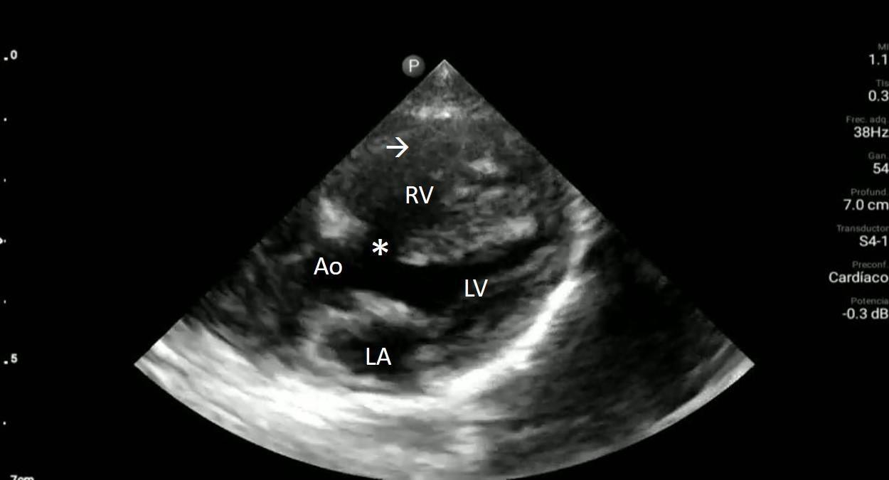
Image 2. Parasternal long-axis view of the heart highlighting the findings of a ventricular septal defect (*) with an overriding aorta (Ao) and right ventricular hypertrophy (arrow). LA indicates left atrium; LV, left ventricle; RV, right ventricle.
assessment of the inferior vena cava showed a noncollapsing vessel.
Treatment was shifted from a focus on sepsis to TOF management. Additional IVF hydration was limited, and the patient was started on propanolol for rate control. Due to the patient’s age, the decision was made to not administer prostaglandins as he was likely not ductus dependent. He was found to improve after these interventions. Cardiology was consulted, and despite not identifying physical exam findings concerning for TOF such as skin discoloration signifying cyanosis, or a systolic thrill and ejection murmur at the left sternal border, once they were shown the POCUS images cardiology consult initiated procedures for referral to the National Institute of Cardiovascular Diseases in Lima for definitive surgical treatment. Four days later a comprehensive echocardiogram confirmed the diagnosis of TOF.
DISCUSSION
The diagnosis of congenital heart disease in the ED can be challenging since many of the presenting symptoms can mimic other more common pathologies, as was the case in our patient whom the clinician initially suspected bronchiolitis. Thus, it is important to maintain a broad differential diagnosis, particularly when working in settings where congenital heart disease is more likely to go undiagnosed due to limited perinatal evaluation for the condition.
POCUS has been demonstrated to improve diagnostic accuracy when evaluating patients in shock.4,8 Consensus guidelines advocate for the use of cardiac POCUS by trained clinicians to help narrow the differential diagnosis and guide clinical management for both adult and pediatric patients presenting with cardiopulmonary instability.5 Previous case reports have similarly demonstrated the role POCUS can play in expediting the diagnosis and treatment course of children suspected of having congenital heart disease.9,10 Although cardiac POCUS is insufficient to rule it out, our case
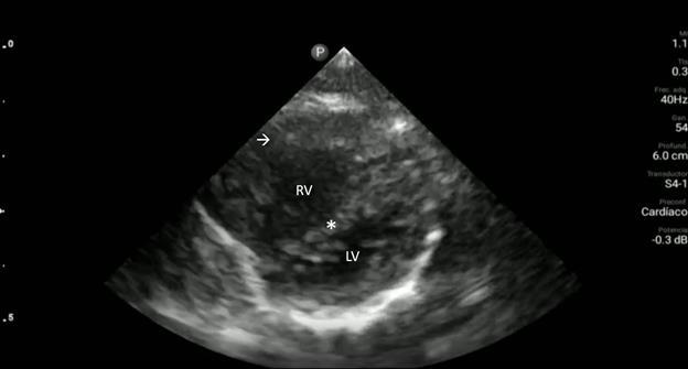
Image 3. Parasternal short-axis view of the heart notable for interventricular septal flattening (*) due to right ventricular pressure overload in the setting of right ventricular hypertrophy (arrow). LV, left ventricle; RV, right ventricle.
demonstrates how POCUS can be used to evaluate for gross abnormalities such as discrepancies in normal anatomy, chamber size or function, which can trigger the need for more comprehensive cardiac evaluation and formal echocardiography. Identifying the specific anatomical abnormalities associated with congenital heart disease can pose a diagnostic challenge, particularly for clinicians who are not experienced with POCUS. Tele-ultrasound may potentially aid in cases where there is diagnostic uncertainty or limited clinical experience using POCUS. A study recently published by Médecins Sans Frontières showed that images obtained by ultrasound-naïve clinicians could be reviewed by pediatric cardiologists using a telemedicine platform to help facilitate the diagnosis and guide management of patients suspected of having congenital heart disease.11 Although clinicians in this study were primarily trained in image acquisition and not interpretation, this study shows promising results for the use of cardiac POCUS to help facilitate the timely diagnosis of congenital heart disease in limited-resource settings where access to pediatric consultative services or comprehensive echocardiography would be otherwise impractical. More broadly speaking, our case also underscores the important role that POCUS has in the ED management of infants presenting with undifferentiated dyspnea or shock in both high- and low-resource settings.
CONCLUSION
POCUS is an invaluable tool for evaluating the undifferentiated patient presenting with dyspnea, particularly when working in a limited-resource setting where there is reduced access to timely laboratory and diagnostic studies. This case highlights the important role of POCUS, particularly in a setting with limited prenatal screening, for congenital cardiac abnormalities and limited access to comprehensive echocardiography or pediatric cardiology consultation services. Here, the POCUS findings of a ventricular septal
Clinical Practice and Cases in Emergency Medicine 282 Volume 6, no. 4: November 2022
Point-of-care Ultrasound Diagnosis of Tetralogy of Fallot Causing Cyanosis: A Case Report
Addepalli et al.
Addepalli et al.
Point-of-care Ultrasound Diagnosis of Tetralogy of Fallot Causing Cyanosis: A Case Report
deficit with an overriding aorta and RVH in the hands of a trained clinician suggested TOF and changed the course of the initial resuscitative efforts leading to ultimate referral to a tertiary care center for definitive surgical treatment.
Video 1. Parasternal long-axis view of the heart. Ventricular septal defect (*); overriding aorta (Ao); right ventricular hypertrophy (arrow); left atrium (LA); left ventricle (LV) right ventricle (RV).
Video 2. Interventricular septal flattening due to right ventricular pressure overload (*); right ventricular hypertrophy (arrow); left ventricle (LV) right ventricle (RV).
The authors attest that their institution does not require Institutional Review Board approval. Patient consent has been obtained and filed for the publication of this case report. Documentation on file.
Address for Correspondence: Aravind Addepalli, MD, Highland Hospital-Alameda Health System. Department of Emergency Medicine, 1411 East 31st Street, Oakland, CA 94602. Email: aaddepalli@alamedahealthsystem.org.
Conflicts of Interest: By the CPC-EM article submission agreement, all authors are required to disclose all affiliations, funding sources and financial or management relationships that could be perceived as potential sources of bias. The authors disclosed none.
Copyright: © 2022 Addepalli et al. This is an open access article distributed in accordance with the terms of the Creative Commons Attribution (CC BY 4.0) License. See: http://creativecommons.org/ licenses/by/4.0/
REFERENCES
1. van der Linde D, Konings EE, Slager MA, et al. Birth prevalence of congenital heart disease worldwide: a systematic review and
meta-analysis. J Am Coll Cardiol . 2011;58(21):2241-7.
2. Diaz-Frias J, Guillaume M. Tetralogy of Fallot. [Updated 2022 Jan 18]. In: StatPearls [Internet]. Treasure Island (FL):StatPearls Publishing. 2022. Available from: https://www.ncbi.nlm.nih.gov/ books/NBK513288/.
3. Mantuani D, Frazee BW, Fahimi J, et al. Point-of-Care Multi-Organ Ultrasound Improves Diagnostic Accuracy in Adults Presenting to the Emergency Department with Acute Dyspnea . West J Emerg Med . 2016;17(1):46-53.
4. Keikha M, Salehi-Marzijarani M, Soldoozi NR, et al. Diagnostic Accuracy of Rapid Ultrasound in Shock (RUSH) Exam; A Systematic Review and Meta-analysis. Bull Emerg Trauma 2018;6(4):271-8.
5. Via G, Hussain A, Wells M, et al. International evidence-based recommendations for focused cardiac ultrasound. J Am Soc Echocardiogr . 2014;27(7):683.e1-683.e33.
6. Spencer KT, Kimura BJ, Korcarz CE, et al. Focused cardiac ultrasound: recommendations from the American Society of Echocardiography. J Am Soc Echocardiogr . 2013;26(6):567-81.
7. Sekar P, and Vilvanathan V. Telecardiology: effective means of delivering cardiac care to rural children. Asian Cardiovasc Thorac Ann . 2007;15(4):320-3.
8. Jones AE, Tayal VS, Sullivan DM, et al. Randomized, controlled trial of immediate versus delayed goal-directed ultrasound to identify the cause of nontraumatic hypotension in emergency department patients. Crit Care Med . 2004;32(8):1703-8.
9. Rosenfield D, Fischer JW, Kwan CW, et al. Point-of-care ultrasound to identify congenital heart disease in the pediatric emergency department. Pediatr Emerg Care . 2018;34(3):223-5.
10. Kehrl T, Dagen CT, Becker BA. Focused cardiac ultrasound diagnosis of cor triatriatum sinistrum in pediatric cardiac arrest. West J Emerg Med . 2015;16(5):753-5.
11. Muhame RM, Dragulescu A, Nadimpalli A, et al. Cardiac point of care ultrasound in resource limited settings to manage children with congenital and acquired heart disease. Cardiol Young 2021;31(10):1651-7.
Volume 6, no. 4: November 2022 283
Medicine
Clinical Practice and Cases in Emergency
Case Report
Recurrent Infantile Hypertrophic Pyloric Stenosis in the Emergency Department: A Case Report
Adeola A. Kosoko, MD Diego Craik Tobar, MD
Section Editor: Melanie Heniff, MD, JD
Submission history: Submitted April 15, 2022; Revision received May 02, 2022; Accepted August 22, 2022 Electronically published October 27, 2022
Full text available through open access at http://escholarship.org/uc/uciem_cpcem DOI: 10.5811/cpcem.2022.8.57140
Introduction: Infantile hypertrophic pyloric stenosis (IHPS) is a common cause of infant vomiting. Emergency department (ED) diagnosis is usually made by pyloric ultrasound and treated by pyloromyotomy.
Case Report: An eight-week-old boy with a history of IHPS about six weeks status post pyloromyotomy presented to the ED with vomiting and failure to thrive, and a critically narrowed pylorus was identified by ultrasound. An upper gastrointestinal series confirmed recurrent pyloric stenosis, necessitating another pyloromyotomy.
Conclusion: Prolonged vomiting after pyloromyotomy should be concerning for recurrent IHPS. Upper gastrointestinal series should augment ultrasound to diagnose recurrent IHPS and determine whether a second pyloromyotomy is warranted. [Clin Pract Cases Emerg Med. 2022;6(4):284–287.]
Keywords: pyloric stenosis; vomiting; surgical failure; case report.
INTRODUCTION
Infantile hypertrophic pyloric stenosis (IHPS) is a welldescribed pediatric surgical emergency often presenting with “projectile” emesis of undigested milk and a hungry baby, not uncommonly with dehydration or even failure to thrive. Ultrasound has generally become more readily available in the emergency setting and has become the mainstay of IHPS diagnosis. With early ultrasound evaluation, it is less common to identify classic clinical findings such as a peristaltic wave, a palpable “olive-shaped” mass, or hypochloremic, hypokalemic metabolic acidosis. Early presentation for vomiting in a young infant, physical exam, and resultant imaging facilitate early surgical intervention and best outcomes. Although an upper gastrointestinal (GI) series may also be used to diagnose IHPS (sensitivity 100% and specificity 100%),1 ultrasound has become the diagnostic imaging of choice in the emergency setting (sensitivity 97-100% and specificity 99-100% with an experienced sonographer).2
Infantile hypertrophic plyroic stenosis is managed by increasing the size of the pyloric canal such that foods may appropriately pass. Procedures are usually curative, and recent
literature suggests that recurrence of pyloric stenosis is exceedingly rare. Although some children receive balloon dilation to alleviate the pathologic obstruction of IHPS, the current mainstay of care is laparoscopic or even open pyloromyotomy.3 With appropriate preoperative preparation (rehydration, electrolyte repletion), pyloromyotomy is considered a relatively minor but effective and curative surgical procedure with excellent survival rates and minimal adverse outcomes.4 Vomiting is the most common complication in the first few days after the procedure but usually resolves with ad libitum feeds.4 We report the case of a young infant who had appropriately previously received surgical intervention for IHPS presenting to the emergency department (ED) with classic signs and symptoms of IHPS, diagnosed with a recurrence of IHPS by findings from an abdominal ultrasound and an upper GI series, necessitating a second pyloromyotomy.
CASE REPORT
An eight-week-old male patient presented to the ED with his parents for a three-week history of frequent episodes of
Clinical
Medicine 284 Volume 6, no. 4: November 2022
Practice and Cases in Emergency
The University of Texas Health Sciences Center at Houston, McGovern Medical School, Houston, Texas
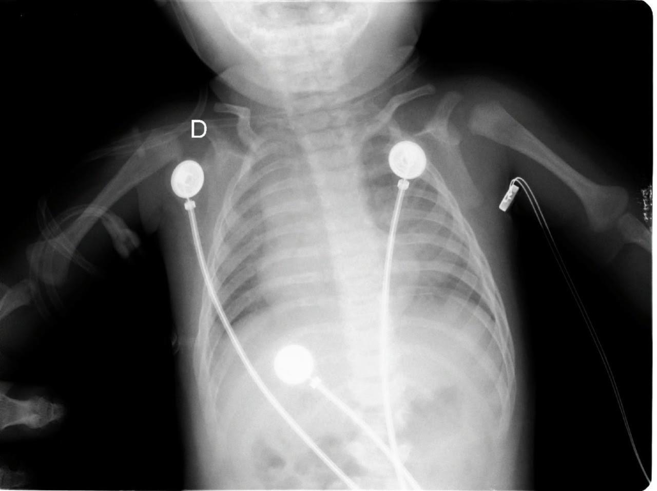
post-prandial non-bloody and non-bilious emesis (Figure 1). The parents were concerned for repeated episodes of emesis for three weeks that were initially intermittent but had become consistent with feeds. Despite being advised by a pediatrician to try a soy-based formula by a telemedicine visit for presumed lactose intolerance a few days prior to this ED visit, the patient’s symptoms persisted. The parents also noticed that the patient was taking less volume when feeding. The mother described the most recent episodes of emesis as “projectile,” large volume, and comprised of what looked like his formula. In addition, he constantly seemed hungry. The parents again took him to his pediatrician where he was noted to have lost weight and seemed lethargic (Figure 2). The pediatrician recommended another ED visit.
On further interview with parents and on chart review, the patient was born at term by vaginal delivery with a birth weight of 3,460 grams and a normal physical exam. Six weeks prior, at 17 days of age, he was taken to the ED after two days of persistent, forceful, non-bloody and non-bilious emesis, which occurred after every feed. During that initial ED visit, the patient’s basic metabolic profile was normal, including a potassium of 4.6 milliequivalents per liter (mEq/L) (reference range: 3.7-5.2 mEq/L) and chloride of 103 mEq/L (reference range: 96-106 mEq/L), and an abdominal ultrasound was concerning for an abnormally large pylorus measuring 2 centimeters (cm) in length and 4 millimeters (mm) in width with no passage of food contents. The patient urgently underwent a successful laparoscopic pyloromyotomy without complication. He was discharged home with his parents with
CPC-EM Capsule
What do we already know about this clinical entity?
Hypertrophic pyloric stenosis is a well-described pathology causing vomiting leading to failure to thrive in infants. Diagnosis is usually made by ultrasound.
What makes this presentation of disease reportable?
This presentation describes a case of a child who had already received a pyloromyotomy with another clinically classic case of pyloric stenosis.
What is the major learning point?
When pyloric stenosis is diagnosed very early in life, despite appropriate surgical intervention, it could possibly recur. An upper gastrointestinal series may help with diagnosis.
How might this improve emergency medicine practice?
The clinical signs and symptoms of pyloric stenosis are generally consistent and require appropriate evaluation even if a child has already what is typically definitive intervention.
an uneventful three-week period including normal oral intake and adequate weight gain for his age.
On evaluation, the patient was lethargic with a weak cry. The child appeared small for stated age with dry mucous membranes and a flat fontanelle. Vital signs on presentation included a temperature of 98.5° Fahrenheit, heart rate 130 beats per minute, respiratory rate 35 breaths per minute, oxygen saturation 98%, and blood pressure 81/48 millimeters of mercury (mm Hg). The patient had clear bilateral tympanic membranes and a normal posterior oropharynx. His chest was clear to auscultation, cardiac exam was grossly normal, and his abdomen was soft, without dilatation, and without palpable masses. He had a three second capillary refill (normal: <2 seconds) and a normal genital exam for his age. The child produced a weak cry when nurses started a peripheral intravenous line.
A basic metabolic panel was significant for a hypokalemic (2.6 mEq/L), hypochloremic (76 mEq/L) metabolic alkalosis. A venous blood gas showed a pH greater than 7.70 (reference range: 7.31-7.41), partial pressure of carbon dioxide 35 mm Hg (reference range: 41-51 mm Hg), and partial pressure of oxygen 53 mm Hg (reference range: 30-40 mm Hg). We were unable to
Volume 6, no. 4: November 2022 285 Clinical Practice
Cases in Emergency Medicine
and
Kosoko et al. Recurrent Infantile Hypertrophic Pyloric Stenosis in the Emergency Department: A Case Report
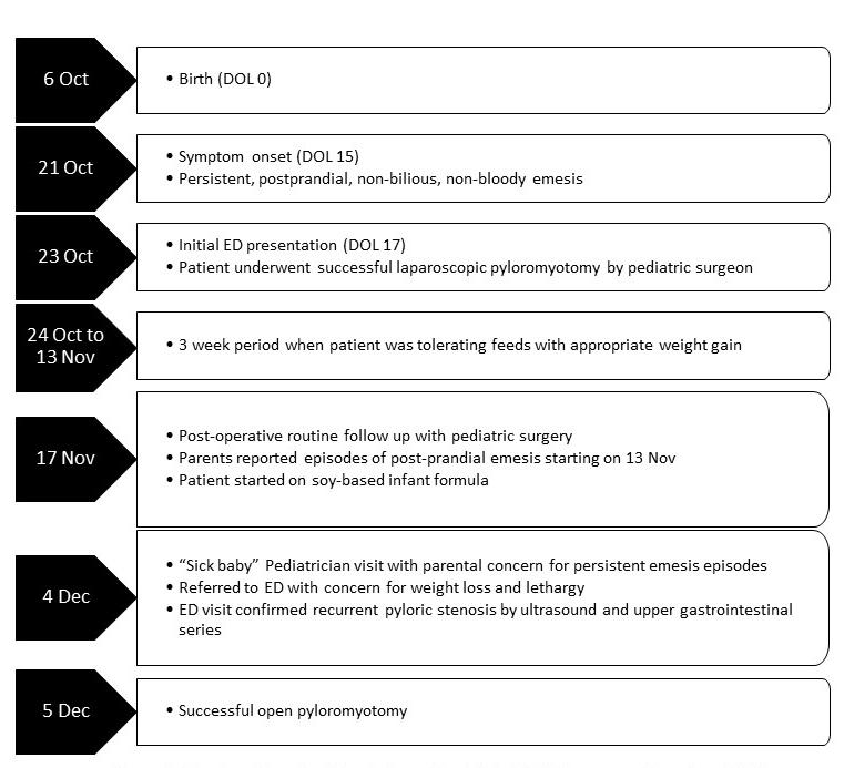
Figure 1. Timeline of events for an infant with recurrent hypertrophic pyloric stenosis. Day of Life (DOL), Emergency department (ED).
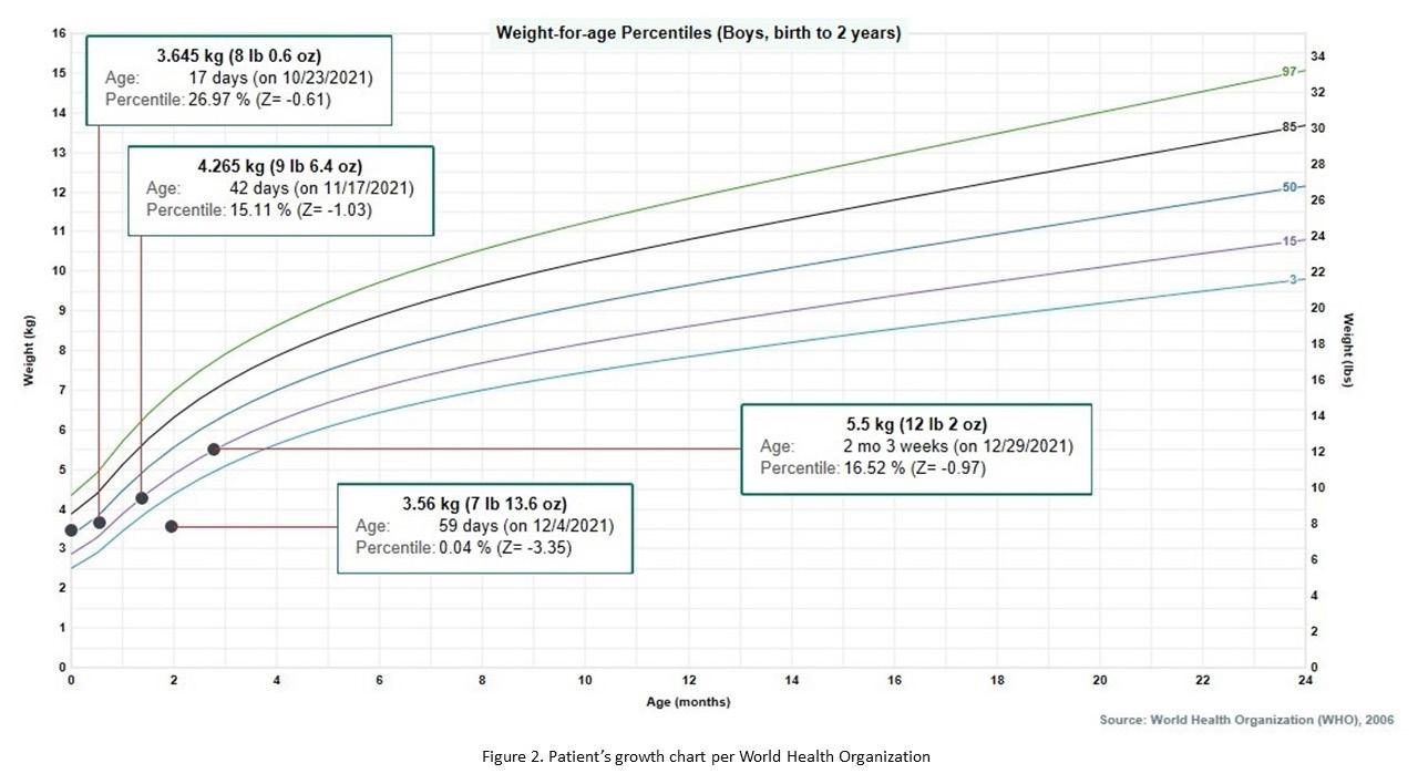
Figure 2. Patient’s weight-for-age growth per the World Health Organization standard statistical distribution describing boys from birth to two years old. Kilogram (kg), pound (lb), ounce (oz), month (mo).
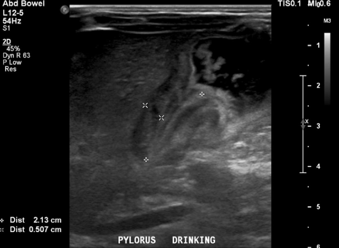
calculate bicarbonate and base excess due to a pH more than 7.70. The patient underwent an abdominal ultrasound in the ED, which suggested IHPS with an enlarged pyloric channel measuring 2.1 cm and a thickened muscle measuring 0.5 cm, again with minimal passage of fluids through the pylorus (Image 1). The surgical team was consulted for the abnormal laboratory and ultrasound findings, but due to the possibility of postoperative edema or residual abnormal external pylorus measurements, the consultant recommended further imaging to conclusively determine pyloric stenosis. The child was admitted for fluid resuscitation and electrolyte replacement. An upper GI series performed the same day confirmed the diagnosis of IHPS when there was lack of contrast passing from the stomach to the duodenum. The patient received a solution of intravenous 5%, dextrose, half normal saline, and 40 mEq potassium chloride at maintenance until electrolytes and intravascular volume
were optimized the next morning. The patient then underwent an uncomplicated open pyloromyotomy by a pediatric surgeon. The patient recovered well from the operative procedure and tolerated ad libitum oral feeds both in the hospital and at discharge. Three months following his open pyloromyotomy, the primary care clinic reported that the child had been tolerating feeds well and gaining weight adequately without any subsequent recurrence or complications of IHPS.
DISCUSSION
The underlying cause of a recurrent hypertrophic pyloric stenosis after a successful pyloromyotomy remains unclear. Emesis after feeds is a common and is the most frequent complaint post pyloromyotomy. It is likely due to pyloric edema, pylorospasm, or reflux.5 Usually postoperative emesis will resolve spontaneously but if it persists for more than five days, incomplete pyloromyotomy should be suspected. Recurrent pyloric stenosis is a rare entity, with an incidence of less than 2% of all children who undergo successful surgery.6 To avoid confusion between recurrence and an incomplete surgical procedure, certain criteria need to be met. True recurrent pyloric stenosis includes complete resolution of symptoms for three or more weeks before recurrence of emesis, weight gain, and evidence of re-stenosis on sonographic or operative confirmation.7 The patient described in this case returned to baseline oral intake, had positive weight gain, and no episodes of emesis for three weeks before symptoms reappeared.
Ankermann et al (2002) described evaluation for recurrent pyloric stenosis. If postprandial projectile emesis occurs after successful operation and a symptom-free interval, the absence of fluid passage through the pyloric channel should be demonstrated radiologically with a swallow of water-soluble contrast before reoperation is considered.8 In cases of recurrent pyloric stenosis, sonographic images have to be interpreted carefully and may not be the image of choice given that the thickness and length of the pyloric channel may remain enlarged after pyloromyotomy and may not return to normal until approximately six weeks9 and up to five months5 after the procedure.8,10
Given that the etiology of recurrent pyloric stenosis after a successful pyloromyotomy is unknown, one consideration is that “recurrence” is actually a result of the progression of the original disease and the initial surgery was probably performed at an early stage after which the pylorus continued undergoing an active process of hypertrophy.
CONCLUSION
Infantile hypertrophic pyloric stenosis is easily identified on abdominal ultrasound as a cause of refractory vomiting. Although rare, despite surgical pyloromyotomy, a minority of infants may develop a recurrence of IHPS, presenting clinically similarly to the sentinel case. Diagnosis can be made
Clinical Practice and Cases in Emergency Medicine 286 Volume 6, no. 4: November 2022
al.
Recurrent Infantile Hypertrophic Pyloric Stenosis in the Emergency Department: A Case Report Kosoko
et
Image. Ultrasound of pylorus with child drinking demonstrating hypertrophy (length 2.13cm and width 0.5cm) with minimum passage of fluids through the pylorus.
et al. Recurrent Infantile Hypertrophic Pyloric Stenosis in the Emergency Department: A Case Report
by repeated ultrasound but should be augmented by upper gastrointestinal series to avoid false positive diagnoses, which might actually be the result of postoperative edema. Management for recurrent cases is a second pyloromyotomy, which usually has good and definitive outcomes.
The authors attest that their institution does not require Institution al Review Board approval. Patient consent has been obtained and filed for the publication of this case report. Documentation on file.
Address for Correspondence: Adeola Kosoko, MD, University of Texas Health Sciences Center at Houston, Department of Emergency Medicine, 6431 Fannin JJL 260G, Houston, TX 77030. Email:adeola.a.kosoko@uth.tmc.edu.
Conflicts of Interest: By the CPC-EM article submission agreement, all authors are required to disclose all affiliations, funding sources and financial or management relationships that could be perceived as potential sources of bias. The authors disclosed none.
Copyright: © 2022 Kosoko et al. This is an open access article distributed in accordance with the terms of the Creative Commons Attribution (CC BY 4.0) License. See: http://creativecommons.org/ licenses/by/4.0/
REFERENCES
1. Olson AD, Hernandez R, Hirschl RB. The role of ultrasonography in the diagnosis of pyloric stenosis: a decision analysis. J Pediatr Surg 1998;33(5):676-81.
2. Hernanz-Schulman M. Pyloric stenosis: role of imaging. Pediatr Radiol. 2009;39 Suppl 2:S134-9.
3. Aspelund G and Langer JC. Current management of hypertrophic pyloric stenosis. Semin Pediatr Surg. 2007;16(1):27-33.
4. Ein SH, Masiakos PT, Ein A. The ins and outs of pyloromyotomy: What we have learned in 35 years. Pediatr Surg Int. 2014;30(5):467-80.
5. Al-Ansari A and Altokhais T. Recurrent pyloric stenosis. Pediatr Int. 2016;58(7):619-21.
6. Van Heurn LW, Vos P, Sie G. Recurrent vomiting after successful pyloromyotomy. Pediatr Surg Int. 15(5-6):385-6.
7. Capiello CD and Strauch E. A rare case of recurrent hypertrophic pyloric stenosis. Pediatr Surg Case Rep. 2014;2:519-21.
8. Ankermann T, Engler S, Partsch CJ. Repyloromyotomy for recurrent infantile hypertrophic pyloric stenosis after successful first pylotomyotomy. J Pediatr Surg. 2002;37(11):E40.
9. Okorie NM, Dickson JA, Carver RA, et al: What happens to the pylorus after pyloromyotomy? Arch Dis Child. 63(11):1339-41.
10. Yoshizawa J, Eto T, Higashimoto Y, et al. Ultrasonographic features of normalization of the pylorus after pyloromyotomy for hypertrophic pyloric stenosis. J Pediatr Surg. 36(4):582-6.
Volume 6, no. 4: November 2022 287 Clinical
Emergency Medicine
Practice and Cases in
Kosoko
Legionnaires’ Disease Causing Severe Rhabdomyolysis and Acute Renal Failure: A Case Report
Andrew Branstetter, MD* Benjamin Wyler, MD, MPH†
Section Editor: Rachel Lindor, MD, JD
* †
Mount Sinai Morningside-West, Department of Emergency Medicine, New York, New York Icahn School of Medicine at Mount Sinai, Department of Emergency Medicine, New York, New York
Submission history: Submitted April 18, 2022; Revision received July 05, 2022; Accepted August 22, 2022 Electronically published October 24, 2022
Full text available through open access at http://escholarship.org/uc/uciem_cpcem
DOI: 10.5811/cpcem.2022.8.57155
Introduction: Legionnaires’ disease is a multisystem disease involving respiratory, gastrointestinal, and neurologic systems. This is a case of a previously healthy 44-year-old man who was diagnosed with Legionella pneumonia causing acute kidney failure and rhabdomyolysis. Case Report: The patient presented with four days of chills, shortness of breath, chest discomfort, diarrhea, and myalgias. Laboratory testing revealed hyponatremia, leukocytosis, elevated inflammatory markers, renal failure, and rhabdomyolysis. He was admitted to the intensive care unit for acute hypoxemic respiratory failure, received a course of antibiotics, and more than two weeks of intermittent hemodialysis with full recovery of renal function. The pathophysiologic mechanisms by which Legionella causes rhabdomyolysis and acute kidney failure are not fully understood, although numerous mechanisms have been proposed including direct invasion of myocytes and renal tubular cells.
Conclusion: Legionnaires’ disease is one of several infections that can cause rhabdomyolysis and kidney failure. Although rarely described in the literature, it is important for emergency physicians to be aware of this clinical entity in order to implement early diagnostic testing and empiric treatment. [Clin Pract Cases Emerg Med. 2022;6(4):288–291.]
Keywords: Legionella; rhabdomyolysis; renal failure; emergency; case report.
INTRODUCTION
We report a case of a previously healthy man who was found to have acute renal failure attributable to rhabdomyolysis in the setting of Legionella pneumonia. He quickly developed respiratory distress due to volume overload, which required treatment with high-flow nasal cannula oxygen and urgent hemodialysis. In this case report we review the patient’s initial presentation and management and highlight rare infectious causes of rhabdomyolysis of which emergency physicians should be aware.
CASE REPORT
A 44-year-old Black man with no known medical history presented to the emergency department (ED) in March 2021
with four days of chills, shortness of breath, chest discomfort, diarrhea, and myalgias. He reported decreased urine output with darkening of his urine. He denied recent travel. He had attended a social gathering the weekend before and was motivated to come to the ED by concern that he might have acquired coronavirus disease 2019 (COVID-19). He reported occasional cannabis use. He worked in construction with a prior history of asbestos abatement work and reported complying with recommended personal protective equipment.
Initial vital signs were blood pressure 150/100 millimeters of mercury, heart rate 86 beats per minute, oxygen saturation of 98% (via 2 liters per minute nasal cannula), and a respiratory rate 20 breaths per minute. Lung exam revealed mildly decreased breath sounds at the bilateral bases without
288 Volume 6,
4: November 2022
Clinical Practice and Cases in Emergency Medicine
no.
Case Report
wheezes or rales. No lower extremity edema was noted. Within a few hours of arrival, the patient subsequently became tachypneic to greater than 40 breaths per minute with increased work of breathing. Point-of-care ultrasound was performed and revealed bilateral B-lines, more prominent in the left lung.
Electrocardiogram showed normal sinus rhythm without signs of ischemia or infarction (Image 1). Chest radiograph demonstrated bilateral infiltrates suggestive of multifocal or viral pneumonia (Image 2). Serum chemistries and complete blood count measured blood urea nitrogen (BUN) 123 milligrams per deciliter (mg/dL) (reference range: 7-20 mg/ dL); creatinine (Cr)14.6 mg/dL (0.7-1.3 mg/dL); sodium 121 millimoles per liter (mmol/L) (135-145 mmol/L); anion gap 25 mmol/L (7-16 mmol/L); glucose 289 mg/dL (60-100 mg/ dL); creatine phosphokinase 36,716 units per liter (U/L) (30-200 U/L); and a white blood cell count 30.6 x 10³ leukocytes per microliter (K/uL) (4.5-11.0 K/uL) with 82% neutrophils (40.0-72.0%).
Other diagnostic tests, including lactate dehydrogenase (LDH), erythrocyte sedimentation rate, C-reactive protein (CRP) and procalcitonin measured 1,010 U/L (100-220 U/L); 114 millimeters per hour (mm/hr) (0-13 mm/hr), 476 milligrams per liter (mg/L) (<5.1 mg/L), and 32.58 nanograms per milliliter (ng/mL) (no reference range provided), respectively. Two sequential reverse transcription polymerase chain reaction severe acute respiratory syndrome coronavirus-2 tests were negative. A Foley catheter was placed with only 50 mL of dark urine output collected in two hours. Urinalysis revealed amber colored, cloudy urine, with protein greater than 500 mg/dL (reference range: negative mg/dL); ketones 150 mg/dL (negative mg/dL); large blood (reference range: negative); and 4 red blood cells (RBC) per high-power field (HPF) (0-3 RBC/HPF).
The patient was administered both high-flow nasal cannula oxygen and empiric broad spectrum intravenous (IV) vancomycin, ceftriaxone, and azithromycin, for presumed
CPC-EM Capsule
What do we already know about this clinical entity?
Legionnaires’ disease involves respiratory, gastrointestinal, and neurologic systems. Rarely, this disease can cause acute renal failure and severe rhabdomyolysis.
What makes this presentation of disease reportable?
This is a rare presentation of a common disease. Previous cases of rhabdomyolysis in Legionnaires’ disease involved older patients with multiple comorbidities.
What is the major learning point? Emergency physicians should include Legionnaires’ disease in the differential for patients with pneumonia and acute kidney dysfunction.
How might this improve emergency medicine practice?
By testing more frequently for Legionnaires’ disease and initiating antibiotics early in the patient emergency room visit, emergency physicians can improve patient outcomes.
bacterial pneumonia. The patient was then admitted to the medical intensive care unit for hypoxemic respiratory failure attributed to the combination of pneumonia and volume overload in the setting of renal failure.
Subsequent testing on hospital day one showed a positive Legionella urine antigen. He was treated with IV levofloxacin until hospital day 3 but required azithromycin due to a skin rash. He completed a two-week course of antibiotic therapy. He received intermittent hemodialysis for 16 days while in the hospital and was discharged home on hospital day 23 with improvement in renal function (BUN 22 mg/dL, Cr 1.88 mg/ dL). At follow-up nephrology visit approximately one and one-half months later, the patient had no residual symptoms and showed further improved kidney function (BUN 15 mg/ dL, Cr 1.14 mg/dL).
DISCUSSION
Legionella is a genus of Gram-negative, aerobic organisms known to cause disease in humans. Legionellosis is classically transmitted through stagnant water, such as cooling towers for air conditioning and pipes for water distribution in
Volume 6, no. 4: November 2022 289 Clinical Practice
Cases in Emergency Medicine
and
Branstetter et al. Legionnaires’ Disease Causing Severe Rhabdomyolysis and Acute Renal Failure
Image 1. Electrocardiogram revealing normal sinus rhythm, heart rate 72 beats per minute, corrected QT interval 477 milliseconds.
large buildings; thus, infection is more prevalent in urban areas than in rural areas.1,2 While this patient had a distant history of construction work, there was no known occupational link to his Legionella infection.
Legionella pneumophila, the most studied species, can cause a spectrum of human diseases that ranges from Legionnaires’ disease—a severe multisystem disease with pneumonia, gastrointestinal, musculoskeletal, and neurologic involvement—to Pontiac fever, a self-limited array of mild flu-like symptoms.1 Because Legionnaires’ disease can present with a wide array of signs and symptoms, clinicians have attempted to risk-stratify patients’ symptoms to determine the likelihood of infection being due to Legionella. Six clinical factors serve as a clinical prediction tool including elevated body temperature; absence of sputum; low serum sodium; high levels of LDH and CRP; and low platelet counts. This tool conferred a negative predictive value of 99.4%, which reliably rules out Legionella infection when fewer than two features are present.3
The Infectious Diseases Society of America and the American Thoracic Society (IDSA/ATS) guidelines recommend Legionella antigen testing for any patient with one of five risk factors: ICU admission; failure of outpatient antibiotics; active alcohol misuse; travel within the prior two weeks; or pleural effusion.4 However, a 2011 retrospective study found that as many as 41% of Legionella cases would have been missed by application of the IDSA/ATS guidelines alone, which suggests that more liberal Legionella testing may be necessary to avoid adverse clinical outcomes.5 The COVID-19 pandemic has further confounded diagnosis of atypical pneumonias such as Legionella, as many clinicians
may focus on the diagnosis of COVID-19 instead of others.6
The first case of rhabdomyolysis attributed to Legionnaires’ disease was reported in 1980.7 Many questions remain regarding the pathogenic mechanisms behind rhabdomyolysis and renal failure in Legionnaires’ disease. Proposed mechanisms include toxin generation leading to microvascular vasoconstriction and muscle ischemia, and direct bacterial invasion of myocytes, as supported by calf muscle biopsies in a 1991 study.8,9 Legionella can also cause acute kidney injury in the absence of respiratory involvement, in contrast to many other pneumonic bacteria.10 Muscle damage with ensuing kidney injury can increase mortality of Legionnaires’ disease by up to 40%, necessitating the early recognition and initiation of IV antibiotics, IV resuscitation, and possible hemodialysis.8 In addition, Legionella infection is believed to cause kidney injury by other mechanisms independent of rhabdomyolysis. A 2000 review of kidney biopsies in Legionella patients found that cytopathology ranged from tubulointerstitial nephritis (TIN) to acute tubular necrosis (ATN) and glomerulonephritis.11 For this reason, steroids may be beneficial to recovery of renal function in the setting of TIN. 8,11 Based on initial urine studies, this patient had a presumed TIN or ATN etiology due to his poor renal function.
Legionella is one of several infectious etiologies of rhabdomyolysis. A 2020 systemic review reported that among organisms causing atypical pneumonia and myositis, the majority were Legionella, with rarer causes including Mycoplasma pneumoniae, Francisella tularensis, Coxiella burnetii, and Chlamydia psittaci 12 Many viruses are also associated with rhabdomyolysis, most commonly influenza, human immunodeficiency virus, and Coxsackie viruses.8,13
To our knowledge, there has been only one previous report of rhabdomyolysis and renal failure complicating Legionella infection discussed in the emergency medicine literature; this involved a patient with several medical comorbidities and also a subacute presentation of signs and symptoms over two to three weeks.14 In contrast, the patient described in this case report developed an acute presentation, lacked comorbidities, and required urgent hemodialysis within 24 hours of ED presentation.
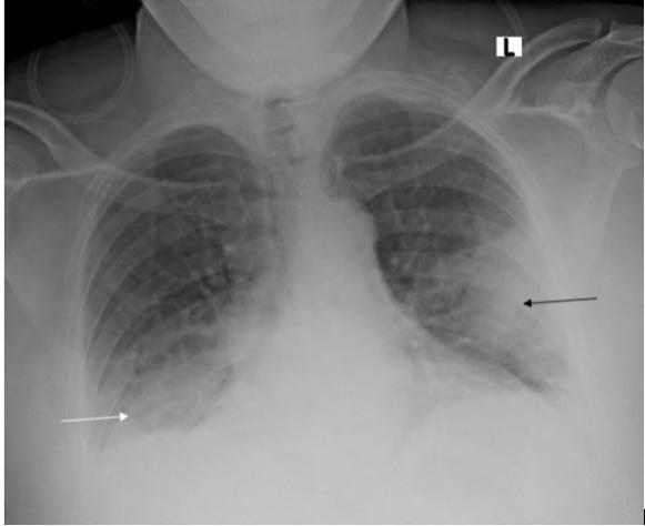
CONCLUSION
Legionnaires’ disease is a rare cause of rhabdomyolysis and renal failure. Case reports have generally described patients with multiple comorbidities unlike the patient described above. Despite a US Centers for Disease Control and Prevention surveillance of all Legionella cases, there is no published data on the incidence of these complications among these patients. Emergency physicians should be cognizant both that Legionella can develop in those without co-morbid conditions and, further, the disease can cause rhabdomyolysis and renal failure. Emergency physicians should have increased
Clinical
Medicine 290 Volume 6, no. 4: November 2022
Practice and Cases in Emergency
Legionnaires’ Disease Causing Severe Rhabdomyolysis and Acute Renal Failure
Branstetter et al.
Image 2. Chest radiograph with bilateral infiltrates involving the left mid to lower lung (black arrow) and the right lung base (white arrow), suggestive of multifocal or viral pneumonia.
Legionnaires’ Disease Causing Severe Rhabdomyolysis and Acute Renal Failure
suspicion for Legionella in patients with hyponatremia, elevated inflammatory markers, pleural effusions, and a history of alcohol misuse.
The authors attest that their institution does not require Institutional Review Board approval. Patient consent has been obtained and filed for the publication of this case report. Documentation on file.
Address for Correspondence : Benjamin Wyler, MD, Icahn School of Medicine at Mount Sinai, Department of Emergency Medicine, 1000 10th Ave, New York, NY 10019. Email: benjamin.wyler@mountsinai.org.
Conflicts of Interest: By the CPC-EM article submission agreement, all authors are required to disclose all affiliations, funding sources and financial or management relationships that could be perceived as potential sources of bias. The authors disclosed none.
Copyright: © 2022 Branstetter et al. This is an open access article distributed in accordance with the terms of the Creative Commons Attribution (CC BY 4.0) License. See: http://creativecommons.org/ licenses/by/4.0/
REFERENCES
1. Fields BS, Benson RF, Besser RE. Legionella and Legionnaires’ disease: 25 years of investigation. Clin Microbiol Rev 2002;15(3):506-26.
2. Graham FF, Hales S, White PS, et al. Review global seroprevalence of legionellosis - a systematic review and meta-analysis. Sci Rep 2020;10(1):7337.
3. Bolliger R, Neeser O, Merker M, et al. Validation of a prediction rule
for legionella pneumonia in emergency department patients. Open Forum Infect Dis. 2019;6(7):268.
4. Mandell LA, Wunderink RG, Anzueto A, et al. Infectious Diseases Society of America; American Thoracic Society. Infectious Diseases Society of America/American Thoracic Society consensus guidelines on the management of community-acquired pneumonia in adults. Clin Infect Dis. 2007;44 Suppl 2:S27-72.
5. Hollenbeck B, Dupont I, Mermel LA. How often is a work-up for legionella pursued in patients with pneumonia? A retrospective study. BMC Infect Dis. 2011;11:237.
6. Cassell K, Davis JL, Berkelman R. Legionnaires’ disease in the time of COVID-19. Pneumonia. 2021;13(1):2.
7. Posner MR, Caudill MA, Brass R, et al. Legionnaires’ disease associated with rhabdomyolysis and myoglobinuria. Arch Intern Med 1980;140(6):848-50.
8. Soni AJ and Peter A. Established association of legionella with rhabdomyolysis and renal failure: a review of the literature. Respir Med Case Rep. 2019;28:100962.
9. Warner CL, Fayad PB, Heffner RR Jr. Legionella myositis. Neurology 1991;41(5):750-2.
10. Yogarajah M and Sivasambu B. Legionnaires’ disease presenting as acute kidney injury in the absence of pneumonia. BMJ Case Rep 2015;2015:bcr2014208367.
11. Nishitarumizu K, Tokuda Y, Uehara H, et al. Tubulointerstitial nephritis associated with Legionnaires’ disease. Intern Med. 2000;39(2):150-3.
12. Simoni C, Camozzi P, Faré PB, et al. Myositis and acute kidney injury in bacterial atypical pneumonia: systematic literature review. J Infect Public Health. 2020;13(12):2020-24.
13. Singh U and Scheld WM. Infectious etiologies of rhabdomyolysis: three case reports and review. Clin Infect Dis. 1996;22(4):642-9.
14. McConkey J, Obeius M, Valentini J, et al. Legionella pneumonia presenting with rhabdomyolysis and acute renal failure: a case report. J Emer Med 2006;30(4):389-92.
Volume 6, no. 4: November
291
Medicine
2022
Clinical Practice and Cases in Emergency
Branstetter et al.
Acute Isolated Thenar Compartment Syndrome in a Patient with Evans Syndrome: A Case Report
Elisabeth
Jiang, MD* Kevin H. Kim, DO† Alan Babigian, MD‡
* † ‡
Section Editors: Christopher San Miguel, MD
University of Connecticut School of Medicine, Emergency Medicine Residency, Farmington, Connecticut University of Connecticut School of Medicine, Hand Surgery Fellowship, Farmington, Connecticut Hartford Hospital, Department of Surgery, Hartford, Connecticut
Submission history: Submitted April 20, 2022; Revision received April 25, 2022; Accepted August 22, 2022 Electronically published November 4, 2022
Full text available through open access at http://escholarship.org/uc/uciem_cpcem
DOI: 10.5811/cpcem.2022.8.57193
Introduction: Acute compartment syndrome of the hand is a rare medical emergency, most often associated with trauma or fracture.
Case Report: Here, we describe a rare case of isolated thenar compartment syndrome of the hand in the absence of major trauma as the presenting symptom of pancytopenia due to Evans syndrome, an uncommon autoimmune hematologic disorder.
Conclusion: In cases of atraumatic compartment syndrome, it is critical to evaluate for underlying coagulopathy in patients presenting to the emergency department. [Clin Pract Cases Emerg Med. 2022;6(4):292–295.]
Keywords : case report, atraumatic compartment syndrome, Evans syndrome, hand compartment syndrome.
INTRODUCTION
Hand compartment syndrome is a rare but critical condition that emergency physicians must recognize. As with any acute compartment syndrome (ACS), it is a true surgical emergency. Missed or delayed diagnosis is likely to result in devastating functional loss to the patient. While most cases of compartment syndrome are associated with bone fractures and major trauma, ACS can also occur in the absence of fracture, and atraumatic ACS is an easily missed diagnosis if the emergency physician does not maintain a high clinical index of suspicion. It is crucial for physicians to understand the anatomy and pathophysiology of hand compartment syndrome to ensure early diagnosis and prompt surgical intervention.
The hand comprises 11 compartments separated by inflexible fascial membranes; an increase in pressure within any of these compartments can result in decreased perfusion, tissue death, and ultimately loss of function of the hand. These compartments include four dorsal interossei compartments, three volar interossei compartments, the thenar compartment, the hypothenar compartment, the
Clinical Practice and Cases in Emergency Medicine
adductor compartment, and the midpalm compartment1 (Figure). The blood supply to the hand is provided by branches of the deep and superficial palmar arches, which are fed by both the radial and ulnar arteries.1
Here, we describe a rare case of isolated thenar compartment syndrome of the hand in the absence of major trauma as the presenting symptom of pancytopenia in a
Figure. Compartments of the hand.
292 Volume 6, no. 4: November 2022 Case Report
previously healthy 17-year-old high school athlete. This is the first reported case in the literature to describe ACS as the presenting clinical manifestation of Evans syndrome.
CASE REPORT
A 17-year-old, previously healthy right-hand dominant male presented to the emergency department (ED) with pain, swelling, and numbness in his left hand. Three days prior to presentation, the patient had injured his left hand while playing football. He could not recall a specific incident leading to his hand injury such as a fall or tackle; rather he noticed pain and swelling in his hand following a football game, which continued to worsen in the following days. He was seen at an outside hospital where imaging was negative for fracture or dislocation. However, he continued to experience worsening pain, paresthesia, and swelling of his thumb and hand predominately located in the thenar compartment.
On physical exam, the patient had exquisite tenderness over the left thenar eminence and thumb with significant swelling in the thenar compartment, without any overlying erythema or open wounds. The patient had severely limited range of motion in the wrist as well as the metacarpophalangeal and interphalangeal joints of the thumb secondary to severe pain. He was unable to oppose his thumb and any passive motion of the thumb caused significant pain. He also reported decreased sensation on the radial aspect of his left thumb. Sensation, capillary refill, strength, and range of motion were normal in digits two through five.
Radiographs were repeated in the ED, again revealing no bony injuries. Despite reassuring imaging, the patient’s clinical presentation, with tense swelling of the left thenar compartment, pain, decreased range of motion, and sensory deficit raised concern for compartment syndrome. Compartment pressures of the thenar compartment of the left hand were obtained using a Stryker needle with readings of 72 and 73 millimeters of mercury (mm Hg), with a diastolic blood pressure of 80 mm Hg. A diagnosis of compartment syndrome was made, and the patient was taken to the operating room for emergent fasciotomy.
Prior to being transported to the operating room (OR), the patient’s bloodwork resulted, revealing pancytopenia with profound neutropenia, thrombocytopenia, anemia, and leukopenia (Table). He denied any known history of bleeding disorder, cancer, recent viral illness, or easy bruising or bleeding. In the OR, emergent fasciotomy of the thenar compartment of the left hand was performed. The surgical team decided to release only the thenar compartment due to bleeding risk and because the patient did not show any signs of compartment syndrome in the other hand compartments. The incision was primarily closed with loosely approximated sutures; we did not use negative pressure device or leave the wound open due to bleeding and infection risk given the patient’s pancytopenic state. Two units of platelets were transfused intraoperatively.
CPC-EM Capsule
What do we already know about this clinical entity?
Compartment syndrome is true medical emergency and early recognition by the emergency physician is critical to prevent devastating functional loss.
What makes this presentation of disease reportable?
Compartment syndrome of the hand is rare and typically associated with fractures, however in this case it occurred in a young patient in the absence of broken bones.
What is the major learning point?
Emergency physicians must maintain a high clinical suspicion for compartment syndrome in order to ensure timely diagnosis and treatment with surgical fasciotomy.
How might this improve emergency medicine practice?
Compartment syndrome should always be a consideration in cases of limb swelling, and further medical workup is needed in cases of atypical compartment syndrome.
Table. Emergency department laboratory results.
Value Reference range
White blood cell count 2,200 per microliter 4,000 - 11,000 per microliter
Hemoglobin 10.7 grams/deciliter 13.0 - 17.7 grams/ deciliter
Hematocrit 31.7% 39.0 - 54.0%
Platelets Less than 2,000 per microliter 150,000 - 450,000 per microliter
Absolute neutrophil count 100 per microliter 2,000 - 7,500 per microliter
The patient was subsequently admitted to the hospital for hematology/oncology workup of pancytopenia. Peripheral blood smear showed no abnormal white blood cells, reduced platelets, few giant platelets, and normal red blood cells without spherocytes. Bone marrow aspirate revealed blasts within normal range and slightly elevated monocytes. Viral workup was negative for human immunodeficiency virus, hepatitis A, B, and C, cytomegalovirus, Epstein-Barr virus, and parvovirus B19. Folate and vitamin B12 levels were
Volume 6, no. 4: November 2022 293 Clinical Practice
Emergency Medicine
and Cases in
Jiang et al. Acute Isolated Thenar Compartment Syndrome in a Patient with Evans Syndrome
within normal limits. Partial thromboplastin time was within normal limits and double-stranded DNA was negative, both reassuring for absence of lupus. Empiric treatment with intravenous immunoglobulin and dexamethasone was initiated for concern of underlying autoimmune process. A bone marrow biopsy was performed, which revealed hypercellular marrow with megakaryocytes and granulocytic left shift. Ultimately, a diagnosis of Evans syndrome was made given the clinical presentation of concomitant autoimmune hemolytic anemia (AIHA) and immune thrombocytopenia (ITP). In this case the patient also presented with autoimmune neutropenia, a less common manifestation of Evans syndrome.2
The patient was discharged from the hospital on postoperative day five, with discharge labs revealing absolute neutrophil count of 810 units per liter (uL), platelets of 115,000/uL, hemoglobin 10.5 grams per deciliter, hematocrit 31.1%, and white blood cell count of 3,000/uL. He was seen for follow-up in the senior author’s office on postoperative day 14. At that time his incision was well healed; however, there was some residual stiffness of metacarpophalangeal and interphalangeal joints, with continued numbness of the radial aspect of the thumb. The patient was discharged on a four-day course of dexamethasone. Upon follow-up with the hematology/oncology service, blood work did reveal persistent pancytopenia, and he was scheduled to begin rituximab infusions to manage his autoimmune pancytopenia.
In this case, the diagnosis of isolated thenar compartment syndrome was likely a blessing in disguise; the patient was instructed to refrain from contact sports due to his high risk of life-threatening bleeding, and he was given appropriate follow-up for his autoimmune pancytopenia.
DISCUSSION
We describe a case of acute thenar compartment syndrome in a previously healthy 17-year-old male who was subsequently diagnosed with Evans syndrome, a rare autoimmune cytopenia syndrome.
Acute compartment syndrome results from increased pressure in any of the body compartments that are contained within an inflexible fascial membrane, causing impairment of local circulation. The incidence of ACS is estimated to be 7.3 per 100,000 in males and 0.7 per 100,000 in females.3 The leg and forearm are the most frequently affected sites; however, it can also involve the arm, hand, foot, and buttock. Incidence is highest in young men and most often occurs after traumatic injuries; 75% of cases of compartment syndrome are associated with bone fractures.3
Compartment syndrome may occur in the setting of traumatic injuries not involving fractures. These include burns, crush injuries, constrictive bandages, splints or casts, penetrating trauma, high pressure injection injuries, vascular injuries, and animal bites and stings. There are also cases of non-traumatic compartment syndrome, which may be seen in the setting of bleeding disorders, anticoagulation use,
nephrotic syndrome, toxic envenomation, massive fluid resuscitation, revascularization procedures, and muscle infection.4 As demonstrated by Hope et al, cases of compartment syndrome not associated with acute fracture often go undiagnosed and there is a risk of delayed treatment with more devastating sequela and functional loss.5 Specifically, Hope et al reported that patients without the presence of fracture were more likely to have muscle necrosis at the time of fasciotomy than patients with ACS in the setting of acute fracture.
Generally, ACS is a clinical diagnosis, and it is often diagnosed based on history and physical examination. However, intracompartmental pressure is sometimes measured to confirm the diagnosis in uncertain situations as these measurements offer the only objective means of diagnosis and may be the only tool to make a diagnosis of ACS in the case of obtunded patients or unusual presentations.1 Diagnosis of ACS is often made by using delta pressure, the difference between diastolic blood pressure and compartment pressure, with a delta less than 30 mm Hg concerning for ACS.6 Other recommendations for emergent fasciotomy are based on an absolute compartment pressures greater than 30 or greater than 45 mm Hg.7
When faced with a swollen hand, the decision to obtain compartment pressures is based on several factors including a detailed history, physical exam, and overall clinical suspicion for ACS. Findings historically associated with ACS (pain, paresthesia, pallor, paralysis, and pulselessness) are often late signs of vascular compromise; so the decision to proceed with compartment pressure measurement and ultimately fasciotomy must often be made in the absence of these clues. Due to the risk of permanent functional loss of the hands, any clinical suspicion for hand compartment syndrome should potentially warrant compartment pressure measurement and surgical consultation. There has been some investigation into the role of laboratory markers as an aid to diagnose compartment syndrome in the ED setting. In a 2020 retrospective study by Weingart et al, several markers were identified that showed correlation with compartment syndrome including creatinine kinase, bicarbonate, ionized calcium, lactic acid, creatinine, blood urea nitrogen, and potassium. This study demonstrated a sensitivity of only 2.9%, indicating that serum markers are not useful in helping to clinically rule out a diagnosis of ACS, limiting their utility in the ED setting.14
Evans syndrome, first described in 1951, is defined as the concomitant or sequential occurrence of immune thrombocytopenia and autoimmune hemolytic anemia.8 It is a rare syndrome, accounting for only 7% of AIHA cases and 2% of ITP cases.2 Evans syndrome may exist as a primary pathology; however, in up to 50% of cases it is secondary to another underlying pathology such as hematologic malignancy, systemic lupus erythematosus, viral infection, or primary immunodeficiency. The first line therapy for treatment of ES is corticosteroids, while IVIG use is typically reserved
Clinical
Medicine 294 Volume 6, no. 4: November 2022
Practice and Cases in Emergency
Acute Isolated Thenar Compartment Syndrome in a Patient with Evans Syndrome Jiang et al.
et al. Acute Isolated Thenar Compartment Syndrome in a Patient with Evans Syndrome
for patients with platelet count less than 20,000. Rituximab and mycophenolate mofetil have also been used in treatment of ES, and there are case reports citing hematopoietic stem cell transplantation and splenectomy.9
CONCLUSION
This report illustrates a rare case of thenar compartment syndrome in a patient with a rare hematologic condition. As demonstrated in previous studies, cases of compartment syndrome not associated with high energy injuries often go undiagnosed, which often results in delayed treatment with more devastating sequela and functional loss.5 Emergency physicians should have a high clinical suspicion for acute compartment syndrome, and if there is no obvious explanation for the patient’s symptoms, a prompt hematological workup is warranted to investigate other predisposing factors for bleeding such as thrombocytopenia, hematologic malignancy, or other coagulopathy.
The authors attest that their institution does not require Institution al Review Board approval. Patient has been obtained and filed for the publication of this case report. Documentation on file.
REFERENCES
1. Reichman EF. Compartment syndrome of the hand: a little thought about diagnosis. Case Rep Emerg Med. 2016;2016;2907067.
2. Audia S, Grienay N, Mounier M, et al. Evans’ syndrome: from diagnosis to treatment. J Clin Med. 2020;9(12):3851.
3. Via AG, Oliva F, Spoliti M, et al. Acute compartment syndrome. Muscles Ligaments Tendons J. 2015;5(1):18-22.
4. Cepkova, J, Ungermann L, Ehler E. Acute compartment syndrome. Acta Medica. 2020;63(3):124-7.
5. Hope MJ and McQueen MM. Acute compartment syndrome in the absence of fracture. J Orthop Trauma. 2004;(4):220-4.
6. Whitesides TE, Haney TC, Morimoto K, et al. Tissue pressure mea surements as a determinant for the need of fasciotomy. Clin Orthop Relat Res. 1975;(113):43-51.
7. Halpern AA and Nagel DA. Compartment syndromes of the forearm: early recognition using tissue pressure measurements. J Hand Surg Am. 1979;(3):258-63.
8. Evan, RS, Takahashi K, Duane RT, et al. Primary thrombocytopenic purpura and acquired hemolytic anemia; evidence for a common etiology. AMA Arch Intern Med. 1951;87(1):48-65.
9. Norton A and Roberts I. Management of Evans syndrome. Br J Haematol. 2006;132(2):125-37.
Address for Correspondence: Alan Babigian, MD, Hartford Hospital, Department of Surgery, 399 Farmington Ave Suite 210, Farmington, CT 06032. Email: Alan.Babigian@hhchealth.org.
Conflicts of Interest: By the CPC-EM article submission agreement, all authors are required to disclose all affiliations, funding sources and financial or management relationships that could be perceived as potential sources of bias. The authors disclosed none.
Copyright: © 2022 Jiang et al. This is an open access article distributed in accordance with the terms of the Creative Commons Attribution (CC BY 4.0) License. See: http://creativecommons.org/ licenses/by/4.0/
10. Desai SS, McCarthy CK, Kestin A, et al. Acute forearm compartment syndrome associated with HIV-induced thrombocytopenia. J Hand Surg Am. 1993;18A(5):865-7.
11. Jennison T, Hardwicke J, Brewster M. An unusual presentation of aplastic anaemia: compartment syndrome. J Surg Case Rep 2014;(2):rjt095.
12. Michel M, Chanet V, Dechartres A, et al. The spectrum of Evans syndrome in adults: new insight into the disease based on the analysis of 68 cases. Blood. 2009;114(15);3167-72.
13. Neth MR. Acute hand pain resulting in spontaneous thenar compartment syndrome. Am J Emerg Med. 2019;37(3):561.e3-561.e4.
14. Weingart GS, Jordan P, Yee KL, et al. Utility of laboratory markers in evaluating for acute compartment syndrome in the emergency department. J Am Coll Emerg Physicians Open. 2020;2(1):e12334.
Volume 6, no. 4: November 2022 295
Medicine
Clinical Practice and Cases in Emergency
Jiang
Tibial Spine Fracture in an Adolescent Male After Minor Injury: A Case Report
Alberto Nunez, BS*
Shayna Sleight, MD†
Zara Khan, MD†
Barbara Blasko, MD*†
Tommy Y. Kim, MD*†
Section Editors: Anna McFarlin, MD
* †
University of California, Riverside School of Medicine, Riverside, California
HCA Healthcare, Riverside Community Hospital, Department of Emergency Medicine, Riverside, California
Submission History: Submitted April 23, 2022; Revision received May 03, 2022; Accepted September 07, 2022
Electronically published October 27, 2022
Full text available through open access at http://escholarship.org/uc/uciem_cpcem
DOI: 10.5811/cpcem.2022.9.57228
Case Presentation: A 13-year-old male presented with right knee pain and swelling from a basketball injury. The right knee exam demonstrated minimal swelling, decreased range of motion secondary to pain, and generalized tenderness. A radiograph of the right knee revealed a tibial spine fracture.
Discussion: Tibial spine fractures are avulsion fractures of the spine of the tibia at the insertion site of the anterior cruciate ligament. The incidence of avulsion fractures is higher in adolescents because the region of the apophyseal growth plate between the soft-tissue attachment site and the body of the bone is weaker in that age group. Tibial spine avulsion fractures are relatively uncommon and occur annually in approximately three per 100,000 children. [Clin Pract Cases Emerg Med. 2022;6(4):296–297.]
Keywords: tibial spine; fracture; avulsion.
CASE PRESENTATION
A 13-year-old male was brought to the emergency department with right knee pain. Prior to the visit, he had been playing basketball and reported 7/10 pain upon jumping. He was unsure whether he had twisted his knee when landing. The right knee exam demonstrated minimal swelling, decreased range of motion, and generalized tenderness. The remaining examination of the right lower extremity, including the hip and ankle, was normal. A radiograph of the right knee revealed a tibial spine fracture (Images 1 and 2). The patient was placed in a knee immobilizer and analgesics were recommended. He was instructed to be non-weight bearing with crutches and was to follow up with orthopedics within the following two weeks.
DISCUSSION
Tibial spine fractures are avulsion fractures of the spine of the tibia at the insertion site of the anterior cruciate ligament. The incidence of avulsion fractures is higher in adolescence because the region of the apophyseal growth plate between the soft tissue attachment site and the body of the bone is weaker in this age group.1 When soft tissues such as tendons and ligaments attached to the bone develop a force capable of overcoming the stress capacity of the
Image 1. Anteroposterior view of right tibial spine avulsion fracture in a teenage boy.
attachment, this force can tear off a portion of the bone leading to an avulsion fracture. Common lower-extremity avulsion fractures in adolescents occur at the ischial tuberosity, anterior superior iliac spine, and the anterior
296 Volume 6, no. 4: November 2022
Clinical Practice and Cases in Emergency Medicine
Case Report
inferior iliac spine. Other less common lower-extremity avulsion fractures occur at the tibial tuberosity, calcaneus, and the greater and lesser trochanters.2
Tibial spine avulsion fractures are relatively uncommon and occur annually in approximately three per 100,000 children. The incidence is generally higher in boys compared to girls.3 Tibial spine fractures typically occur during an athletic activity or trauma. The mechanism leading to injury is a combination of hyperextension and internal rotation at the knee. Associated soft tissue injuries such as meniscal or ligament tears and/or entrapment of the meniscus occurs in up to 59% of patients, which may influence the management plan of the fracture between immobilization vs surgery.4
Standard anteroposterior and lateral knee radiographs have conventionally been used to diagnose tibial spine fractures. Computed tomography can provide further detail about the fracture (such as the extent of displacement), and magnetic resonance imaging can allow assessment of the menisci, cartilage, and ligaments. Classifications of tibial spine fractures are based on severity and displacement: Type I fractures are non-displaced; Type II fractures are displaced anteriorly with an intact posterior hinge; Type III fractures are completely displaced; and Type IV fractures are those that are completely displaced and comminuted.3 Generally, non-operative management with immobilization is indicated for isolated Type I and mild Type II fractures.3 Operative management is generally indicated for Types III and IV fractures, which leads to better functional outcomes and patient satisfaction.3
The authors attest that their institution does not require Institutional Review Board approval. Patient consent is on file.
CPC-EM Capsule
What do we already know about this clinical entity? Avulsion fractures are common in the adolescent years. Tibial spine fractures are relatively uncommon compared to other lower extremity avulsion fractures.
What makes this presentation of disease reportable? Avulsion fracture of the tibial spine occurred in an adolescent after a minor knee injury.
What is the major learning point? Although uncommon, avulsion fractures do occur in adolescents with knee injuries often associated with ligamentous injuries.
How might this improve emergency medicine practice?
Practitioners should thoroughly evaluate x-rays for associated avulsion fractures such as tibial spine fractures in any adolescent with a joint injury.
Address for Correspondence: Tommy Y. Kim, MD, Riverside Community Hospital, Department of Emergency Medicine, 4445 Magnolia Ave, Riverside, CA 92501. Email: tommy.kim@ hcahealthcare.com.
Conflicts of Interest: By the CPC-EM article submission agreement, all authors are required to disclose all affiliations, funding sources and financial or management relationships that could be perceived as potential sources of bias. This research was supported (in whole or in part) by HCA Healthcare and/or an HCA Healthcare affiliated entity. The views expressed in this publication represent those of the author(s) and do not necessarily represent the official views of HCA Healthcare or any of its affiliated entities.
Copyright: © 2022 Nunez et al. This is an open access article distributed in accordance with the terms of the Creative Commons Attribution (CC BY 4.0) License. See: http://creativecommons.org/ licenses/by/4.0/
REFERENCES
1. McCoy JS, Nelson R. Avulsion Fractures. [Updated 2022 Aug 8]. In: StatPearls [Internet]. Treasure Island (FL): StatPearls Publishing; 2022 Jan-. Available from: https://www.ncbi.nlm.nih.gov/books/NBK559168/.
2. Schiller J, De-Froda S, Blood T. Lower extremity avulsion fractures in the pediatric and adolescent athlete. J Am Acad Orthop Surg 2017;25(4):251-9.
3. Adams AJ, Talathi NS, Gandhi JS, et al. Tibial spine fractures in children: evaluation, management, and future directions. J Knee Surg. 2018;31(5):374-81.
4. Edmonds EW, Fornari ED, Dashe J, et al. Results of displaced pediatric tibial spine fractures: a comparison between open, arthroscopic, and closed management. J Pediatr Orthop. 2015;35(7):651-6.
Volume 6, no. 4: November 2022 297 Clinical Practice and Cases in Emergency Medicine
Nunez
et al.
Tibial Spine Fracture in an Adolescent Male
Image 2. Lateral view of right tibial spine avulsion fracture showing anterior displacement.
Life-Threatening Complications Associated with Bladder Decompression: A Case Report
Barry J. Knapp, MD Lauren Apgar, MD Kirstin Pennell, MD
Section Editors: Christopher Sampson, MD
Submission History: Submitted July 12, 2022; Revision received August 4, 2022; Accepted September 1, 2022 Electronically published November 4, 2022
Full text available through open access at http://escholarship.org/uc/uciem_cpcem DOI: 10.5811/cpcem.2022.9.57956.
Introduction: The clinical course of patients who present to the emergency department (ED) with urinary retention is usually uneventful. In this case, we explore the life-threatening complications of urinary retention and bladder decompression.
Case Report: We report the case of a 57-year-old man who presented to the ED with difficulty voiding. A urinary catheter was placed. The patient had severe post-obstructive diuresis. He developed hematuria and became hypotensive. After aggressive resuscitation, including blood products, the patient required operative intervention for hemorrhage control.
Conclusion: Clinicians should be aware of and be able to manage the rare but life-threatening complications associated with urinary retention. [Clin Pract Cases Emerg Med. 2022;6(4):298-301.]
Keywords: urinary retention; bladder decompression; hematuria; hypotension; case report.
INTRODUCTION
Urinary retention is a common problem for men who present to the emergency department (ED). It is estimated that one in three men in their 80s will develop an episode of urinary retention.1 Emergency department management is usually straightforward with bladder decompression accomplished after placement of a urinary catheter. Although not common, severe complications associated with urinary retention and bladder decompression can occur. Acute renal failure, electrolyte abnormalities, post-obstructive diuresis, gross hematuria, and hypotension are well documented complications in the literature.
Severe complications associated with urinary retention and bladder decompression are uncommon and usually self-limiting. Reports of patients requiring life-saving interventions are rare. We present the case of a patient with urinary retention who suffered multiple severe complications after bladder decompression requiring aggressive resuscitation and, ultimately, operative intervention. With this report we aim to increase clinician awareness of these uncommon but potentially life-threatening diagnoses.
CASE REPORT
A 57-year-old man with a history of an unknown previous urologic surgery as a child presented to the ED because of difficulty voiding for approximately 10 days. He reported dribbling when trying to urinate, which later progressed to urinary incontinence. Seven days prior to presentation, he also developed left lower extremity swelling. On ED arrival, he had a blood pressure of 187/111 millimeters of mercury (mm Hg) and a heart rate of 104 beats per minute.
His physical exam was notable for a minimally tender, distended abdomen. The entire left lower extremity was moderately swollen compared to the right, although wellperfused. Routine labs were sent, and the emergency physician performed a point-of-care abdominal ultrasound (POCUS). Normal sonographic abdominal anatomy in the right and left upper quadrants was challenging to identify with several large areas of hypoechoic fluid noted. Owing to the historical concern for urinary retention, bladder catheterization was initiated in the ED. The catheter insertion was atraumatic with an immediate return of one liter of urine. The nurse clamped the catheter to prevent further rapid bladder decompression.
Clinical Practice and
Medicine 298 Article in Press
Cases in Emergency
Case Report
Eastern Virginia Medical School, Department of Emergency Medicine, Norfolk, Virginia
Laboratory studies were notable for an initial creatinine of 12.4 milligrams per deciliter (mg/dL) (reference range: 0.7-1.3 mg/dL), blood urea nitrogen of 77 mg/dL (6-24 mg/dL), potassium of 5.9 milliequivalents per liter (mEq/L) (3.5-5.0 mEq/L), and bicarbonate of 14 mEq/L (23-29 mEq/L). The patient’s initial hemoglobin (Hgb) was 10.1 grams per deciliter (g/dL) (14-18 g/dL). The urinalysis showed 10-20 white blood cells and 50-100 red blood cells (RBC) per high power field.
A computed tomography (CT) of the abdomen and pelvis was ordered owing to the abnormal initial POCUS (Images 1 and 2). The CT images were acquired after approximately 1,200 milliliters (mL) of urine output from the bladder. The radiologist’s interpretation was: “Severe bilateral obstructive uropathy. Diffuse bladder wall thickening may relate to chronic cystitis, but underlying malignancy is not excluded. Bladder diverticula are noted.”
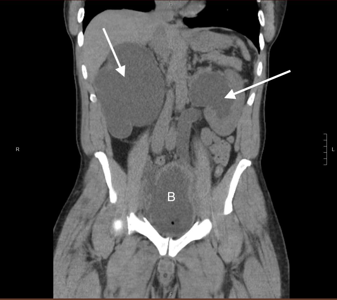
A urologist was consulted in the ED and requested the bladder catheter remain unclamped. After the clamp was removed, the patient rapidly drained over seven liters of urine. The urine output became grossly bloody during the latter portion of the bladder decompression. During decompression, the patient developed diaphoresis and fatigue. At that time, his blood pressure was found to be 70/52 mm Hg. An intravenous (IV) fluid bolus was initiated. The patient ultimately received a total of three liters of normal saline while in the ED.
Owing to the degree of hematuria, serial Hgbs were sent during his ED and hospital stays. Eight and one-half hours after the initial presentation, the patient’s Hgb dropped to 5.7 mg/dL, a drop in 4.4 mg/dL from the initial level. Additional volume resuscitation was initiated with packed RBCs. A total
CPC-EM Capsule
What do we already know about this clinical entity?
Complications of bladder decompression for patients experiencing urinary retention include hemorrhage and post-obstructive diuresis. These complications are usually self-limiting.
What makes this presentation of disease reportable?
We report the rare case of a 57-year-old man with post-decompression hemorrhage, diuresis and hemodynamic collapse requiring aggressive resuscitation and operative intervention.
What is the major learning point?
Life-threatening complications associated with bladder decompression are rare though can occur.
How might this improve emergency medicine practice?
Awareness of the potentially severe complications associated with bladder decompression will allow the clinician to anticipate and intervene quickly when they do occur.
Volume 6, no. 4: November 2022 299 Clinical Practice and Cases in Emergency Medicine
Knapp et al. Life-threatening Complications Associated with Bladder Decompression
Image 1. Computed tomography (CT) of the abdomen and pelvis without contrast (coronal view). Note the severe bilateral hydronephrosis (arrows) and bladder distention (indicated by the letter B). The patient had approximately 1,200 milliliters of urine output at the time of the CT.
Image 2. Computed tomography of the abdomen and pelvis without contrast (axial view). Severe bilateral hydronephrosis is seen (arrows). The large areas of fluid are likely responsible for confusing point-of-care ultrasound findings.
of four units of packed RBCs were transfused. The patient was admitted in the intensive care unit and treated further with continuous bladder irrigation (CBI).
Owing to continued hemodynamic instability and hemorrhage, the patient required operative intervention. He underwent cystoscopy, bladder biopsy, clot evacuation, and fulguration. Postoperatively the patient had continued bladder hemorrhage and was treated with tranexamic acid administered through the urinary catheter along with steroid infused-CBI.
The patient’s course was complicated by the recognition of bilateral deep vein thrombosis (DVT) while in the intensive care unit. He was started on IV heparin. He subsequently redeveloped gross hematuria and required further blood transfusion. Additionally, the patient underwent bilateral percutaneous nephrostomy (PCN) placement to divert urine away from the bladder. The patient was eventually discharged on day five with an inferior vena cava filter and bilateral PCN. Upon discharge, the patient’s creatinine was 1.8 mg/dL. The cause of the patient’s initial urinary retention was never definitively determined.
DISCUSSION
The presentation of acute urinary retention is usually straightforward severe abdominal pain associated with the inability to void. Clinicians must be aware that the presentation of chronic urinary retention can be more insidious. As was the case in our patient, abdominal pain may not be a historical feature. Emergency physicians need to recognize urinary retention in its various forms and understand the uncommon though serious complications that can occur.
The normal bladder can hold 450-500 milliliters (mL) of urine. An obstruction of urinary outflow can be precipitate acute renal failure. Our patient presented in acute renal failure with an initial creatinine of 12.4 mg/dL. Fortunately, the patient’s potassium was not severely elevated (5.9 mEq/dL). In most cases of post-obstructive renal failure, relieving the obstruction will enable the return of baseline renal function. The patient’s creatine returned to its baseline level during his hospital course without the need for dialysis.
Our patient decompressed over seven liters of urine during the first five hours of his ED stay. Patients who put out more than 1,500 mLs of urine immediately after bladder catheterization are thought to be at higher risk of developing post-obstructive diuresis,2 which is seen in up to 52% of patients with urinary retention.3 It is defined as an output of ≥200 mLs of urine for ≥two hours after the initial decompression, or greater than 3,000 mLs in the first 24 hours.2 Post-obstructive diuresis is primarily a problem with chronic, not acute, urinary retention and usually represents an appropriate attempt to excrete excess fluid retained during the period of obstruction.4
Although rare, post-obstructive diuresis can lead to hypotension and hemodynamic collapse. This was the case for our patient. Volume replacement should be initiated early in these patients with a recommendation that no more than 75% of the average hourly urine output be replaced to avoid stimulation for further diuresis.5
Hematuria is an additional recognized complication of bladder decompression. Hematuria is reported to occur in 2-16% of patients.6 The mechanism of hemorrhage postdecompression is not clearly understood, although it is thought to be related to bladder stretch injury. Bladder hemorrhage is usually self-limited and rarely requires aggressive or invasive intervention. It was previously thought that complications associated with bladder decompression could be avoided by gradually releasing urine over a period of hours. More recent literature supports the practice of rapid bladder decompression as it has not been shown to increase complication rates.
Etafy et al randomized two groups with acute urinary retention to receive either rapid or gradual bladder decompression. Of the 31 patients in each cohort, no significant complications in either group were noted.7 Similarly, Boettcher et al randomized 294 patients into rapid and gradual decompression groups. Their study differed in that it included patients with both acute and chronic urinary retention. They found no statistically significant difference in complication rates between gradual and rapid bladder emptying. They concluded that gradual emptying did not reduce the risk of hematuria or circulatory collapse and that there is no need to prefer gradual over rapid emptying.8
Although they may not be influenced by the rate of bladder decompression, complications including hematuria, post-obstructive diuresis, and hypotension do occur. When these complications happen, they are rarely clinically significant.3 When they are, clinicians must be ready to intervene. Owing to persistent hemorrhage and hemodynamic instability despite IV fluids, our patient required transfusion of multiple units of packed RBCs. As his bladder hemorrhage was not self-limited, hemorrhage control in the operating room by the urologist was required.
Our patient was subsequently diagnosed with bilateral DVTs. An interesting association between urinary retention and DVT exists. Lower extremity clot formation is likely related to direct compression of the iliac veins, which creates stasis in the venous system. Deep vein thrombosis associated with urinary retention is rare, although it has been reported in the literature.9 The venous stasis and resulting thrombotic complications caused by urinary retention should prompt clinicians to perform a detailed physical examination, including the identification of leg swelling. For patients with associated shortness of breath, an investigation for pulmonary embolism should be undertaken. The anticoagulation required for the treatment of an acute DVT further complicated the management of our patient’s bladder hemorrhage.
Identifying patients who will develop severe complications after bladder decompression is not straightforward. Patients who develop persistent hypotension and gross hematuria will benefit from urologic consultation and hospital admission. As chronic urinary retention with high volumes of urine output (greater than 1,500 mLs) are at greater risk for post-obstructive diuresis, it is reasonable to observe those patients for 24 hours in the hospital
Clinical Practice and Cases in Emergency Medicine 300 Volume 6, no. 4: November 2022
Life-threatening Complications Associated with Bladder Decompression Knapp et al.
setting. For others, it is recommended that patients be monitored for a minimum of four hours for significant hourly urinary output (>200 mL per hour over intake) after the initial return. If this degree of output continues, the patient should be admitted with appropriate volume replacement.1
CONCLUSION
Understanding the complications associated with urinary retention and bladder decompression is essential for clinicians. Emergency physicians must ensure they are adequately prepared to recognize and manage these rare but potentially life-threatening problems.
The authors attest that their institution requires neither Institutional Review Board approval nor patient consent for publication of this case report. Documentation on file.
Address for Correspondence: Barry J. Knapp, MD, FACEP, RDMS, Eastern Virginia Medical School, Department of Emergency Medicine, 600 Gresham Drive Ste. 304, Norfolk, VA 23507. Email: knappbj@evms.edu.
Conflicts of Interest: By the CPC-EM article submission agreement, all authors are required to disclose all affiliations, funding sources and financial or management relationships that could be perceived as potential sources of bias. The authors disclosed none.
Copyright: © 2022 Knapp et al. This is an open access article distributed in accordance with the terms of the Creative Commons Attribution (CC BY 4.0) License. See: http://creativecommons.org/ licenses/by/4.0/
REFERENCES
1. Manthey DE and Story DJ. (2020). Acute urinary retention. In: Tintinalli JE, Ma O, Yealy DM, Meckler GD, Stapczynski J, Cline DM, Thomas SH. eds. Tintinalli’s Emergency Medicine: A Comprehensive Study Guide, 9e. McGraw Hill; New York, NY .
2. Halbgewachs C and Domes T. Postobstructive diuresis: Pay close attention to urinary retention. Can Fam Physician. 2015;61(2):137-42.
3. Nyman MA, Schwenk NM, Silverstein MD. Management of urinary retention: rapid versus gradual decompression and risk of complications. Mayo Clin Proc. 1997;72(10):951-6.
4. Barrisford GW and Steele GS. Acute urinary retention. 2022. Available at: https://www.uptodate.com/contents/acuteurinaryretention?search=urinary%20retention&source=search_re sult&selectedTitle=1~150&usage_type=default&display_rank=1. Accessed August 29, 2022.
5. Leslie SW, Sajjad H, Sharma S. Postobstructive diuresis. [Updated 2022 Sep 19]. In: StatPearls [Interent].Treasure Island (FL): StatPearls Publishing; 2022. Available from: https://www.ncbi.nlm.nih. gov/books/NBK459387/.
6. Gabriel C and Suchard JR. Hematuria following rapid bladder decompression. Clin Pract Cases Emerg Med. 2017;1(4):443-5.
7. Etafy MH, Saleh FH, Ortiz-Vanderdys C, et al. Rapid versus gradual bladder decompression in acute urinary retention. Urol Ann. 2017;9(4):339-42.
8. Boettcher S, Brandt AS, Roth S, et al. Urinary retention: benefit of gradual bladder decompression - myth or truth? A randomized controlled trial. Urol Int. 2013;91(2):140-4.
9. Kawada T, Yoshioka T, Araki M, et al. Deep vein thrombosis and pulmonary embolism secondary to urinary retention: a case report. J Med Case Rep. 2018;12(1):78.
Volume
6, no. 4: November 2022 301 Clinical Practice and Cases in Emergency Medicine
Knapp et al.
Life-threatening Complications Associated with Bladder Decompression
Acute Brachial Artery Occlusion on Point-of-Care Ultrasound in the Emergency Department: A Case Report
Anisa Y. Mughal, MD* Patrick Kishi, MD†
* †
Section Editor: Shadi Lahham, MD
Creighton University School of Medicine, Department of Emergency Medicine, Phoenix, Arizona Mayo Clinic, Department of Emergency Medicine, Phoenix, Arizona
Submission History: Submitted June 9, 2022; Revision received September 08, 2022; Accepted September 09, 2022 Electronically published November 5, 2022 Full text available through open access at http://escholarship.org/uc/uciem_cpcem DOI: 10.5811/cpcem.2022.9.57482
Introduction: This is a case report of an acute right brachial artery occlusion found on point-of-care ultrasound in the emergency department (ED) that illustrates the developing role of ultrasound in rapid differentiation and identification of acute vascular emergencies.
Case Report: An 87-year-old male with a past medical history of coronary artery bypass graft presented to the ED with acute right upper extremity pain, with point-of-care ultrasound (POCUS) findings consistent with acute right brachial artery occlusion.
Conclusion: Arterial occlusions are vascular emergencies that can be rapidly identified on POCUS. [Clin Pract Cases Emerg Med. 2022;6(4):302–304.]
Keywords: case report; brachial artery occlusion; point-of-care ultrasound; vascular ultrasound.
INTRODUCTION
Acute arterial occlusion is a medical emergency caused by a disruption in arterial flow. The occlusion may occur from thrombosis or embolus. Clinical presentation and symptoms vary based on the artery affected. These occlusions ultimately are responsible for a significant incidence of disability and death (eg, myocardial infarction, stroke).
Acute limb ischemia affects approximately 10-14 patients per 100,000 annually and more commonly occurs in the lower extremities.1,2 The brachial artery is located in the upper extremities and is commonly used to measure blood pressure. Acute embolization of the brachial artery is most commonly cardiac in origin.2 Trauma, aneurysms, and iatrogenic injuries following cardiac catheterization may also cause acute thrombosis.3,4 Several studies have identified upper extremity arterial occlusions after coronary catheterization; however, there are few reports of unprovoked brachial artery occlusion.1,5,6
When considering brachial artery occlusion in patients, the diagnostic workup should include a thorough physical exam, point-of-care ultrasound (POCUS), and coagulation laboratory studies.7 A computed tomography (CT) angiogram
can be considered if POCUS and clinical exam are insufficient to provide this time-sensitive diagnosis.7 In this case report we describe an acute brachial arterial occlusion in an elderly male presenting with right upper extremity pain.
CASE REPORT
A left-handed, 87-year-old male unvaccinated for COVID-19 with a past medical history significant for a four-vessel coronary artery bypass graft in 2017, not on any anti-coagulation medications, and ischemic cardiomyopathy presented to our emergency department with acute-onset right upper forearm and right-hand pain. The patient reported severe, excruciating pain in his right upper extremity starting at the level of the elbow extending to his distal fingertips upon waking approximately two hours prior to presentation. He also reported paresthesias that were most prominent in his right hand. Prior to arrival, the patient took two full-strength aspirin tablets (325 milligrams), which did not provide any relief. The review of systems was otherwise unremarkable.
The patient’s initial vital signs were notable for temperature 36.6° Celsius, heart rate 89 beats per minute,
Clinical Practice and Cases in Emergency Medicine 302 Article in Press Case
Report
respiratory rate 16 breaths per minute, blood pressure 189/82 millimeters of mercury, and oxygen saturation of 100% on room air. On physical exam, he appeared uncomfortable but was not toxic-appearing or diaphoretic. There was no palpable pulse in the right brachial artery, the right radial artery, or in the right ulnar artery. Examination of the right upper extremity demonstrated a cold and pale limb from the level of the elbow to the hand. All compartments were soft. Motor strength and sensation were intact.
Due to the concerning physical exam findings, a POCUS was performed by the emergency physician. The ultrasound findings demonstrated an arterial occlusion in the right brachial artery, with visible clot burden (Image, Video 1). Color Doppler revealed no evidence of blood flow from the point of the occlusion in the antecubital fossa and distally (Video 2). Vascular surgery was immediately consulted, and a heparin drip was ordered, along with analgesic medications and intravenous fluids. While laboratory studies were pending, vascular surgery evaluated the patient, and a decision was made to take the patient to the operating room for emergent repair with a right brachial artery thrombectomy. He also tested positive for severe acute respiratory syndrome coronavirus 2 (SARS-CoV-2) but did not report any related symptoms.
CPC-EM Capsule
What do we already know about this clinical entity?
Upper extremity arterial occlusions occur less commonly than lower extremity arterial occlusions and are often traumatic or iatrogenic in origin.
What makes this presentation of disease reportable?
We describe an unprovoked brachial artery occlusion in an elderly male patient with asymptomatic coronavirus disease.
What is the major learning point?
Point-of-care ultrasound can quickly identify and diagnose acute arterial occlusions in the upper extremity.
How might this improve emergency medicine practice?
Point-of-care ultrasound can lead to rapid diagnosis of vascular occlusions and facilitates rapid treatment to prevent irreversible ischemic pathology.
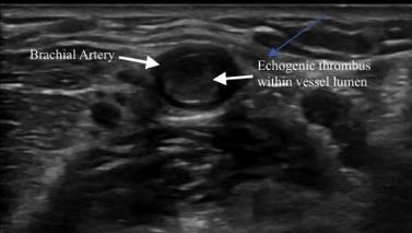
Ultrasound was used intraoperatively to identify the proximal extent of the thrombus within the brachial artery. The brachial artery appeared clear of thrombus and embolus approximately three finger breadths proximal to the antecubital fossa. The clot was removed, and at the conclusion of the procedure the patient had palpable radial and ulnar pulses. Physical exam indicated the patient had Doppler signals in the right palmar arch after thrombectomy. Postoperatively, he reported that his pain and paresthesias had resolved.
The patient underwent further workup during hospitalization, which included a transthoracic
echocardiogram demonstrating an apical left ventricular thrombus that was pedunculated and mobile. Ejection fraction was calculated to be 29%. A CT angiogram of the chest demonstrated no evidence of an ascending aortic or great vessel thrombus to explain embolization. The patient was continued on a heparin drip and subsequently transitioned to oral warfarin with a goal international normalized ratio (INR) of 2.0-3.0. He was discharged on post-operative day two with a 10-day course of therapeutic enoxaparin as a bridge until he reached a therapeutic INR. Chart review demonstrated no evidence of complications after discharge.
DISCUSSION
Our report highlights a unique presentation of acute brachial artery occlusion in which POCUS led to a rapid identification and expedited treatment of this critical, acute vascular emergency. Clinical signs of arterial occlusion follow the common mnemonic of the six Ps: pulselessness, pain, pallor, poikilothermia, paresthesia, and paralysis.7 Sonographic signs of arterial occlusion include visible clot
Volume 6, no. 4: November 2022 303 Clinical Practice
Cases in Emergency Medicine
and
Mughal et al. Acute Brachial Artery Occlusion on POCUS in the ED
Image. Point-of-care ultrasound image of proximal radial artery with an occlusive, non-compressible thrombus.
on POCUS in the ED
burden or a non-compressible vessel, and absence of color Doppler flow of the affected vessel.
The prevalence of acute arterial occlusion varies by anatomical location, risk factors, and gender.2 Upper arterial occlusion occurs less frequently than lower extremity arterial occlusion.2 Modifiable risk factors include smoking, obesity, high cholesterol, diabetes, and hypertension.7 Increasing age correlates with a higher incidence of arterial occlusion.8 COVID-19 may be a new risk factor in the development of arterial occlusion. In 2021, a nine-patient retrospective analysis was performed, demonstrating that patients with underlying conditions presenting with elevated inflammatory markers or D-dimers in the setting of SARSCoV-2 infection were at a higher risk of developing acute arterial occlusion.9
Most cases of radial artery occlusion are studied in the context of a transradial catheterization approach and document strategies to reduce iatrogenic or post-procedural radial arterial occlusion.6 In this case, the extremity clot was likely a result of embolization from the ventricular thrombus demonstrated on formal echocardiography. A ventricular thrombus can form in vivo, or secondary to recent myocardial infarction, and is often the source of peripheral arterial occlusions.10 In patients presenting to the ED with arterial occlusion, it may be beneficial to perform a POCUS echocardiogram to evaluate for a thrombotic source.
CONCLUSION
Point-of-care ultrasound can lead to rapid diagnosis of vascular occlusions. This is particularly important in identifying peripheral arterial occlusions that otherwise can lead to irreversible ischemic pathology. It is important to include acute thrombosis in the differential diagnoses in patients with recent or active SARS-CoV-2 infections, as it may lead to a hypercoagulable state.
Video 1. Point-of-care ultrasound of the right brachial artery with an occlusive thrombus.
Address for Correspondence: Anisa Y. Mughal, MD, Creighton University School of Medicine, Department of Emergency Medicine, 2601 E. Roosevelt St., Phoenix, AZ, 85008. Email: aym30633@creighton.edu.
Conflicts of Interest: By the CPC-EM article submission agreement, all authors are required to disclose all affiliations, funding sources and financial or management relationships that could be perceived as potential sources of bias. The authors disclosed none.
Copyright: © 2022 Mughal et al. This is an open access article distributed in accordance with the terms of the Creative Commons Attribution (CC BY 4.0) License. See: http://creativecommons.org/ licenses/by/4.0/
REFERENCES
1. Ologun GO, Bohan C, Lau T, et al. Acute brachial artery occlusion in an elderly patient with acute myocardial ischemia. Cureus 2017;9(9):e1700.
2. Bae M, Chung SW, Lee CW, et al. Upper limb ischemia: clinical experiences of acute and chronic upper limb ischemia in a single center. Korean J Thorac Cardiovasc Surg. 2015;48(4):246-51.
3. Mehrabi Nasab E, Heidarzadeh S, Yavari B, et al. Acute upper limb ischemia in a patient with COVID-19: a case report. Ann Vasc Surg 2021;77:83-85.
4. Ibrahim IG and Tahtabasi M. Doppler ultrasound diagnosis of brachial artery injury due to blunt trauma: a case report. Radiol Case Rep 2020;15(8):1207-10.
5. Nijhuis HH and Müller-Wiefel H. Occlusion of the brachial artery by thrombus dislodged from a traumatic aneurysm of the anterior humeral circumflex artery. J Vasc Surg. 1991;13(3):408-11.
6. Rashid M, Kwok CS, Pancholy S, et al. Radial artery occlusion after transradial interventions: a systematic review and meta-analysis. J Am Heart Assoc. 2016;5(1):e002686.
7. Smith DA and Craig JL. Acute arterial occlusion. PubMed. 2022. Available at: pubmed.ncbi.nlm.nih.gov/28722881/. Accessed May 30, 2022.
Video 2. Point-of-care ultrasound demonstrating absence of color Doppler flow in the brachial artery, indicating an occlusive thrombus.
8. Howard DP, Banerjee A, Fairhead JF, et al. Population-based study of incidence, risk factors, outcome, and prognosis of ischemic peripheral arterial events: implications for prevention. Circulation 2015;132(19):1805-15.
9. Ogawa M, Doo FX, Somwaru AS, et al. Peripheral arterial occlusion due to COVID-19: CT angiography findings of nine patients. Clin Imaging. 2021;73:43-7.
The authors attest that their institution requires neither Institutional Review Board approval nor patient consent for publication of this case report. Documentation on file.
10. Agarwal KK, Douedi S, Alshami A, et al. Peripheral embolization of left ventricular thrombus leading to acute bilateral critical limb ischemia: a rare phenomenon. Cardiol Res. 2020;11(2):134-7.
Clinical Practice
Emergency Medicine 304 Volume 6, no. 4: November 2022
and Cases in
Acute Brachial Artery Occlusion
Mughal et al.
Cells Gone Wild: A Case Report on Missed Acute Leukemia and Subsequent
Disseminated
Intravascular Coagulation in the Emergency Department
Onyinyechukwu Okorji, DO Rachael Kern, DO Shaylor Klein, DO, MSEd Brian Jordan, DO Kuljit Kaur, DO
Section Editor: R Gentry Wilkerson, MD
Submission History: Submitted June 18, 2022; Revision received September 15, 2022; Accepted September 20, 2022 Electronically published November 11, 2022 Full text available through open access at http://escholarship.org/uc/uciem_cpcem DOI: 10.5811/cpcem.2022.9.57811
Introduction: Emergency physicians must maintain a broad differential when seeing patients in the emergency department (ED). Occasionally, a patient may have an undiagnosed, life-threatening medical condition not related to the presenting chief complaint. It is imperative to review all ordered laboratory tests and any available previous laboratory values to assess for any abnormalities that may warrant further evaluation.
Case Report: This case report is regarding the missed diagnosis of acute leukemia and subsequent disseminated intravascular coagulation in a 27-year-old male who presented to multiple EDs with the unrelated chief complaint of finger ring entrapment. This patient ultimately succumbed to his illness.
Conclusion: When evaluating patients in the ED, it is important to review any prior available test results for abnormalities, even if the results do not specifically correlate with the chief complaint. Emergency physicians must remain vigilant to avoid missing a critical diagnosis. [Clin Pract Cases Emerg Med. 2022;6(4):305–309.]
Keywords: case report; acute leukemia; hyperleukocytosis; disseminated intravascular coagulation.
INTRODUCTION
In the emergency department (ED), the breadth of possible diagnoses requires physicians to maintain a broad differential. Potentially missed diagnoses can result in negative patient outcomes; therefore, it is imperative to fully review all available test results, even if not related to the chief complaint. In this case, we discuss a patient presenting with a chief complaint of finger ring entrapment who was subsequently diagnosed with acute leukemia based on his initial complete blood count (CBC) with cell differential. Chart review revealed multiple recent ED visits with abnormal CBCs that had not been addressed. This patient went on to develop disseminated intravascular coagulation (DIC) and ultimately succumbed to his illness.
CASE PRESENTATION
This case is regarding a 27-year-old male with a medical history of intravenous (IV) opioid use disorder who was brought to the ED by emergency medical services following an assault with a complaint of right-hand pain. No further history was provided by the patient. Upon arrival, he was ill-appearing with vital signs of temperature 36.5o Celsius, heart rate 104 beats per minute, blood pressure 114/58 millimeters of mercury (mmHg), respiratory rate 24 breaths per minute, and oxygen saturation 100% on room air. Physical exam revealed bilateral periorbital ecchymosis, anterior neck ecchymosis, petechial rash on his chest, anasarca, and widespread necrotic wounds. An embedded ring was noted at the base of his right fourth finger with significant surrounding edema, erythema, and necrosis.
Article in Press 305 Clinical Practice and Cases in Emergency Medicine Case Report
Jefferson Health - Northeast, Department of Emergency Medicine, Philadelphia, Pennsylvania
Upon chart review of the prior two months, we found that the patient had presented to multiple surrounding EDs with the same chief complaint of right-hand pain with ring entrapment. His first presentation to an outside hospital consisted of unremarkable lab work. The patient left against medical advice (AMA) prior to ring removal. He presented an additional nine times with similar outcomes: lab work would be obtained and the patient would leave AMA prior to ring removal. Thirteen days prior to this case presentation, the patient’s ED workup revealed leukocytosis of 68.2 x10⁹ cells per liter (L) with 19% blasts and thrombocytopenia of 15 x 109/L, which were not addressed due to the patient leaving AMA.
On this current presentation (day zero), a radiograph of his right hand was significant for “tourniquet syndrome” of his right fourth digit with extensive circumferential periostitis (Image 1). The admission CBC was remarkable for a white blood cell count of 157.8 x 10⁹ cells/L (reference range: 4.0-11.0 x 10⁹ cells/L) with blasts of 61% and a subsequent pathology report consistent with acute myelogenous leukemia (AML) (Image 2). See Table 1 for hospitalization laboratory results.


The patient was admitted to the intensive care unit for sepsis secondary to osteomyelitis of the right fourth digit requiring broad spectrum IV antibiotics. Shortly after admission, he developed a change in mental status requiring

Image 1. Radiograph of right hand with marked fourth digit swelling distal to the patient’s ring suspicious for tourniquet syndrome. The blue arrow shows soft tissue swelling and the red arrow shows periosteal elevation. Periosteal reaction involving proximal fourth digit phalanges suspicious for osteomyelitis (both arrows).
CPC-EM Capsule
What do we already know about this clinical entity?
Patients presenting with blast crisis due to acute leukemia require emergent hematology consultation from the Emergency Department in order to initiate treatment and prevent a delay in care.
What makes this presentation of disease reportable? This is a case of an unusual clinical presentation of a medical disease masked by a different chief complaint.
What is the major learning point?
To encourage emergency physicians to review and compare current test results with previously available data, despite the urge to only focus on a chief complaint.
How might this improve emergency medicine practice?

If physicians review and compare curresnt test results with previously available data, patients can have an appropriate disposition and less delay of care.
endotracheal intubation. Computed tomography of the brain did not reveal any acute abnormalities.

Hematology/oncology was consulted and emergently reviewed the peripheral smear, which revealed blast cells, increased cytoplasmic granules without Auer rods, occasional schistocytes, and a negative Coombs test. Results were

Clinical
in
Medicine 306 Volume 6, no. 4: November 2022
Practice and Cases
Emergency
Missed Acute Leukemia and Subsequent Disseminated Intravascular Coagulation in the ED Okorji et al.
Image 2. Peripheral blood smear with marked leukocytosis and numerous circulating blasts with monocytic features consistent with acute myeloid leukemia (arrows).
Table 1. Patient’s laboratory values during hospitalization.
Laboratory values
Dec. 192 Jan. 18 Jan. 29 Jan. 303 Jan. 31 Feb. 1 Feb. 2
Hemoglobin (g/dL)1[14.0-17.0] 15.3 10.8 5.1 4.6 7.2 6.9 6.7
White blood cell count (109 cells/L) [4.0-11.0] 9.2 68.2 135.9 157.8 132.8 71.5 60.2
Platelets (109 cells/L) [140-400] 217 15 13 7 21 5 34
Blasts (%) [<0] 19 82.9 56 29 38 26
Urea-nitrogen (mg/dL) [10-22] 14 22 51 50 54 69 84
Creatinine (mg/dL) [0.7-1.40] 0.85 1.04 1.98 1.68 2.39 2.68 2.86
Potassium (mmol/L) [3.5-5.0] 3.8 4.3 4.8 4.5 4.4 5.3 5.9
Phosphate (mg/dL) [2.5-4.5] 4.7 7.6 11.7 15.3
Calcium (mg/dL) [8.6-10.0] 9.5 8.6 8.5 7.5 7.7 7.3 6.7
International normalized ratio (INR) [0.82-1.13] 1.42 1.75 1.76 1.82 1.87 2.03
Prothrombin time (PT) (seconds) [9.4-13.0] 16.4 20.2 21.6 21 21.6 21.8 D-dimer (ng/mL) [<243] 4539 6562 10567
Fibrinogen (seconds) [200-393] 121 109 163 156 137
Lactic acid (mmol/L) [0.5-2.0] 2.9 2 5
Lactate dehydrogenase (IU/L) [135-225] 1040 904 911 918
Urate (mg/dL) [3.4-7.0] 16.5 13.1 13.7 15.3
Haptoglobin (mg/dL) [30-200] 39
Reticulocytes (%) [0.5-2.5] 6.2
1Reference ranges for units 2Initial presentation with baseline laboratory values obtained 3Day of presentation of this case presentation (day 0) g/dL, grams her deciliter; mL, milliliter; mg/dL, milligram per deciliter; mmol/L, millimole per liter; IU/L, international units per liter; ng/mL, nanograms per milliliter.
consistent with AML, DIC, and auto-tumor lysis syndrome (TLS) due to abnormalities all noted in Table 1. He was emergently treated with rasburicase 6 mg intravenously, hydroxyurea 2 g orally every eight hours, allopurinol 50 mg orally daily, and IV fluids. For treatment of DIC, he received a total of eight units of packed red blood cells, five units of platelets, and one unit of cryoprecipitate. On day three of hospitalization, the patient developed hypotension requiring vasopressor support. He was transferred to an outside hospital for advanced treatment of his AML and succumbed to his illness shortly thereafter.
DISCUSSION
Leukocytosis is a term broadly used to describe an abnormal elevation of certain cells that originate from the bone marrow. To help guide diagnosis and treatment, one must first distinguish which cell type is elevated. The main cell types measured in the leukocyte count are neutrophils, lymphocytes, eosinophils, monocytes, and basophils, of which neutrophils are the most abundant. Neutrophilia has a wide range of diagnoses ranging from benign to malignant. This is due to upregulation of bone marrow production and/or demargination of existing neutrophils from the endothelium.
Acute elevations can be linked to recent emotional or physical stress, infection, medications, trauma, and smoking. Chronic elevations are the result of inflammatory diseases such as rheumatic disease, inflammatory bowel disease, chronic hepatitis, vasculitis, steroid use, bone marrow stimulation, and congenital diseases. Infection and other forms of acute inflammation will result in leukocytosis with cell counts as high as 25 x 109 cells/L.1
When analyzing leukocytosis, cell maturity and the degree of elevation must be taken into consideration. The presence of immature granulocytes and precursors such as blasts and myelocytes are significant abnormal findings and should raise concern for possible malignancy. The morphology of the blast cell may help determine the type of malignancy, which can be AML, promyelocytic leukemia, or other precursor neoplasms.2
Hyperleukocytosis is seen with levels greater than 100 x 109 cells/L and should always prompt immediate evaluation for a malignant cause.1 Two important lab abnormalities that may be seen with hyperleukocytosis are reverse pseudohyperkalemia and spurious hypoxemia. Although not seen in this patient, reverse pseudohyperkalemia is an in vitro phenomenon in which the potassium concentration is elevated on plasma testing yet normal on serum testing.3 This occurs due to intracellular release of potassium as cells undergo lysis due to fragile membranes.4
Volume 6, no. 4: November 2022 307 Clinical Practice and Cases in Emergency Medicine
Okorji et al. Missed Acute Leukemia and Subsequent Disseminated Intravascular Coagulation in the ED
Spurious hypoxemia, which was seen in this patient, is a low partial pressure of arterial oxygen (PaO2) noted on blood gas in the presence of normal oxygen saturation. The increased presence of leukocytes results in increased consumption of oxygen, which reflects on the PaO2 5 In addition, a delay in blood gas processing will result in worsening spurious hypoxemia. This phenomenon is referenced in Table 2. Understanding these lab abnormalities may help mitigate inappropriate or unnecessary treatment of hyperkalemia and hypoxemia. Acute leukemia has a high mortality due to complications including leukostasis, TLS, and DIC.
Tumor lysis syndrome is an oncologic emergency that occurs when the lysis of tumor cells leads to a massive release of intracellular ions such as potassium, phosphorus, and nucleic acids that metabolize to uric acid. This condition is most often diagnosed in patients with leukemia and an exceedingly high white blood cell count.6 In this condition, kidney damage occurs via uric acid obstructive uropathy; potassium and phosphorus build-up can cause fatal arrhythmias and hypocalcemia, respectively.
In high-risk patients, allopurinol may be started prior to initiation of chemotherapy to help prevent TLS as it decreases the production of uric acid. Treatment of TLS begins with crystalloid fluids to help expand intravascular volume and to help improve the glomerular filtration rate. Recombinant urate oxidase (rasburicase) may be used to treat hyperuricemia in cases that involve leukemia, lymphoma, and solid tumor malignancy. When possible, it is important to check for glucose-6-phosphate dehydrogenase deficiency prior to administration of this medication. Physicians may also consider additional treatment modalities including sodium bicarbonate infusions for urine alkalinization, hemodialysis, calcium repletion, and febuxostat (xanthine oxidase inhibitor).
Table 2. Patient’s arterial blood gas on day of admission and expiration.
Laboratory values1 Day 03 Day 3
pH2 [7.35-7.45] 7.47 7
PaCO2 (mm Hg) [34-45] 38 53
PaO2 (mm Hg) [83-103]4 173 75
Oxygen saturation (%) [95-99] 99.5 94
Bicarbonate (mmol/L) [22-27] 28 13
Base excess (mmol/L) [0-2.4] 3.6 17
1Laboratory values are as follows: grams her deciliter (g/dL), mL (milliliter), milligram per deciliter (mg/dL), millimole per liter (mmol/L), international units per liter (IU/L), nanograms per milliliter (ng/mL)
2Reference range for units.
3Day of presentation of this case study (day 0).
4Expected PaO2 (mmHg) on 100% fraction of inspired oxygen (FiO2) is greater than 400.
PaCO2, partial pressure of carbon dioxide in arterial blood; PaO2, partial pressure of oxygen in arterial blood; mm Hg, millimeters of mercury.
Disseminated intravascular coagulation is defined as widespread intravascular fibrin formation in response to increased blood protease activity that overcomes the natural anticoagulant mechanisms.7 Vascular endothelial insult results in the formation of systemic microthrombi, which leads to shearing of red blood cells and consumption of coagulation factors and platelets, and can result in hemorrhage. Certain diseases such as promyelocytic leukemia, as noted in this patient, may result in a severe hyper-fibrinolytic state in addition to dysfunction of the coagulation pathway.7 Clinical features of DIC include bleeding, thrombosis, shock, acute renal failure, acute liver failure, central nervous system involvement, and respiratory dysfunction. In cases of DIC related to severe sepsis, as seen in this patient, purpura fulminans may be present and are described as large purpuric lesions that result from extensive tissue thrombosis and hemorrhagic skin necrosis.
Testing for DIC consists of coagulation tests, such as platelet count, prothrombin time (PT), D-dimer, fibrinogen, and a peripheral blood smear to assess for the presence of schistocytes. Patients with severe liver disease may exhibit similar lab findings; therefore, lab work should be repeated in 6-8 hours as DIC will exhibit rapid and dramatic changes over a short time frame.8
Treatment for DIC begins with determining and correcting the underlying cause. Platelet replacement should be considered when platelet levels are below 50 x109 cells/L of blood with active bleeding or below 20 x 109 cells/L without evidence of bleeding. Fresh frozen plasma may be transfused to a goal PT and partial prothrombin time less than 1.5 times the normal limit. Cryoprecipitate repletion may be given to maintain a fibrinogen goal of 100-150 mg/dL. Prothrombin complex concentrates and tranexamic acid are generally contraindicated in DIC as they may increase the risk of thrombotic complications.9 Extensive thrombosis may warrant anticoagulation therapy with heparin infusion, and those without active bleeding may receive venous thromboembolism prophylaxis with unfractionated heparin or low molecular weight heparin. Finally, activated protein C concentrate should be reserved for cases of severe sepsis with DIC exhibiting purpura fulminans.10
CONCLUSION
When evaluating patients in the ED, it is important to review any prior available test results for abnormalities, even if the results do not specifically correlate with the chief complaint. Our case highlights a patient who presented with finger ring entrapment and a missed diagnosis of leukemia who unfortunately succumbed to his medical illness. We hope this case presentation drives home the point for all physicians to acknowledge and act on all available results in the ED.
The authors attest that their institution requires neither Institutional Review Board approval nor patient consent for publication of this case report. Documentation on file.
Clinical Practice
Cases in Emergency Medicine 308 Volume 6, no. 4: November 2022
and
Missed Acute Leukemia and Subsequent Disseminated Intravascular Coagulation in the ED Okorji et al.
Okorji et al. Missed Acute Leukemia and Subsequent Disseminated Intravascular Coagulation in the ED
Address for Correspondence: Onyinyechukwu Okorji, DO, Jefferson Health-Northeast, Department of Emergency Medicine, 10800 Knights Rd, Philadelphia, PA, 19114. Email: oxo028@jefferson.edu.
Conflicts of Interest: By the CPC-EM article submission agreement, all authors are required to disclose all affiliations, funding sources and financial or management relationships that could be perceived as potential sources of bias. The authors disclosed none.
Copyright: © 2022 Okorji et al. This is an open access article distributed in accordance with the terms of the Creative Commons Attribution (CC BY 4.0) License. See: http://creativecommons.org/ licenses/by/4.0
REFERENCES
1. Mank V and Brown K. Leukocytosis. 2022. In: StatPearls [Internet]. Treasure Island (FL): StatPearls Publishing; 2022. Available from: https://www.ncbi.nlm.nih.gov/books/NBK560882/
2. George TI. Malignant or benign leukocytosis. Hematology Am Soc Hematol Educ Program. 2012;2012:475-84.
3. Asirvatham JR, Moses V, Bjornson L. Errors in potassium measurement: a laboratory perspective for the clinician. N Am J Med
Sci. 2013;5(4):255-9.
4. Colussi G and Cipriani D. Pseudohyperkalemia in extreme leukocytosis. Am J Nephrol. 1995;15(5):450-2.
5. Van de Louw A, Desai RJ, Schneider CW, et al. Hypoxemia during extreme hyperleukocytosis: how spurious? Respir Care 2016;61(1):8-14.
6. Cairo MS and Bishop M. Tumour lysis syndrome: new therapeutic strategies and classification. Br J Haematol. 2004;127(1):3-11.
7. Costello RA and Nehring SM. Disseminated intravascular coagulation. 2022 Jul 12. In: StatPearls [Internet]. Treasure Island (FL): StatPearls Publishing; 2022. Available from: https://www.ncbi. nlm.nih.gov/books/NBK441834
8. Jameson JL. (2018). Acute Myeloid Leukemia. In: Harrison’s Principles of Internal Medicine Volume 1, 20th ed. New York, New York: McGraw Hill Education.
9. Walker J and Bonavia A. To clot or not: HELLP syndrome and disseminated intravascular coagulation in an eclamptic patient with intrauterine fetal demise. Case Rep Anesthesiol. 2020;2020:9642438.
10. Kroiss S and Albisetti M. Use of human protein C concentrates in the treatment of patients with severe congenital protein C deficiency Biologics. 2010;4:51-60.
Practice and Cases in Emergency
Volume 6, no. 4: November 2022 309 Clinical
Medicine
Anthony
Consideration of Acute Porphyria in an Emergency Department Patient: A Case Report and Discussion of Common Pitfalls
Rios, BS*
Lisa Kehrberg, MD
†
‡
Hillary E. Davis, MD, PhD
†
* † ‡
Section Editors: R. Gentry Wilkerson, MD
University of Tennessee, Department of Biochemistry, Knoxville, Tennessee University of Tennessee Medical Center, Department of Emergency Medicine, Knoxville, Tennessee Family Medicine, Lincolnshire, Illinois
Submission History: Submitted May 26, 2022; Revision received September 14, 2022; Accepted September 20, 2022. Electronically published November 7, 2022
Full text available through open access at http://escholarship.org/uc/uciem_cpcem
DOI: 10.5811/cpcem.2022.9.57507
Introduction: Porphyria refers to a group of disorders associated with defects in heme synthesis. They can be associated with severely debilitating features, including abdominal pain, psychiatric symptoms, neurological defects, and cardiovascular irregularities. Although these diseases are rare, patients with attacks often do present to the emergency department (ED) where consideration of porphyria is generally not included in the differential.
Case Report: Here, we examine a case of a 16-year-old male who presented to our ED for evaluation of recurring abdominal pain and auditory hallucinations in which porphyria was considered by the emergency physician.
Discussion: Not considering acute porphyria in patients with recurring neurovisceral symptoms in the ED can lead to missed opportunities for diagnosing such pathologies. [Clin Pract Cases Emerg Med. 2022;6(4):310-313.]
Keywords: acute hepatic porphyria; porphobilinogen; aminolevulinic acid; recurring abdominal pain; case report.
INTRODUCTION
Porphyria refers to a group of rare metabolic disorders caused by deficiencies in crucial enzymes in the biosynthesis of heme resulting in the accumulation of toxic precursors, deltaaminolevulinic acid (ALA) and porphobilinogen (PBG).1 These disorders can be broken down into two types clinically: acute (neurovisceral) or non-acute (cutaneous).2 The acute types of porphyria include acute intermittent, hereditary coproporphyria, variegate, and ALA-dehydratase deficient. Acute porphyria can present with severe but non-specific symptoms including abdominal pain, neurological changes, psychiatric disturbances, and cardiovascular irregularities. In addition, acute porphyria attacks can be triggered by medications, alcohol, infection, endogenous hormones, illicit drugs, low caloric intake, or stress that require an increased demand for heme.1
Patients with porphyria who present to the emergency departments (ED) with acute attacks are often misdiagnosed. Emergency physicians do not naturally consider porphyria in differential diagnoses given their unfamiliarity with the disorder and the non-specific and variable symptoms with which these patients present. Further, on the rare occasion that the disorder is considered, emergency physicians are often unfamiliar with ordering and obtaining the correct screening tests.
Here we present a case in which the emergency physician did consider the possibility of acute porphyria in a patient with recurring neurovisceral symptoms. We identify and discuss several pitfalls in the hope that these patients can be more readily identified and treated in the acute care setting.
Clinical Practice and
Medicine 310 Volume 6, no. 4: November 2022
Cases in Emergency
Case Report
CASE REPORT
A 16-year-old Black male presented to our ED at a 710-bed academic institution with two days of auditory hallucinations and epigastric abdominal pain. The patient had a history of psychosis and had required an inpatient psychiatric hospitalization for both auditory and visual hallucinations at the age of 12. He did not experience hallucinations at baseline and was doing well in school. The patient’s family had a significant history of mental illness: the patient’s uncle at the time was institutionalized in a psychiatric hospital, and the patient’s brother, later diagnosed with schizophrenia, had presented similarly with auditory hallucinations at the age of 13. For the prior two years, the patient had repeated episodes of upper abdominal pain that were associated with nausea and vomiting. He was followed by a pediatric gastroenterologist and had undergone both a colonoscopy and esophagogastroduodenoscopy just three months prior with no remarkable findings. On review of external charts, we learned he had presented to varying EDs on 12 separate occasions for abdominal pain and had been prescribed multiple antibiotics for presumed colitis on several occasions.
Upon arrival, the patient was afebrile (36.6°C) with a heart rate of 81 beats per minute, respiratory rate of 18 breaths per minute, blood pressure of 128/83 millimeters of mercury (mm Hg), and an oxygen saturation of 99% on room air. Physical exam was notable for a soft, non-distended abdomen with epigastric pain that was not replicated by palpation. He had no focal neurological deficits. He appeared anxious and tearful but was organized in thought. Although he reported auditory hallucinations, he did not actively seem to be responding to internal stimuli.
Screening tests typical for psychiatric patients at our ED were ordered, including a nine-panel urine drug screen, a complete blood count, serum chemistries, liver function tests, an ethanol level, and a urinalysis. The ethanol level was not elevated, and the urine drug screen was unremarkable. An elevated total bilirubin of 2.0 milligrams per deciliter (mg/dL) (reference range: 0.3-1.0 mg/dL) was detected while all other lab values were within the normal range. The patient was subsequently placed in a monitored psychiatric room and referred out to inpatient psychiatric facilities.
After 24 hours, the patient was evaluated by a different emergency physician. Given the patient’s continued abdominal pain, an abdominal ultrasound was ordered, which demonstrated no sonographic abnormality. After reviewing the patient’s medical history and being concerned about the possibility of porphyria, the physician ordered a spot urine porphyria assay. No amber or light-sensitive collection tubes were available in the hospital; consequently, the sample was collected in a test tube wrapped in foil. The hospital laboratory subsequently sent the specimen to an outside facility for analysis. The patient was sent to an inpatient psychiatric facility the following day.
The urine porphyria assay results were available five days after initial collection and were as follows: urine
CPC-EM Capsule
What do we already know about this clinical entity?
Porphyria attacks cause a high degree of morbidity but are under-recognized in emergency departments secondary to lack of understanding of the diseases and their associated confirmatory tests.
What makes this presentation of disease reportable?
We consider a rare case in which the physician was concerned about porphyria in an emergency department patient but unfortunately ordered the wrong test secondary to lack of familiarity.
What is the major learning point?
When a clinical suspicion arises for porphyria, initiating the workup in the emergency department is ideal. Physicians need to be cognizant of the timing and types of confirmatory diagnostics.
How might this improve emergency medicine practice?
More patients with porphyria may be identified leading to a subsequent decrease in repetitive and costly generalized workups and better porphyria patient outcomes.
uroporphyrins of 24 micrograms per deciliter (mcg/dL) (reference range: 0-2.0 mcg/dL), urine pentacarboylporphyrins of 5 mcg/dL (0-2.0 mcg/dL), urine heptacaroxylporphyrins of 8 mcg/dL (0-2.0 mcg/dL), urine coproporphyrin I of 46 mcg/ dL (0-2.0 mcg/dL), and urine coproporphyrin III of 134 mcg/ dL (0-2.0 mcg/dL). Given the notable elevations, the emergency physician contacted the patient’s primary care physician for further follow-up, as the phones of the patient’s family had been disconnected. On review of external records, it was found that the patient had three more ED visits for abdominal pain during the following four months.
DISCUSSION
Initiating a clinical suspicion for porphyria is difficult as the condition may present with broad and non-specific symptoms. Patients with porphyria often undergo expensive and broad workups prior to their ultimate diagnosis.3 Further, when there is a concern for the disease, the diagnosis may be elusive secondary to clinicians’ lack of familiarity with confirmatory tests, and the timing of the tests themselves.
Volume 6, no. 4: November 2022 311 Clinical Practice
Cases in Emergency Medicine
and
Rios et al. Consideration of Acute Porphyria in an ED Patient: Common Pitfalls
Consideration of Acute Porphyria in an ED Patient: Common Pitfalls
In the case we describe, porphyria was appropriately included in the differential diagnosis; however, the incorrect screening test was ultimately obtained. In acute exacerbations, the levels of porphyrin precursors generally increase. These levels can either normalize or remain elevated in an asymptomatic state.4 Here, a urine porphyrin assay was attained as opposed to the precursor assay: the urine porphobilinogen (PBG). During acute porphyria attacks, a substantial elevation in PBG or ALA, a porphyrin precursor that becomes PBG in the second step of heme biosynthesis, occurs.5 Urine porphyrins, alternatively, are produced in the later stages of the heme biosynthesis pathway. These are non-specific for porphyria disorders; they are elevated in acute exacerbations but also in common hepatocellular and biliary pathologies, oral contraceptive use, and alcohol consumption.6-8 Urine porphyrins aid in the classification of porphyria, but they are not used alone to diagnose the disorder. However, given that urine porphyrins may persist longer between attacks, they can increase the sensitivity of testing and should be collected simultaneously.
Ordering the correct test is not straightforward because not only is the precursor molecule desired, but there are also several assays to measure urine PBG along with different aliases. Some of the most common include the following: “PBG quantitative,” “porphobilinogen,” “PBG trace kit,” “Watson-Schwartz test,” and the “Hoesch test.”4 Samples can be collected either by “spot” (single-occurrence) or 24-hour collection, with the latter being slightly more sensitive. Recently, experts have advocated for obtaining only spot samples as 24-hour collections can delay necessary therapeutic measures.4 Given the time constraints of patient interactions and limited available resources, spot collections lend themselves to being more feasible in acute care settings.
Spot collections should be obtained from preferably symptomatic patients using freshly voided urine, while excluding first-voided morning samples, samples after significant fluid intake, and samples voided after 8 pm. 9 At least one milliliter is necessary, but ideally three milliliters should be collected to allow for repeat tests if indicated. Additionally, if ALA is to be measured, 0.5 mL of 30% glacial acetic acid must be added to the sample to obtain the correct acidity for measurement.9 Samples should be collected in a light-protected or amber plastic tube (Image) and subsequently frozen for transport. Freeze-thaw cycles should be avoided as these can result in decreased PBG concentrations.10 The turnaround time for commercial lab tests, which commonly use liquid chromatography-tandem mass spectrometry, ranges from 3-5 days.4,9,10
A fourfold increase from the reference ALA and PBG levels is used to identify several types of acute hepatic porphyria and further suspicion of others.11 Confirmatory and classification testing can use a variety of other means, including measurement of urine porphyrins, erythrocyte porphyrins, fecal porphyrins, and genetic analysis; however, these are best suited for the non-acute setting, commonly by specialists at porphyria centers.4,10,11
Image. Appropriate sample tubes to collect and freeze urine samples for urine porphobilinogen assays. The amber tube (right) is ideal; however, if none can be obtained, a plain test tube may be covered in aluminum foil (left).
If urine PBG is elevated, or if a patient already has a confirmed diagnosis of porphyria, treatment should initiate in the ED. Precipitating or exacerbating factors should be withdrawn including environmental stressors, medications, or drops in caloric intake. Carbohydrate-loading prevents further upregulation of ALA.11 Symptomatic control should be obtained using only medications deemed “safe” for acute porphyria; one such database is readily available on the United Porphyrias Association website (www.porphyria.org).12 Hemin therapy, which reduces the production of hepatic porphyrin precursors, should be infused intravenously. Of importance, hemin treatment can be initiated at any stage during an acute attack. Recently, the US Food and Drug Administration approved givosiran, a smallinterfering RNA (siRNA) that ultimately reduces the enzyme responsible for synthesizing ALA, as a new treatment option for a subset of patients with acute hepatic porphyria.13,14 However, this therapy is not yet readily available at many institutions given its significant cost compared to hemin.
A wide breadth of pathologies presents to the ED. Clinicians in this setting should consider acute porphyria in patients with
Clinical Practice and Cases in Emergency Medicine 312 Volume 6, no. 4: November 2022
al.
Rios et
neurovisceral symptoms and whose previous evaluations have been unrevealing. Many porphyria patients report years of ED visits before receiving an ultimate diagnosis, resulting in irreversible neurological damage and significant allocations of healthcare resources.3,15 Given that undiagnosed porphyria patients are often symptomatic when they seek acute care, the ED may be an ideal place to initiate competent screening.
CONCLUSION
We present an unusual case in which acute porphyria was considered in the diagnosis of an ED patient with neurovisceral symptoms. Regrettably, the wrong screening test was ordered by the emergency physician resulting in a missed opportunity for identification. Given the high morbidity associated with acute porphyria, it is imperative that those who work in acute care settings be familiar with this collection of diseases and their screening and treatment.
The authors attest that their institution requires neither Institutional Review Board approval nor patient consent for publication of this case report. Documentation on file.
Address for Correspondence: Hillary E. Davis, MD, University of Tennessee Medical Center. Department of Emergency Medicine, 1924 Alcoa Highway, Knoxville, TN 37920. Email: hillary.e.davis@gmail.com
Conflicts of Interest: By the CPC-EM article submission agreement, all authors are required to disclose all affiliations, funding sources and financial or management relationships that could be perceived as potential sources of bias. The authors disclosed none.
Copyright: © 2022 Rios et al. This is an open access article distributed in accordance with the terms of the Creative Commons Attribution (CC BY 4.0) License. See: http://creativecommons.org/ licenses/by/4.0/
REFERENCES
1. Bissell DM, Anderson KE, Bonkovsky HL. Porphyria. N Engl J Med 2017;377(9):862-72.
2. Puy H, Gouya L, Deybach J-C. Porphyrias. Lancet 2010;375(9718):924-37.
3. O’Malley R, Rao G, Stein P, et al. Porphyria: often discussed but too often missed. Pract Neurol. 2018;18(5):352-8.
4. Di Pierro E, De Canio M, Mercadante R, et al. Laboratory diagnosis of porphyria. Diagnostics (Basel).2021;11(8):1343.
5. Gonzalez-Mosquera LF, Sonthalia S. Acute intermittent porphyria. StatPearls. 2022.
6. de Rover CM, Rukmini C, Tulpule PG. Urinary porphyrin pattern in liver damage. Food Chem Toxicol. 1984;22(3):241-3.
7. Schoenfeld N, Mamet R, Leibovici L, et al. Alcohol-induced changes in urinary aminolevulinic acid and porphyrins: unrelated to liver disease. Alcohol. 1996;13(1):59-63.
8. Ochiai T, Morishima T, Kondo M. Symptomatic porphyria secondary to hepatocellular carcinoma. Br J Dermatol. Jan 1997;136(1):129-31.
9. Porphobilinogen (PBG), Quantitative, Random Urine. Laboratory Corporation of America Holdings and Lexi-Comp Inc. 2021. Available at: https://www.labcorp.com/tests/003053/porphobilinogen-pbgquantitative-random-urine. Accessed May 13, 2022.
10. Woolf J, Marsden JT, Degg T, et al. Best practice guidelines on first-line laboratory testing for porphyria. Ann Clin Biochem. 2017;54(2):188-98.
11. Stölzel U, Doss MO, Schuppan D. Clinical guide and update on porphyrias. Gastroenterology. 2019;157(2):365-81.e4.
12. Drug Safety Database for AHP. 2022. Available at: https://www. porphyria.org/patient-resources/drug-safety-database-for-ahp/ Accessed May 25, 2022.
13. Gonzalez-Aseguinolaza G. Givosiran — Running RNA interference to fight porphyria attacks. New Engl J Med. 2020;382(24):2366-7.
14. Syed YY. Givosiran: a review in acute hepatic porphyria. Drugs 2021;81(7):841-8.
15. Suh Y, Gandhi J, Seyam O, et al. Neurological and neuropsychiatric manifestations of porphyria. Int J Neurosci. Dec 2019;129(12):1226-33.
Volume 6, no. 4: November 2022 313
Emergency Medicine
Clinical Practice and Cases in
Rios et al. Consideration of Acute Porphyria in an ED Patient: Common Pitfalls
Ultrasound-Guided Erector Spinae Plane Block in Emergency Department for Abdominal Malignancy Pain: A Case Report
Henry Ashworth, MD, MPH
Noah Sanders, MD
Daniel Mantuani, MPH, MD
Arun Nagdev, MD
Section Editor: R. Gentry Wilkerson, MD
Alameda Health System, Highland Hospital, Department of Emergency Medicine, Oakland, California
Submission history: Submitted December 21, 2021; Revision received February 24, 2022; Accepted March 9, 2022
Electronically published November 5, 2022
Full text available through open access at http://escholarship.org/uc/uciem_cpcem
DOI: 10.5811/cpcem.2022.3.55752
Introduction: Severe breakthrough pain is a common occurrence in patients with cancer and is responsible for thousands of emergency department (ED) visits each year. While opioids are the current mainstay of treatment, they have multiple limitations including inadequate control for a quarter of patients with cancer. The ultrasound-guided erector spinae plane block (ESPB) has been used in the ED to effectively treat pain for pathologies such as acute pancreatitis, since it provides somatic and visceral analgesia.
Case Report: In this case report we describe the use of an ESPB to treat breakthrough pain safely and effectively in a 54-year-old female with a history of metastatic colon cancer.
Conclusion: The ESPB may have utility in addressing well documented disparities in pain treatment in the ED, but additional research is needed to understand side effects, duration of pain control, and clinical outcomes of the ESPB. [Clin Pract Cases Emerg Med. 2022;6(4):314–317.]
Keywords: ultrasound-guided nerve block; erector spinae plane block, breakthrough pain, cancer pain; case report.
INTRODUCTION
Severe breakthrough pain is a common occurrence in patients with cancer, leading to thousands of emergency department (ED) visits for symptom control.1,2 Patients with chronic abdominal pain from cancer are particularly susceptible to breakthrough pain, with over half experiencing severe breakthrough pain episodes.2
Opioids are the mainstay of treatment for breakthrough pain. Unfortunately, opioids have several limitations and adverse effects. First, they do not provide relief for up to a quarter of chronic cancer pain patients.3 Second, opioids can cause respiratory depression, delirium, hypotension, and constipation. Third, patients often develop tolerance with long-term use.4 Finally, when the primary oncology or pain team cannot be reached, many emergency physicians may be hesitant to add additional opioids to a patient’s current regimen. Overall, research is limited on how to effectively
provide non-opioid treatment for acute and breakthrough cancer pain in the ED.
Nerve blocks, a mainstay in the World Health Organization analgesic ladder for pain management, offer a short-term analgesic solution without the side effects of opioids 4 In acute on chronic pain treatment, ultrasoundguided nerve blocks may also break the “wind-up phenomenon” where severe pain is felt when a stimulus is repeated above a critical rate.5 Blocking this hyperexcitable, central sensitization in chronic and acute pain patients may allow for improved longer term pain control. The ultrasoundguided erector spinae plane block (ESPB) has been shown to effectively treat pain in the ED for a number of pathologies including acute pancreatitis, appendicitis, and rib fractures 6,7 The ESPB is especially useful in treating abdominal pain as it provides somatic analgesia at the dorsal and ventral rami of the spinal nerves, and visceral analgesia by blocking the
Clinical Practice and Cases in Emergency Medicine 314 Article in Press Case
Report
sympathetic fibers. 8 Here we present the first description of a lower thoracic ESPB to successfully manage breakthrough pain due to an abdominal malignancy in the ED.
CASE REPORT
A 54-year-old female with a history of metastatic colon cancer presented to the ED with persistent right-sided abdominal pain refractory to her prescribed home pain regimen. The patient had numerous recent ED visits and admissions for persistent abdominal pain. She had been placed on methadone 35 milligrams (mg) daily for opiod use disorder yet continued to have breakthrough pain after a prescribed oral protocol (from the outpatient pain management service) of hydromorphone 4 mg every six hours in conjunction with gabapentin 300 mg every eight hours. All her other labs were similar to her previous visit five days earlier except for an elevated alkaline phosphatase of 490 units per liter (U/L), which trended up from 275 U/L (reference range: 44-147 U/L). Three days prior to the ED visit, a computed tomography of the abdomen had shown numerous hepatic metastatic lesions. After four intravenous doses of 8 mg of morphine and one intravenous dose of 25 mg of ketamine (over a three-hour ED visit), the patient’s pain persisted.
The emergency medicine team planned for an inpatient admission for persistent, intractable abdominal pain. Because of the patient’s severe and refractory pain, a right-sided ultrasoundguided ESPB was offered to her. After consenting her for the procedure and placing her on a cardiac monitor, a 13-6 megahertz linear transducer was placed longitudinally and paraspinal on the right lower thorax. The patient remained in a seated position at the edge of the bed, and ultrasound was used to locate the transverse process at the ninth thoracic vertebra (T9). The area was disinfected with chlorhexidine, and a wheal of local anesthesia was placed. A 21-gauge x 100-millimeter SonoBlock II Facet S block needle (The Pajunk Group, Geisingen, BadenWürttemberg, Germany) was advanced in-plane from cranial to caudal direction until the needle tip touched the superior portion of the transverse process of the T9 (Image).
To ensure correct placement of local anesthetic, 10 milliliters (mL) of normal saline was used to hydrodissect the dorsal fascial plane of the erector spinae muscle overlying the transverse process. After negative aspiration, 20 mL of 0.5% bupivicaine was gradually injected in 3-5 mL aliquots. After the anesthetic was gradually placed in the fascial plane below the erector spinae muscle, 10 mL of normal saline was again injected to flush the line as well as dilute the anesthetic.
Thirty minutes after the ESPB was performed the patient reported complete resolution of her pain. She was able to sleep comfortably and reported 1/10 pain when awakened by the admitting medicine attending. The patient was evaluated by the inpatient internal medicine service who contacted her oncologist. She was discharged with an increased oral pain protocol and a 24-hour follow-up with her oncologist. There were no reported complications from the ESPB.
CPC-EM Capsule
What do we already know about this clinical entity?
This is the first reported case of the erector spinae plane block being used in the emergency department for breakthrough chronic pain from malignancy.
What makes this presentation of disease reportable?
The erector spinae plane block is a safe and effective block that can be utilized for acute on chronic abdominal pain in the emergency department (ED).
What is the major learning point?
By providing effective pain management without serious side effects to the thousands of chronic pain patients who visit the ED annually.
How might this improve emergency medicine practice?
The erector spinae plane block can offer safe and effective treatment of somatic and visceral pain for patients in the emergency department.
Image. A) A representative patient is in seated position with the ultrasound screen in clear line of sight. A blunt-tip block needle is used in-plane cranial to caudal. B) At approximately the T9 level, note the block needle clearly visualized and aiming for the transverse process (TP). The goal is to spread anesthetic under the erector spinae muscle.
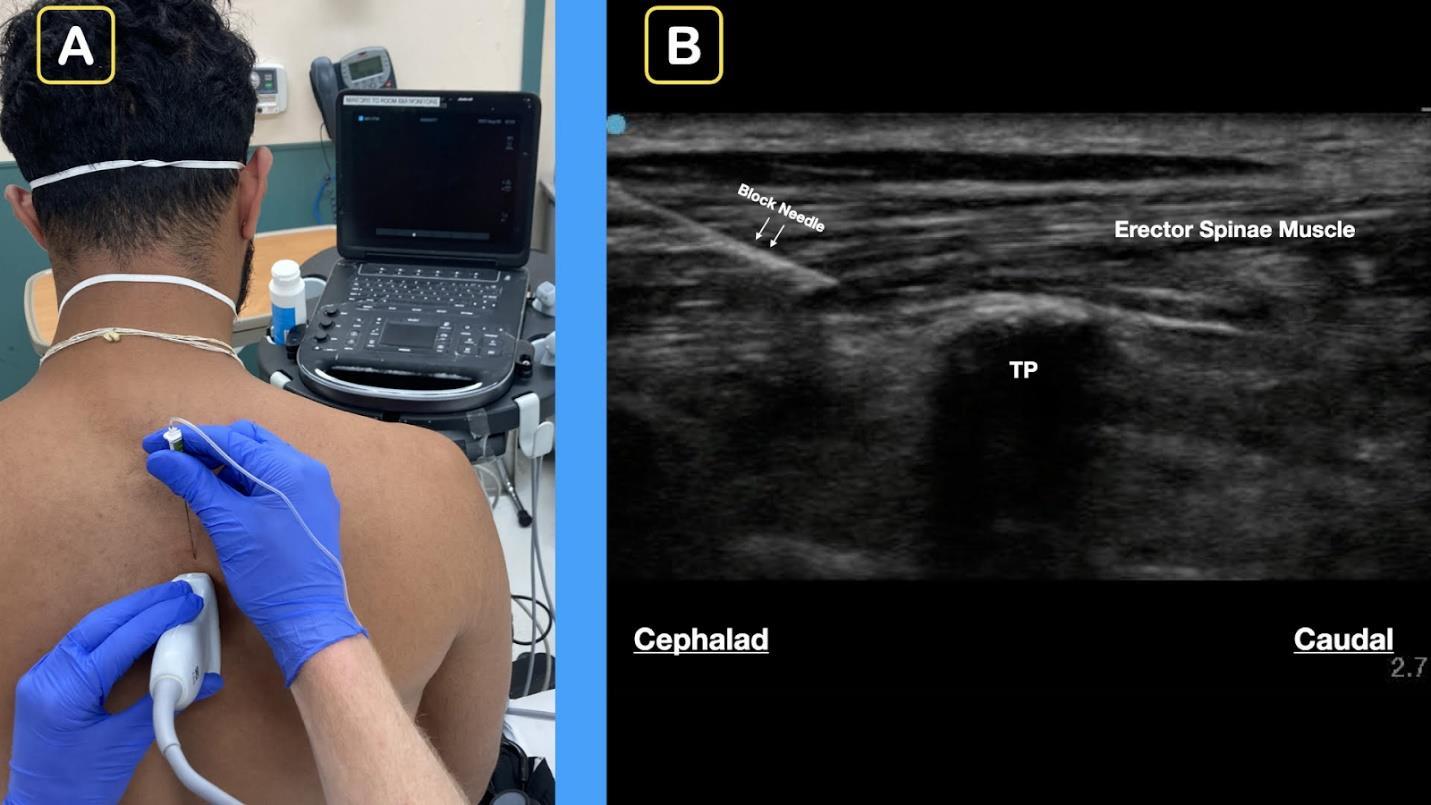
DISCUSSION
This case adds to the body of literature that indicates ESPB is safe, simple, and efficacious in treating thoracoabdominal pain in the ED.6,7 The ESPB is uniquely
Articles in Press 315 Clinical Practice and Cases in Emergency Medicine
Ashworth et al.
Ultrasound-guided ESPB in ED for Abdominal Malignancy Pain
Pain Ashworth
effective as it can treat both somatic and visceral pain without the harmful side effects of opioids.3,8-10 To our knowledge, this is the first case of an ESPB for the treatment of acute on chronic abdominal pain due to metastatic abdominal disease in the ED. As abdominal pain from metastatic disease is a common reason why patients present to the ED, this block offers a promising solution to treat their pain.11
The ESPB may have utility in addressing several well documented disparities in pain treatment in the ED. As already discussed, ESPB could have the ability to provide adequate pain relief for under-treated pain from cancer.10 An aversion to prescribing opioids and the evidence of racial bias in prescribing patterns, as well as a lack of pain management specialists, have led to poor treatment of acute pain in all patients, but particularly in racial minorities.10,12 Patients of color experience great disparities in opioid prescriptions in the ED due to racial bias and the social stigma related to opioid use.13,14 In the ED, Black patients are prescribed opioids less often than Whites across all levels of socioeconomic status.13 Addressing this disparity in treatment has no simple solution and requires addressing systemic, interpersonal, and internalized discrimination. Removing the social stigma and bias related to opioids with increased utilization of blocks such as the ESPB could be a singular step in improving equitable and more effective treatment in the ED.
While our group has not experienced any complications related to the ESPB (such as pneumothorax, anesthetic systemic toxicity, or lower extremity weakness) complication rates in the ED should be explored. The impacts of increasing use of ultrasound-guided nerve blocks for pain management in the ED could parallel outcomes seen in the inpatient setting such as reduced opioid use, decreased admission time, lower cost to patients, and improved recovery.15 As the evidence continues to grow for the successful use of ESPB in the ED for severe pain, there is a need for more systematic studies to investigate side effects, duration of pain control, and clinical outcomes.
CONCLUSION
As EDs continue to face challenges in adequately treating patients with acute on chronic pain from cancer, nerve blocks such as the ESPB offer a solution. Blocks such as the ESPB also offer several advantages over opioids and could lead to more equitable treatment of pain. While this case report adds to the growing levels of evidence that the ESPB may help treat acute pain for abdominal pathologies in the ED, more research is needed to understand the large-scale efficacy and adverse effects of the ESPB.
The authors attest that their institution requires neither Institutional Review Board approval, nor patient consent for publication of this case report. Documentation on file.
Address for Correspondence: Henry Ashworth, MD, MPH, Alameda Health System, Highland Hospital, Department of Emergency Medicine, 1411 E 31st St, Oakland, CA 94602. Email: hcashwor@gmail.com.
Conflicts of Interest: By the CPC-EM article submission agreement, all authors are required to disclose all affiliations, funding sources and financial or management relationships that could be perceived as potential sources of bias. The authors disclosed none.
Copyright: © 2022 Ashworth et al. This is an open access article distributed in accordance with the terms of the Creative Commons Attribution (CC BY 4.0) License. See: http://creativecommons.org/ licenses/by/4.0/
REFERENCES
1. Tsai SC, Liu LN, Tang ST, et al. Cancer pain as the presenting problem in emergency departments: incidence and related factors. Support Care Cancer. 2010;18(1):57-65.
2. Mercadante S, Adile C, Giarratano A, et al. Breakthrough pain in patients with abdominal cancer pain. Clin J Pain. Jun 2014;30(6):510-4.
3. Bennett M, Paice JA, Wallace M. Pain and opioids in cancer care: benefits, risks, and alternatives. Am Soc Clin Oncol Educ Book. 2017;37:705-13.
4. Stjernswärd J. WHO cancer pain relief programme. Cancer Surv 1988;7(1):195-208.
5. Herrero JF, Laird JM, López-García JA. Wind-up of spinal cord neurones and pain sensation: much ado about something? Prog Neurobiol. 2000;61(2):169-203.
6. Mantuani D, Josh Luftig PA, Herring A, et al. Successful emergency pain control for acute pancreatitis with ultrasound guided erector spinae plane blocks. Am J Emerg Med. 2020;38(6):1298.e5-7.
7. Luftig J, Mantuani D, Herring AA, et al. Successful emergency pain control for posterior rib fractures with ultrasound-guided erector spinae plane block. Am J Emerg Med. 2018;36(8):1391-6.
8. Forero M, Adhikary SD, Lopez H, et al. The erector spinae plane block: a novel analgesic technique in thoracic neuropathic pain. Reg Anesth Pain Med. 2016;41(5):621-7.
9. Kurita GP, Sjøgren P, Klepstad P, et al. Interventional techniques to management of cancer-related pain: clinical and critical aspects. Cancers (Basel). 2019;11(4):443.
10. Scarborough BM and Smith CB. Optimal pain management for patients with cancer in the modern era. CA Cancer J Clin 2018;68(3):182-96.
11. van den Beuken-van Everdingen MH, de Rijke JM, Kessels AG, et al. Prevalence of pain in patients with cancer: a systematic review of the past 40 years. Ann Oncol. 2007;18(9):1437-49.
12. Anderson KO, Green CR, Payne R. Racial and ethnic disparities in pain: causes and consequences of unequal care. J Pain 2009;10(12):1187-204.
13. Tulgar S, Selvi O, Senturk O, et al. Ultrasound-guided erector
Clinical Practice and Cases in Emergency Medicine 316 Articles in Press
et al.
Ultrasound-guided ESPB in ED for Abdominal Malignancy
Ashworth et al.
spinae plane block: indications, complications, and effects on acute and chronic pain based on a single-center experience. Cureus 2019;11(1):e3815.
14. Pletcher MJ, Kertesz SG, Kohn MA, et al. Trends in opioid prescribing by race/ethnicity for patients seeking care in US
Ultrasound-guided ESPB in ED for Abdominal Malignancy Pain
emergency departments. JAMA. 2008;299(1):70-8.
15. Cecchi F. Can peripheral nerve blocks improve patients’ outcomes in adults with hip fracture?: A Cochrane Review summary with commentary on implications for practice in rehabilitation. Am J Phys Med Rehabil. 2021;100(10):e139-41.
Articles in Press 317 Clinical Practice and Cases in Emergency Medicine
Case Report
A Case Report of a Lebanon Viper (Montivipera bornmuelleri) Envenomation in a Child
Faysal Tabbara, MD*
Sarah S Abdul Nabi, MD*
Riad Sadek, PhD‡
Ziad Kazzi, MD†
Tharwat El Zahran, MD*
Section Editor: Rachel Lindor, MD, JD
* † ‡
American University of Beirut, Department of Emergency Medicine, Beirut, Lebanon Emory University, Department of Emergency Medicine, Atlanta, Georgia American University of Beirut, Department of Biology, Beirut, Lebanon
Submission History: Submitted January 21, 2022; Revision received May 10, 2022; Accepted May 12, 2022 Electronically published August 6, 2022
Full text available through open access at http://escholarship.org/uc/uciem_cpcem
DOI: 10.5811/cpcem.2022.2.56176
Introduction: Snake envenomation is a serious public health concern. In the Middle East little is known about snakebite envenomation, which raises several challenges for emergency physicians caring for these patients.
Case report: We report the case of a five-year-old boy bitten by a rare snake, Montivipera bornmuelleri, who presented to an emergency department in Lebanon. We also discuss the proper management of snake envenomation.
Conclusion: This case is unique as snakebites in Lebanon are poorly studied, and little is known about the epidemiology and clinical manifestations of local snakebites. [Clin Pract Cases Emerg Med. 2022;6(4):318-322.]
Keywords: Lebanon viper; Montivipera bornmuelleri; envenomation; case report.
INTRODUCTION
In 2017, snake envenomation was listed by the World Health Organization (WHO) as a neglected tropical disease.1 Globally, snake envenomation represents a serious public health concern with approximately five million snakebites occurring every year. An estimate of 81,000 to 138,000 people die yearly because of snakebites, and approximately three times as many amputations and other disabilities are caused by snakebites annually.2,3 Unfortunately, in the Middle East there is insufficient data describing snakebite envenomation, which constitutes a challenge for clinicians who care for these patients in the emergency department (ED). Indeed, the lack of knowledge about these snakes and the lack of timely appropriate antivenom at the Lebanese hospitals are two of the main causes of morbidity. One of the few studies available has described the types of snakes in Lebanon, incidence, clinical outcomes, and available antivenoms.4 We report the case of a five-year-old child who was bitten by a rarely encountered Lebanese snake, Montivipera bornmuelleri, which is also known as the Lebanon viper.
CASE REPORT
A five-year-old boy presented to the ED 13 hours after sustaining a snakebite to his right middle finger. The incident occurred in the Cedars (Arez) area (northern Lebanon) at around 2000 meters above sea level. Prior to arrival to our institution, the patient was taken to another hospital where he was treated with dexamethasone and promethazine. The parents killed the snake and stored it in a bottle filled with an isopropyl alcohol-based solution (Image 1). Beside it is a photograph of Montivipera bornmuelleri taken in nature (Image 1). The parents obtained the polyvalent anti-venom Antivenom-2 from the Lebanese Ministry of Public Health. Antivenom-2 is manufactured in Syria [Scientific Studies and Research Center, Damascus]) (Image 2).
Upon arrival to the ED, the patient was complaining of mild pain in his right middle finger. He denied vomiting, abdominal pain, shortness of breath, loss of consciousness, headache, or other complaints. The patient was hemodynamically stable with a blood pressure of
Clinical Practice
Medicine 318 Volume 6, no. 4: November 2022
and Cases in Emergency
105/63 millimeters of mercury, temperature 36.5°C, heart rate 88 beats per minute, and oxygen saturation 99% on room air. Pertinent physical examination findings included a well-appearing and comfortable child with a normal rate and rhythm and normal pulses. He had good bilateral air entry on lung auscultation, and his neurologic exam was unremarkable. On musculoskeletal exam, his right middle finger was swollen with ecchymosis of the middle phalanx (Image 3). The finger had intact range of motion over the proximal and distal interphalangeal joints, with intact sensation. There was no erythema or warmth, and the capillary refill was less than two seconds.
Initial laboratory studies revealed a complete blood count (CBC) with a white blood cell count (WBC) of 9.7 thousand cells per cubic millimeters. (K/mm3) (reference range 5 to 17 K/mm3), hemoglobin (Hb) 13.9 grams per deciliter (g/ dL) (10-15 g/dL), platelets (plt) 346,000 /µL (150 to 400,000/ µL), creatinine 0.4 milligrams/dL (mg/dL) (0.6 to 1.2 mg/dL), D-dimer 210 nanograms per milliliter (ng/mL) (normal less than 255 ng/mL), prothrombin time 11.9 seconds (control 11.4 seconds), activated partial thromboplastin time 32.1 seconds (control 32.5 seconds), international normalized ratio (INR) 1.0 (0.9 to 1.2), fibrinogen 2.38 grams per liter (g/L) (1.7 to 4 g/L) and a creatinine phosphokinase (CPK) 712 international units per kilogram (IU/kg) (20 to 205 IU/kg).
The snake was identified by the consulting medical toxicology team as Montivipera bornmuelleri. A digital plethysmography was done by the vascular surgery consultant, which showed normal digital pressures in both hands as well as patent radial and ulnar arteries with normal triphasic flow pattern. Due to the swelling and the concern for additional progression, the child was given 5 mL of Antivenom-2
Repeat laboratory studies taken eight hours later (20 hours after the bite) revealed a WBC of 15,400 K/mcL. Hb of 12.5 g/ dL, plt 340,000/µL , D-dimer 182 ng/mL, INR 1.0, fibrinogen 2.05 g/L, and CPK 849 IU/L. No systemic toxicity developed. The swelling did not extend beyond the middle phalanx, and
CPC-EM Capsule
What do we already know about this clinical entity?
Montivipera bornmuelleri snake is rare and was shown in experimental studies to exhibit hemodynamic, hemolytic, pro-inflammatory and neurotoxic properties.
What makes this presentation of disease reportable?
We detail the clinical presentation, management and the use of the polyvalent anti-venom specific for rare snakes inhabiting the Middle East area.
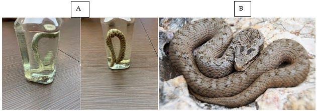
What is the major learning point?
The major learning point from this case report is to highlight the clinical manifestation and management of this rare envenomation.
How might this improve emergency medicine practice?
It would benefit the emergency physicians as a first guide to the presentation and management of this rare case of snake envenomation.
ecchymosis decreased. Repeated laboratory studies including coagulation profile remained normal. The increase in WBC was attributed either to being stress-induced or due to the corticosteroids taken at the outside hospital. The patient was successfully discharged home from the ED and instructed to obtain repeat CBC, coagulation profile, and CPK the next day and to follow up with the toxicology team. Follow-up images of
Volume 6, no. 4: November 2022 319 Clinical Practice and Cases in Emergency Medicine
Tabbara et al. Lebanon Viper (Montivipera bornmuelleri) Envenomation in a Child
Image 1. A Lebanon viper (Montivipera bornmuelleri) snake killed at 2000-meter altitude in northern Lebanon (A) and an image of the Montivipera bornmuelleri taken in nature (B).
the right middle phalanx were obtained the next day (Image 3b) and three days after discharging the patient (Image 3c).
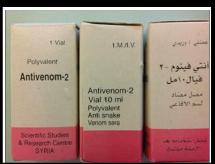
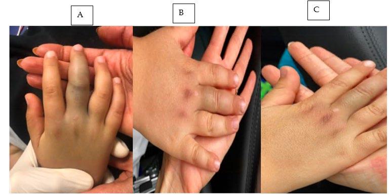
DISCUSSION
Snakebites in Lebanon have been poorly studied, and little is known about the epidemiology or clinical manifestations of local snakebites.5 Lebanon has around 25 species of snakes, three of which are venomous vipers: Daboia palaestinae, Macrovipera lebetina, and M. bornmuelleri. One study4 found that most cases of envenomation were attributed to M. lebetina and D. palaestinae. The clinical manifestations of
these snakebites include tachycardia (33.3%), hypotension (20.8%), anaphylaxis (12.5%), headache (4.2%), and nausea, vomiting, and abdominal pain (4.2% each). Hematological abnormalities include leukocytosis (37.5%), and coagulopathy and thrombocytopenia (12.5% each). Among the reported complications are dizziness or impaired consciousness, compartment syndrome, deep venous thrombosis, acute respiratory distress syndrome, sepsis, cellulitis, upper gastrointestinal bleeding, vaginal bleeding, and congestive heart failure.
Although little is known about the biology of this rare species, it is likely to share aspects with other members of the Viperidae family and the subfamily Viperinae. This species is endemic to high altitudes (above 1800 meters) and is mainly found in Lebanon, specifically in Mount Lebanon, and less abundantly in Palestine. Members of the Montivipera genus usually have a short tail and can grow to a maximum of 75 centimeters in length.6
M. bornmuelleri viper venom is known to reduce blood pressure by displaying vasorelaxant effects that act synergistically on different pathways. It can act on endothelial cells and induce the release of the vasoactive mediator, nitric oxide, reduce calcium ion influx through voltagedependent calcium channels, and inhibit contraction induced by angiotensin I.7 The venom has been shown to exhibit strong antibacterial, hemolytic, anticoagulant, and proinflammatory activities.8 Recent in vivo studies have explored some potential anti-cancer and immunomodulator properties that the M. bornmuelleri viper venom might exhibit.8,9 It is well known that viper venom can act on the nervous system.7,8,9 However, no studies exist outlining the effect of M. bornmuelleri venom on the nervous system. Some researchers
Clinical Practice
Cases in Emergency Medicine 320 Volume 6, no. 4: November 2022
and
Lebanon Viper (Montivipera bornmuelleri) Envenomation in a Child Tabbara et al.
Image 2. Images of the polyvalent anti-venom (Antivenom-2) manufactured by the Scientific Studies and Research Center in Damascus, Syria, which was obtained by the child’s parents from the Lebanese Ministry of Public Health.
Image 3. A five-year-old patient with a Montivipera bornmuelleri envenomation resulting in swelling over his right middle phalanx (A) with follow-up after anti-venom administration the next day (B) and three days later (C).
suggest that this neurologic effect might be attributed to the effect that this venom has on calcium/potassium and sodium channel blockage.7 Future studies are needed to highlight other potential biological and clinical manifestations of this rare viper’s envenomation.
To our knowledge, few or no reported cases exist in the literature describing the clinical manifestations or mortality caused by envenomation by the M. bornmuelleri as it is considered one of the rarest snakes inhabiting high altitudes in the Middle East.10 In fact, Abi-Rizk et al, in a study performed on mice, reported that the median lethal dose (LD50) of M. bornmuelleri venom injected intramuscularly LD50 is 5.93 mg/kg and intraperitoneally (IP) LD50 is around 1.93. The lethality of the M. bornmuelleri venom is similar to that of D. palestinae with an experimental IP LD50 of 1.9 mg/kg.9
Luckily our patient developed only minor local signs and symptoms without any systemic toxicity. The role of the antivenom that was administered by the treating physician is unknown. This patient received the antivenom in this case because it is not a common envenomation; however, it is one of the toxic envenomations that can lead to coagulopathy and death. Furthermore, it is the first snakebite treated at our institution. Antivenom-2 is manufactured from the serum of horses that were immunized with venom from the snakes D. palaestinae, Macrovipera lebetinus, Vipera xanthina, Vipera amodytes, and Cerastes cerastes. This antivenom had formerly been adopted by the Lebanese Ministry of Public Health and distributed free of charge to hospitals. Due to the war in Syria and the resulting difficulties in procuring this antivenom, it was recently substituted by the Menaven/Biosnake antivenom manufactured in India by VINS Bioproducts Ltd, Hyderabad.11
While this antivenom is listed to be active specifically against some of the Lebanese snakes, M. lebitinus and D. palaestinae, it is not listed for use in cases of bites from the Lebanon viper. In our clinical experience in Lebanon, other products are occasionally used that are not listed to be active specifically against any of the snakes indigenous to Lebanon. A geographically specific and comprehensive database of snakes and antivenoms is urgently needed to address these important gaps in the appropriate management of snakebites, which the WHO has deemed a neglected tropical disease with an asymmetrical impact on developing countries. Of note, the dosing of the antivenom is not dependent upon age or weight but it does vary based on the severity of the envenomation.11 For more information about the antivenoms available, we are attaching below in Appendix A the leaflet of the Syrian antivenom along with its translation and in Appendix B the leaflet of the Indian antivenom.
CONCLUSION
The scenario we report describes an interesting case of a five-year-old male patient who presented after a snakebite envenomation on his right finger, secondary to the M. bornmuelleri. He was successfully treated with polyvalent
antivenom, after which the swelling did not extend beyond the middle phalanx and the ecchymosis decreased. The patient was discharged home after observation at our ED. This case is unique as snakebites in Lebanon are poorly studied, and little is known about the epidemiology and clinical manifestations of local snakebites, in particular those caused by envenomation by the M. bornmuelleri, given that it is considered one of the rarest snakes inhabiting high altitudes in the Middle East.
Author Contributions: FT, SSAN, ZK, RS, and TEZ made a substantial contribution to the conception of this manuscript. ZK and TEZ were directly involved in patient care. FT and SSAN equally wrote and contributed to the paper. All authors read and approved the final manuscript.
Documented patient informed consent and/or Institutional Review Board approval has been obtained and filed for publication of this case report.
Address for Correspondence: Tharwat El Zahran, MD, American University of Beirut, Department of Emergency Medicine, Riad El Solh, Beirut 1107 2020, Lebanon. Email: te15@aub.edu.lb.
Conflicts of Interest: By the CPC-EM article submission agreement, all authors are required to disclose all affiliations, funding sources and financial or management relationships that could be perceived as potential sources of bias. The authors disclosed none.
Copyright: © 2022 Tabbara et al. This is an open access article distributed in accordance with the terms of the Creative Commons Attribution (CC BY 4.0) License. See: http://creativecommons.org/ licenses/by/4.0/
REFERENCES
1. World Health Organization (WHO). Snakebite envenoming. [Internet] 2019; 2019. Geneva: WHO. Available from: https://www.who.int/ news-room/fact-sheets/detail/snakebite-envenoming. Accessed March 18, 2022.
2. Chippaux JP. Snake-bites: appraisal of the global situation. Bull World Health Organ. 1998;76(5):515-524.
3. Kasturiratne A, Wickremasinghe AR, de Silva N, et al. The global burden of snakebite: a literature analysis and modelling based on regional estimates of envenoming and deaths. PLoS Med 2008;5(11):e218.
4. El Zahran T, Kazzi Z, Chehadeh AA, et al. Snakebites in Lebanon: a descriptive study of snakebite victims treated at a tertiary care center in Beirut, Lebanon. J Emerg Trauma Shock. 2018;11(2):119-24.
5. Attar S and Nassif R. Snake bite and snakes in Lebanon. J Med Liban. 1953;6(6):366-75.
6. Rima M, Alavi Naini SM, Karam M, et al. Vipers of the Middle East: a rich source of bioactive molecules. Molecules. 2018;23(10):2721.
Volume 6, no. 4: November 2022 321 Clinical Practice
Emergency Medicine
and Cases in
Tabbara et al. Lebanon Viper (Montivipera bornmuelleri) Envenomation in a Child
7. Accary C, Hraoui-Bloquet S, Sadek R, et al. The relaxant effect of the Montivipera bornmuelleri snake venom on vascular contractility. J Venom Res. 2016;7:10-5.
8. Yacoub T, Rima M, Sadek R, et al. Montivipera bornmuelleri venom has immunomodulatory effects mainly up-regulating pro-inflammatory cytokines in the spleens of mice. Toxicol Rep. 2018;5:318-23.
9. Abi-Rizk A, Rima M, Bloquet SH, et al. Lethal, hemorrhagic,
and necrotic effects of Montivipera bornmuelleri venom. Current Herpetology. 2017; 58-62.
10. Amr ZS, Abu Baker MA, Warrell DA. Terrestrial venomous snakes and snakebites in the Arab countries of the Middle East. Toxicon 2020;177:1-15.
11. Jabbour, E. Snakebite in Lebanon: the painful reality. Med J Emerg Med. 2020;28:64-8.
Clinical Practice
Medicine 322 Volume 6, no. 4: November 2022
and Cases in Emergency
Lebanon Viper (Montivipera bornmuelleri) Envenomation in a Child Tabbara et al.
Pancreatitis with a Normal Serum Lipase, a Rare Post-esophagogastroduodenoscopy Complication: A Case Report
Molly Sturlis, BS* Karen McGrane, MD†
Section Editor: Christopher San Miguel, MD
Submission History: Submitted December 15, 2021; Revision received February 25, 2022; Accepted March 9, 2022 Electronically published July 27, 2022
Full text available through open access at http://escholarship.org/uc/uciem_cpcem
DOI: 10.5811/cpcem.2022.3.55706
Introduction: Pancreatitis after esophagogastroduodenoscopy (EGD) is not a common occurrence, particularly in the setting of a normal serum lipase. The lack of commonality may delay diagnosis and treatment in some patients presenting to the emergency department (ED) with abdominal pain after an otherwise uncomplicated procedure.
Case Report: A patient with a history of gastroesophageal reflux disease presented to the ED with a complaint of abdominal pain and fever three days after an uncomplicated EGD. The patient was ultimately diagnosed with pancreatitis after a computed tomography showed pancreatic head inflammation, despite having a normal serum lipase. There were no other identified risk factors for pancreatitis in this case.
Conclusion: This case serves to bring awareness to this potential procedural complication and the possibility of pancreatitis with a normal serum lipase. [Clin Pract Cases Emerg Med. 2022;6(4):323-326.]
Keywords: case report; pancreatitis; esophagogastroduodenoscopy; lipase.
INTRODUCTION
Upper gastrointestinal endoscopy, also known as esophagogastroduodenoscopy (EGD), is a well tolerated, common diagnostic and therapeutic procedure with typical complications related to sedation, bleeding, perfor-ation, and infection.1 There have been fewer than an estimated 10 case reports of pancreatitis as a complication of post-upper endoscopies that do not involve ampullary cannulation; therefore, it is not routinely considered.2-4 We present a case of acute pancreatitis diagnosed in the emergency department (ED) three days after uncomplicated EGD. This case presented with an atypical location of pain, normal lipase level, and a lack of common risk factors.
CASE REPORT
A 41-year-old White patient with a history of hypertension and gastroesophageal reflux disease (GERD) presented to the ED with fever and right mid to low abdominal pain after
undergoing an uncomplicated esophagogastroduodenoscopy (EGD), with biopsies taken from the lesser curvature of the stomach, three days prior as part of the workup for persistent GERD. The pain began the night of the endoscopy and was originally in the mid to periumbilical region. The symptoms spontaneously improved the next day before progressively worsening, with the pain migrating to the right lower quadrant and reported fever of 38.9°C. The patient noted some nausea over the preceding days but had not vomited and had a normal appetite. The patient denied having diarrhea and stated they had only one bowel movement since the endoscopy, which was abnormally pellet-like, and they had not had a bowel movement or flatus in the prior 24 hours. The patient denied any previous abdominal surgeries. Current medications were bupropion, lisinopril, and omeprazole.
On physical examination, the patient was well appearing and afebrile, hypertensive to 147/102 millimeters of mercury, normal heart rate and respiratory rate, and in no acute
Volume 6, no. 4: November 2022 323 Clinical Practice and Cases in Emergency Medicine Case Report
Midwestern University Arizona College of Osteopathic Medicine, Glendale, AZ Madigan Army Medical Center, Department of Emergency Medicine, Tacoma, WA * †
Pancreatitis with a Normal Serum Lipase
respiratory distress. The abdomen was soft and non-distended with hypoactive bowel sounds. There was mild tenderness and voluntary guarding in the right lower quadrant, and the area of maximal tenderness was at McBurney’s point. No rebound tenderness, Rovsing’s, or Murphy’s signs were present.
Laboratory findings were notable for a white blood cell count of 11.5 x 10³ per microliter (103/µL) (reference range 4.5 – 13.0 103/µL) with 74.1% neutrophils (reference range 38.5 – 76.5%), with the remainder of the complete blood count and complete metabolic panel within normal limits. Lipase level was also within normal limits at 36 units per liter (U/L) (reference range 13-60 U/L). A computed tomography of the abdomen and pelvis with intravenous contrast was obtained and indicated slight heterogeneity and enlargement of the pancreatic head with adjacent inflammatory fat stranding indicative of acute pancreatitis (Images 1 and 2).
There was no evidence of appendicitis. On re-evaluation, the patient stated that their pain had mildly improved after receiving ketorolac 15 milligrams intravenously and a normal saline fluid bolus and that the pain was now located periumbilically, similarly to how it felt at initial onset. The patient no longer had tenderness in the right lower quadrant, and there was no guarding or rebound tenderness.
A right upper quadrant ultrasound was obtained to assess for gallstones as a possible cause of acute pancreatitis in this patient. It indicated no cholelithiasis with poor visualization of the pancreas due to bowel gas. The patient denied alcohol use. Prior record review indicated the patient had a history of borderline hypertriglyceridemia, with a level of 204 milligrams per
CPC-EM Capsule
What do we already know about this clinical entity? While there have been some case reports of pancreatitis following uncomplicated esophagogastroduodenoscopy, it remains an uncommon complication.
What makes this presentation of disease reportable? This case report features two uncommon occurrences - pancreatitis following uncomplicated esophagogastroduodenoscopy (EGD) in the setting of a normal lipase level. The two findings in conjunction make this an exceedingly rare event.
What is the major learning point?
Index of suspicion for pancreatitis should remain elevated in the setting of normal lipase enzyme levels. Consider pancreatitis in patients presenting with abdominal pain following EGD.
How might this improve emergency medicine practice?
This case identifies a rare cause of pancreatitis and may guide the clinician’s decision making process when seeing patients presenting with concerns following EGD.
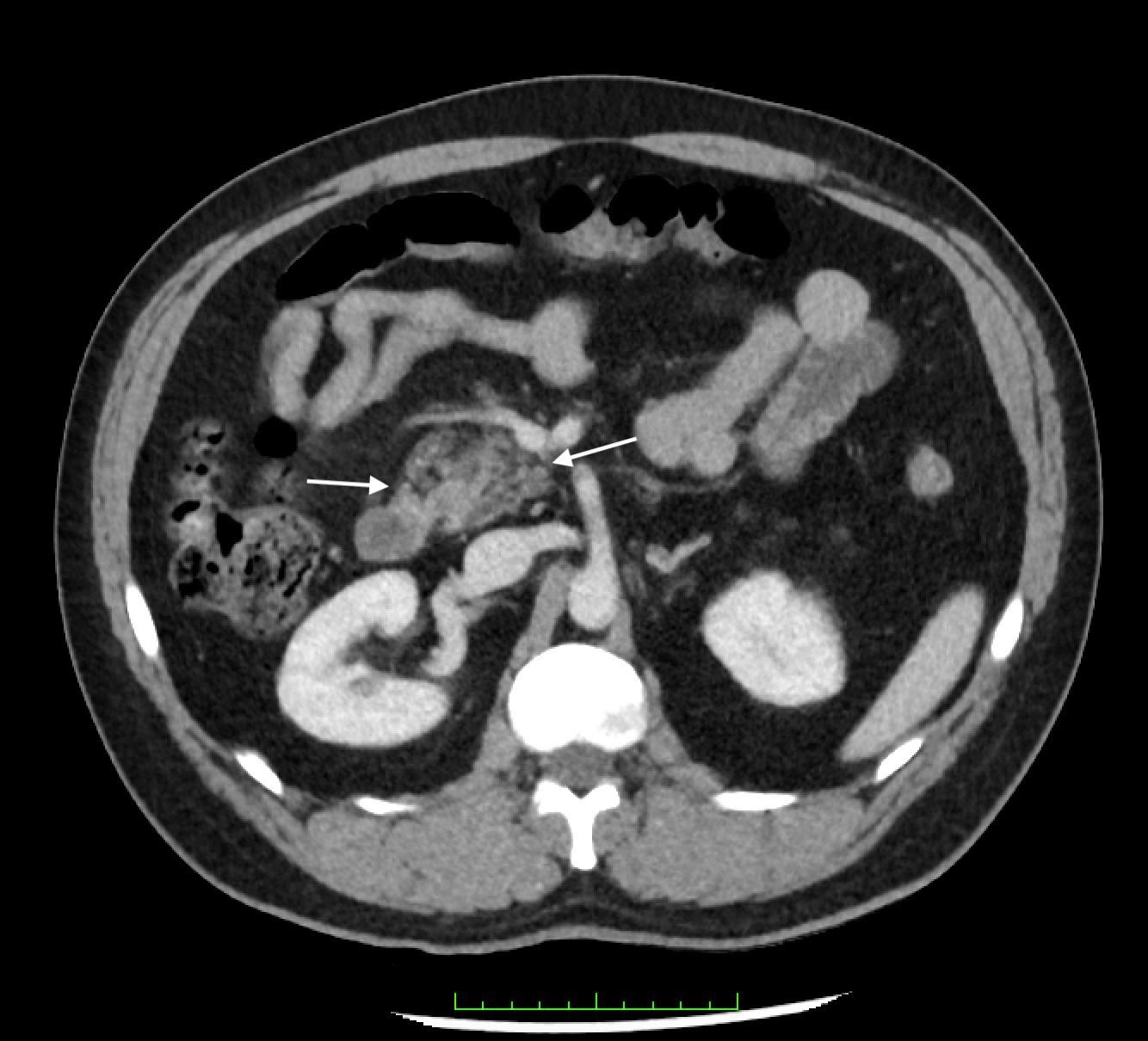
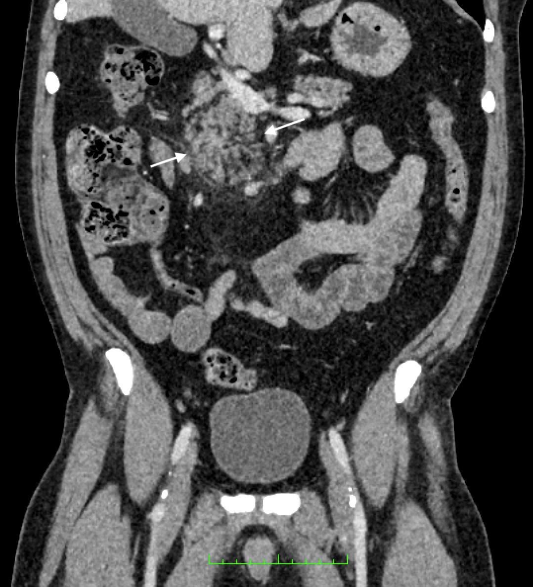
Clinical Practice and Cases in Emergency Medicine 324 Volume 6, no. 4: November 2022
Sturlis et al.
Image 1. Axial computed tomography view showing inflammatory stranding of the pancreatic head (arrows).
Image 2. Coronal computed tomography view showing inflammatory stranding of the pancreatic head (arrows).
et al.
deciliter (mg/dL) (reference range 0 mg/dL to 200 mg/dL) just three months prior to this presentation.
Given the improvement of symptoms and mild disease course, the patient was discharged home in stable condition and given strict return precautions as well as instruction to follow up with the gastroenterologist. On chart review, telephonic follow-up conducted by the patient’s gastroenterologist revealed improved symptoms, and biopsies from the EGD were normal, with no additional potential etiologies discovered for the clinical presentation and radiographic findings consistent with pancreatitis.
DISCUSSION
In the absence of other plausible etiologies of acute pancreatitis, including alcoholism, cholelithiasis, hypertriglyceridemia, endoscopic retrograde cholangiopancreatography (ERCP), or offending medications, it is reasonable to explore a correlation between the recent upper endoscopy and findings consistent with pancreatitis on imaging.5 Pancreatitis is a well documented complication of ERCP, which involves ampullary cannulation but is not considered to be a complication of EGD.6 There have been few case reports of acute pancreatitis following EGD described in the literature. The mechanism by which uncomplicated EGD may cause acute pancreatitis is not well understood; it is theorized to be due to mechanical trauma or over insufflation during the procedure that causes local inflammation and ultimately may lead to development of acute pancreatitis within several days of the procedure.
This patient initially experienced periumbilical pain that migrated to the right lower quadrant, along with fever and some associated nausea, most suggestive of acute appendicitis. However, the CT did not support this diagnosis but instead indicated an acute pancreatitis. After pain control with ketorolac, the patient’s pain did again localize to the periumbilical region. The non-classic location of pain, the normal lipase level, and the lack of common risk factors for pancreatitis make its diagnosis a peculiar one; however, according to the guidelines put forth by American College of Gastroenterology, the diagnosis of acute pancreatitis should still have been made in this case. The guidelines suggest that to make the diagnosis, two of the three following criteria must be met: 1) abdominal pain; 2) serum amylase and/or lipase three times greater than the upper limit; and 3) characteristic findings on abdominal imaging.7
While this patient had normal lipase levels, they did present with abdominal pain and had a CT indicative of acute pancreatitis. The sensitivity of lipase in acute pancreatitis is reported as 64-100%, with a higher likelihood of a normal lipase either early in the presentation or later in the clinical course. Studies have shown the lipase levels rise within three to six hours of onset of pancreatitis and peak within 24 hours. Lipase levels may be persistently elevated for up to two weeks following acute pancreatitis.8 This patient’s
symptoms began more than 24 hours prior to presentation, making a pre-elevation sample unlikely. There have been case reports in the past supporting the possibility of normal lipase levels, particularly in cases of acute on chronic pancreatitis, hypertriglyceridemia induced or in late presentations, none of which fit this clinical scenario.9-13
CONCLUSION
Making the association between EGD and acute pancreatitis is important, since as it stands, pancreatitis is not considered a post-EGD complication. This fact may delay diagnosis and treatment in some patients presenting to the ED with abdominal pain after an otherwise uncomplicated procedure. This case report serves to bring awareness to this potential procedural complication.
The authors attest that their institution requires neither Institutional Review Board approval nor patient consent for publication of this case report. Documentation on file.
Address for Correspondence : Karen McGrane, MD, Madigan Army Medical Center, Department of Emergency Medicine, 9040 Jackson Avenue, McChord, WA 98431. Email: Karen.m.mcgrane.mil@mail.mil.
Conflicts of Interest: By the CPC-EM article submission agreement, all authors are required to disclose all affiliations, funding sources and financial or management relationships that could be perceived as potential sources of bias. The authors disclosed none. The views expressed here are those of the authors and do not reflect the official policy of the Department of the Army, the Department of Defense, or the U.S. Government.
Copyright: © 2022 Sturlis et al. This is an open access article distributed in accordance with the terms of the Creative Commons Attribution (CC BY 4.0) License. See: http://creativecommons.org/ licenses/by/4.0/
REFERENCES
1. Cohen J and Greenwald DA. Overview of upper gastrointestinal endoscopy (esophagogastroduodenoscopy). 2020. Available at: https://www.uptodate.com/contents/ overview-of-upper-gastrointestinal-endoscopyesophagogastroduodenoscopy?search=Overview%20of%20 upper%20gastrointestinal%20endoscopy%20&source=search_re sult&selectedTitle=1~150&usage_type=default&display_rank=1 Accessed October 24, 2021.
2. Fadaee, N and De Clercq, S. Gastroscopy-induced pancreatitis: a rare cause of post-procedure abdominal pain. J Med Cases. 2019;10(5)126-8.
3. Ahmad Y, Anwar K, Dassum SR, et al. Acute pancreatitis after a routine esophagogastroduodenoscopy (EGD). Chest 2018;154(4)286A.
Volume 6, no. 4: November 2022 325 Clinical
Emergency Medicine
Practice and Cases in
Sturlis
Pancreatitis with a Normal Serum Lipase
Pancreatitis with a Normal Serum Lipase Sturlis et
4. Nwafo NA. Acute pancreatitis following oesophagogastroduodenoscopy. BMJ Case Rep. 2017;2017:bcr2017-222272.
5. Nevins AB and Keeffe EB. Acute pancreatitis after gastrointestinal endoscopy. J Clin Gastroenterol. 2002;34(1):94-5.
6. Vege SS. Etiology of acute pancreatitis. 2021. Available at: https://www.uptodate.com/contents/etiology-of-acutepancreatitis?search=Etiology%20of%20Acute%20 Pancreatitis&source=search_result&selectedTitle=1~150&usag e_type=default&display_rank=1. Accessed October 24, 2021.
7. Tringali A, Loperfido S, Costamagna G. Post-endoscopic retrograde cholangiopancreatography (ERCP) pancreatitis. 2021. Available at: https://www.uptodate.com/contents/post-endoscopic-retrogradecholangiopancreatography-ercp-pancreatitis?search=Postendoscopic%20retrograde%20cholangiopancreatography%20 (ERCP)%20pancreatitis&source=search_result&selectedTitle=1~150
al.
&usage_type=default&display_rank=1. Accessed October 26, 2021.
8. Ismail O and Bhayana V. Lipase or amylase for the diagnosis of acute pancreatitis? Clin Biochem. 2017:50(18):1275-80.
9. Yadav D, Agarwal N, Pitchumoni CS. A critical evaluation of laboratory tests in acute pancreatitis. Am J Gastroenterol. 2002;97(6):1309-18.
10. Wang YY, Qian ZY, Jin WW, et al. Acute pancreatitis with abdominal bloating and distension, normal lipase and amylase. Medicine (Baltimore). 2019;98(15):e15138.
11. Shafqet MA, Brown TV, Sharma R. Normal lipase drug-induced pancreatitis: A novel finding. Am J Emerg Med. 2015;33(3):476.e5-6.
12. Agrawal A, Parikh M, Thella K, et al. Acute pancreatitis with normal lipase and amylase: an ED dilemma. Am J Emerg Med. 2016;34(11):P2254.E3-2254.E6.
13. Limon O, Sahin E, Kantar F, et al. A rare entity in ED: normal lipase level in acute pancreatitis. Turkish J Emerg Med. 2016:16(1):32-4.
326 Volume
November 2022
Clinical Practice and Cases in Emergency Medicine
6, no. 4:
19-Year-Old with Sudden Onset Left Testicular Pain
Elan Small, MD Nicholas Ashenburg, MD Kimberly Schertzer, MD
Section Editors: Joel Moll, MD
Stanford University School of Medicine, Department of Emergency Medicine Residency, Palo Alto, California
Submission history: Submitted March 20, 2022; Revision received March 26, 2022; Accepted July 02, 2022 Electronically published October 24, 2022
Full text available through open access at http://escholarship.org/uc/uciem_cpcem
DOI: 10.5811/cpcem.2022.7.56747
Case Presentation: A previously healthy 19-year-old man presented to the emergency department with severe, sudden onset of left testicular pain. Physical exam revealed a left high-riding, horizontally oriented testicle without cremasteric reflex. Point-of-care ultrasound was used to confirm the diagnosis of testicular torsion, as well as to guide manual detorsion, verifying return of blood flow after reduction.
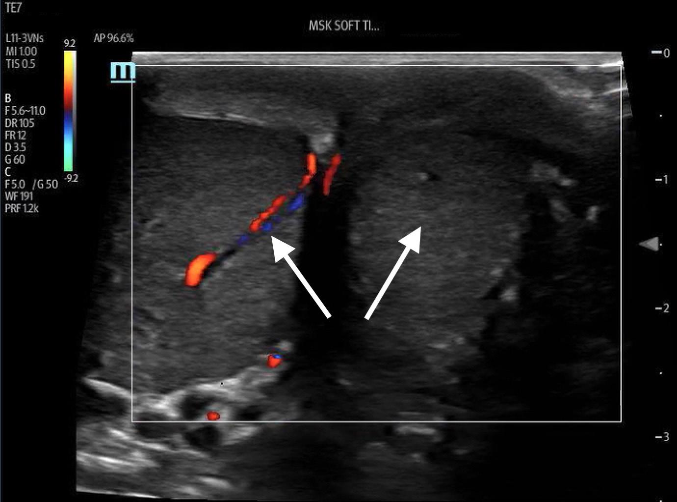
Discussion: Testicular torsion is a urologic emergency in which testicular viability is time dependent. Point-of-care ultrasound can be an important and helpful tool to not only confirm suspicion but help guide adequacy of blood flow return after manual detorsion in conjunction with comprehensive ultrasound. [Clin Pract Cases Emerg Med. 2022;6(4):327–329.]
Keywords: point-of-care ultrasound; testicular torsion; testicle pain.
CASE PRESENTATION
A 19-year-old man was brought by ambulance to the emergency department with left-sided testicular pain. He reported sudden severe, non-radiating left testicular pain that woke him from sleep 30 minutes prior to arrival. He presented in severe pain, tremulous from discomfort. His exam revealed a high-riding, firm, left testicle in a horizontal lie with absent cremasteric reflex. Urology was consulted and a comprehensive ultrasound ordered. The patient’s testicles were immediately examined with point-of-care ultrasound (POCUS) (Image 1). After receiving 100 micrograms of fentanyl, manual detorsion was conducted, and repeat ultrasonography was performed (Image 2). The patient was then transported for comprehensive ultrasonography (Image 3).
DISCUSSION
Testicular torsion is a urologic emergency in which the testicle rotates 180-720 degrees, compromising venous and ultimately arterial circulation, leading to ischemia, necrosis, and nonviability. The literature has demonstrated good survivability at six hours, although it is time dependent with improved outcomes at quicker intervention.1 Classic history and exam features include sudden onset of pain, and a firm, horizontally oriented testicle with absent cremasteric reflex.2 Conventional
teaching invokes early urologic consultation in highly suspicious cases prior to comprehensive ultrasonography.
Article in Press 327 Clinical Practice and Cases in Emergency Medicine
Images in Emergency Medicine
Image 1. Side-by-side view of initial testicular assessment using point-of-care ultrasound. The testicle on the image-right corresponds to the patient’s painful left testicle which was found to be enlarged with surrounding edema and absent Doppler flow. Arrows highlight presence of flow in unaffected testicle and absence of flow in affected testicle.
Image 2. Side-by-side view of testicular assessment with pointof-care ultrasound after manual detorsion. The testicle on the image-right corresponds to the patient’s left testicle, with bilateral appreciable Doppler flow highlighted by the included arrow.
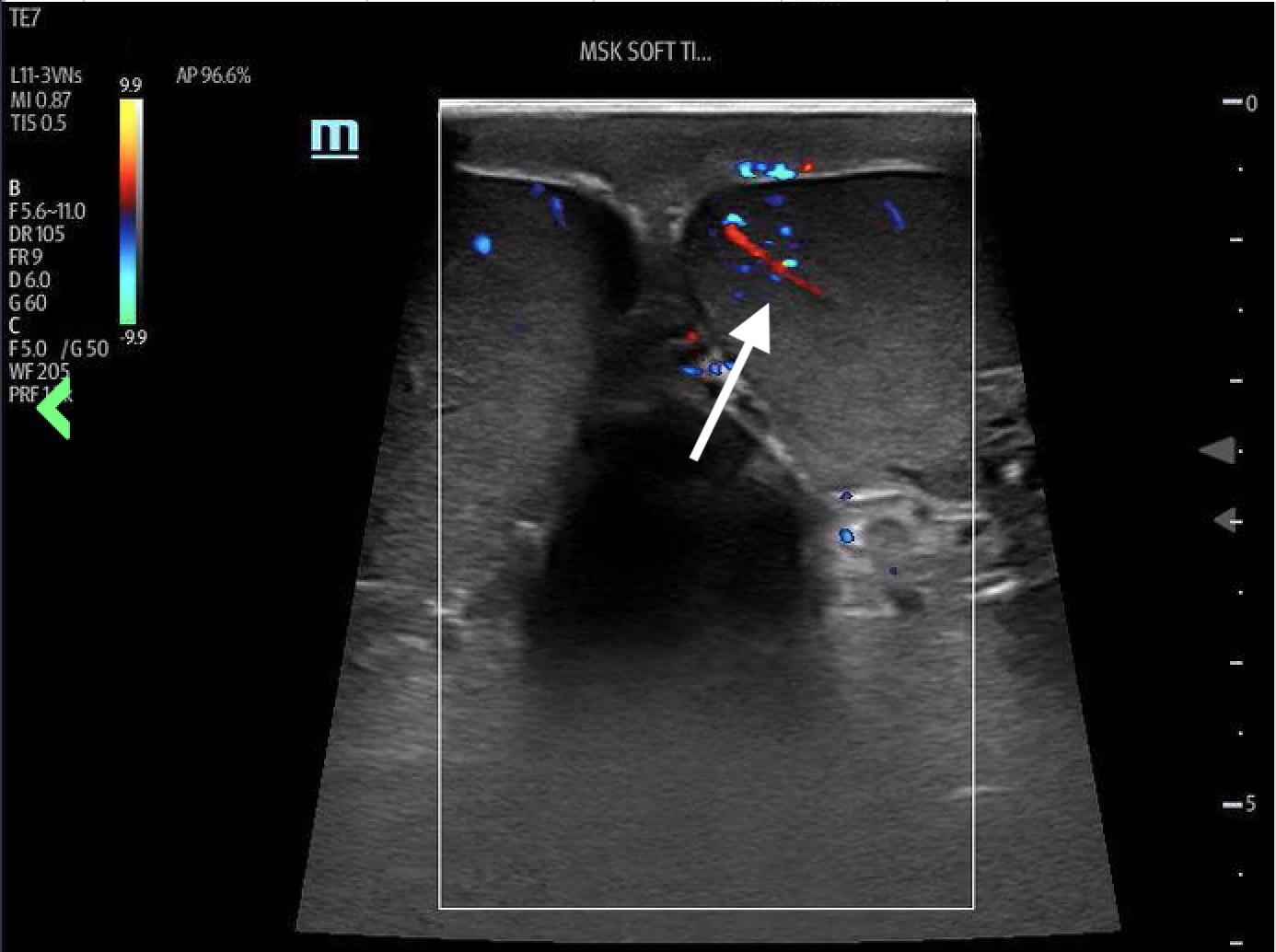
Image 3. Comprehensive ultrasonographic side-by-side view of testicles after manual detorsion. The image-right corresponds to the patient’s left testicle, which demonstrated hyperemia likely related to detorsion event highlighted by the included arrow. Surrounding edema over affected testicle is also seen.
POCUS has emerged as an important diagnostic tool, with up to 100% sensitivity reported in small-sample studies for both fellowship- and non-fellowship trained emergency physicians.3,4 Ultrasonographic features can include the following: an enlarged edematous hypoechoic testicle without Doppler flow; a whirlpool sign (spiral twist of the spermatic cord at the external inguinal ring or scrotal sac); and epidydimal enlargement without hyperemia.5 This patient underwent bedside manual detorsion with return of flow after approximately two lateral rotations. Comprehensive ultrasound demonstrated a hyperemic testicle consistent with recent detorsion. Depending on availability of resources and timing, POCUS can be an important tool in identifying testicular torsion and
CPC-EM Capsule
What do we already know about this clinical entity?
Testicular torsion is a time-sensitive emergent condition for which point-of-care ultrasound has emerged as a potential tool for both identification and management.
What is the major impact of the image(s)?
These images illustrate classic ultrasound findings of testicle edema, and absence of Doppler flow and demonstrate return of Doppler flow after attempted detorsion.
How might this improve emergency medicine practice?
Depending on resources, providers should consider point-of-care ultrasound, paired with comprehensive studies, to identify testicular torsion and confirm detorsion.
helping guide adequacy of reduction in conjunction with comprehensive ultrasonography.
The authors attest that their institution does not require Institution al Review Board approval. Patient consent has been obtained and filed for the publication of this case report. Documentation on file.
Address for Correspondence: Elan Small, MD, Stanford University, Department of Emergency Medicine, 900 Welch Road Ste 350, Palo Alto, CA 94034. Email: esmall17@stanford.edu.
Conflicts of Interest: By the CPC-EM article submission agreement, all authors are required to disclose all affiliations, funding sources and financial or management relationships that could be perceived as potential sources of bias. The authors disclosed none.
Copyright: © 2022 Small et al. This is an open access article distributed in accordance with the terms of the Creative Commons Attribution (CC BY 4.0) License. See: http://creativecommons.org/ licenses/by/4.0/
REFERENCES
1. Blaivas M, Batts M, Lambert M. Ultrasonographic diagnosis of testicular torsion by emergency physicians. Am J Emerg Med 2000;18(2):198-200.
2. Germann C, Holmes, Jeffery. 2017. Selected urologic disorders
Clinical Practice and Cases in Emergency Medicine 328 Volume 6, no. 4: November 2022 19-Year-Old
Small et al.
with Sudden Onset Left Testicular Pain
Small et al.
In: Rosen’s Emergency Medicine: Concepts and Clinical Practice Amsterdam, Netherlands. Elsevier.
3. Friedman N, Pancer Z, Savic R, et al. Accuracy of point-of-care ultrasound by pediatric emergency physicians for testicular torsion. J Pediatr Urol. 2019;15(6):608.e1-608.e6.
4. Blaivas M, Sierzenski P, Lambert M. Emergency evaluation of patients
19-Year-Old with Sudden Onset Left Testicular Pain
presenting with acute scrotum using bedside ultrasonography. Acad Emerg Med. 2001;8(1):90-3.
5. Lee DY, Lee SH, Yoon JA, et al. Detection of testicular torsion with preserved intratesticular flow by point of care ultrasound in the emergency department: a case report. J Emerg Med 2022;62(4):e88-e90.
Volume 6, no. 4: November 2022 329 Clinical Practice and Cases in Emergency Medicine
Spontaneous Tension Hemothorax in a Patient with Asbestosis
Toshinao Suzuki, MD* Toshihiko Takada, MD, PhD†
Section Editor: Steven Walsh, MD
* † Teikyo
Submission History: Submitted April 04, 2022; Revision received April 12, 2022; Accepted June 03, 2022
Electronically published October 27, 2022
Full text available through open access at http://escholarship.org/uc/uciem_cpcem
DOI: 10.5811/cpcem.2022.6.57031
Case Presentation: A 75-year-old man with a history of asbestosis presented to the emergency department with sudden-onset dyspnea and hemoptysis, triggered by coughing. The patient was hemodynamically unstable and in respiratory distress. Computed tomography revealed a massive hemothorax on the left side and compression of the descending thoracic aorta. He underwent emergency surgical exploration after decompression by chest tube insertion. The hemothorax was caused by tears in the pleural adhesions due to asbestosis and induced by coughing.
Discussion: Spontaneous hemothorax is a rare subtype of hemothorax. There have been only a few case reports of spontaneous tension hemothorax. In addition to its typical findings, compression of the thoracic descending aorta was observed in our patient. We hypothesize that severely diminished pulmonary compliance contributed to the extremely high intrathoracic pressure, which led to this unusual finding. [Clin Pract Cases Emerg Med. 2022;6(4):330-332.]
Keywords: thoracic cavity; hemorrhage; shock.
CASE PRESENTATION
A 75-year-old man with a history of asbestosis and cirrhosis with thrombocytopenia presented to the emergency department with sudden-onset dyspnea and hemoptysis triggered by coughing. He had a history of asbestos exposure during his tenure as an air-conditioning engineer 30 years prior. He had no trauma history and was not on anticoagulant or antiplatelet therapy. The patient was hemodynamically unstable and in respiratory distress with a blood pressure of 100/84 millimeters of mercury (mm Hg), pulse rate 116 beats per minute, respiratory rate 24 breaths per minute, and oxygen saturation level 90% on room air. Breath sounds were substantially decreased on the left side, and jugular vein distension was observed. Upon laboratory testing, the patient had a hemoglobin level of 12.4 grams per deciliter (g/dL) (reference range: 13.5-17.5 g/dL), platelet count of 42,000 per microliter (µL) (13,000-37,000/ µL), prothrombin time of 16.5 seconds (10-13 seconds), and activated partial thromboplastin time of 26.3 seconds (20.0-38.0 seconds).
Chest radiography revealed bilateral pleural plaques, complete opacification of the left thorax, and mediastinal shift toward the right (Image 1). Computed tomography (CT) revealed massive hemothorax on the left and compression of the
descending thoracic aorta (Image 2A). While undergoing CT, the patient’s systolic blood pressure dropped to 60 mm Hg. After draining 1,200 milliliters of blood by a chest tube insertion into the left thorax (Image 2B), his blood pressure increased to 98/62 mm Hg.
After transfusion of 20 units of platelets, surgical exploration was performed. Pleural adhesion tears and bleeding were detected. Histopathological examination of the torn parietal pleura with plaques revealed fibrinous pleuritis with massive fibrinosanguineous exudate. During postoperative mechanical ventilation, a severe reduction in pulmonary compliance was observed. After 26 days with delayed ventilator withdrawal and treatment of a urinary tract infection that developed during hospitalization, the patient fully recovered and was discharged. At one-year follow-up he was doing well without recurrence.
DISCUSSION
Spontaneous hemothorax is a rare subtype of hemothorax, characterized by accumulation of blood within the pleural space, with no associated trauma or underlying cause. Conditions such as vascular malformations and neoplasms have been reported as potential causes.1 In our patient, hemothorax was caused by coughing-induced tears
Clinical Practice and Cases in Emergency Medicine 330 Article in Press Images
Emergency Medicine
in
University Chiba Medical Center, Interventional Radiology Center, Chiba, Japan Fukushima Medical University, Department of General Medicine, Shirakawa Satellite for Teaching And Research (STAR), Fukushima, Japan
completely opaque left thorax (black arrowheads), and mediastinal shift toward the right (black arrow).
intrathoracic hemorrhage (arrowheads), and a drainage tube (black arrow) within the left-sided pleural effusion.
in the pleural adhesions due to asbestosis. This entity has been reported previously only in a patient with chronic obstructive pulmonary disease.2
The hemodynamic instability of our patient was attributed to obstructive shock due to tension hemothorax. Tension hemothorax is typically caused by major chest trauma or ruptured thoracic aortic aneurysm, and it rarely
CPC-EM Capsule
What do we already know about this clinical entity?
Spontaneous hemothorax is a rare subtype of hemothorax with no associated trauma or underlying cause.
What is the major impact of the image(s)? In addition to typical findings of tension hemothorax, computed tomography revealed compression of the descending thoracic aorta in a patient with asbestosis.
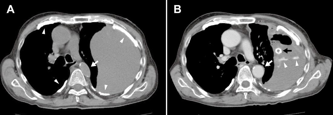
How might this improve emergency medicine practice?
In patients with decreased lung compliance, tension hemothorax could be caused not only by major trauma or ruptured aneurysm but also by spontaneous hemothorax. occurs spontaneously.3 In addition to the typical findings of tension hemothorax, such as opacification of the affected thorax and mediastinal shift,4 compression of the thoracic descending aorta was observed in our patient. Ideally, CT should not be performed before stabilization of the patient in such cases. In patients with asbestosis, asbestos fibers inhaled deep into the lungs get lodged in the tissues, eventually resulting in diffuse alveolar and interstitial fibrosis, leading to decreased lung compliance.5
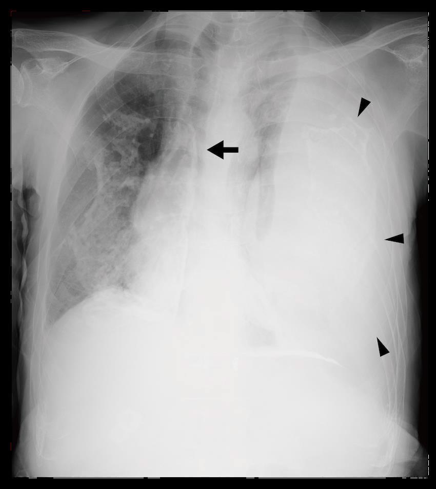
Documented patient informed consent a has been obtained and filed for publication of this case report.
Address for Correspondence: Toshinao Suzuki, MD, Teikyo University Chiba Medical Center, Interventional Radiology Center, 3426-3 Anesaki, Ichihara, Chiba, Japan, 299-0111. Email: toshinao24@gmail.com.
Conflicts of Interest: By the CPC-EM article submission agreement, all authors are required to disclose all affiliations, funding sources and financial or management relationships that could be perceived as potential sources of bias. The authors disclosed none.
Copyright: © 2022 Suzuki et al. This is an open access article distributed in accordance with the terms of the Creative Commons Attribution (CC BY 4.0) License. See: http://creativecommons.org/ licenses/by/4.0/
Volume 6, no. 4: November 2022 331
Medicine
Clinical Practice and Cases in Emergency
Suzuki et al.
Spontaneous Tension Hemothorax in a Patient with Asbestosis
Image 2. Computed tomography (CT) at the level of the tracheal bifurcation (A) before and (B) after chest drainage. (A) Plain CT at the level of the tracheal bifurcation shows mediastinal shift toward the right, compression of the descending thoracic aorta (arrow), massive left hemothorax, and multiple pleural plaques (arrowheads). (B) Contrast-enhanced CT after decompression of the left thorax shows improvement in the aortic compression (white arrow), a contrast blush indicating
Spontaneous Tension Hemothorax in a Patient with Asbestosis Suzuki
REFERENCES
1. Patrini D, Panagiotopoulos N, Pararajasingham J, et al. Etiology and management of spontaneous haemothorax. J Thorac Dis. 2015;7(3):520-6.
2. Luo X, Shi W, Zhang X, et al. Multiple-site bleeding at pleural adhesions and massive hemothorax following percutaneous coronary intervention with stent implantation: a case report. Exp Ther Med . 2018;15(3):2351-5.
3. Jacobsen GH, Brandt B, Ellesøe SG. Spontaneous tension hemothorax in a young male with a Nuss implant. J Pulm Respir Med. 2017;6:394.
4. Ho ML and Gutierrez FR. Chest radiography in thoracic polytrauma. Am J Roentgenol. 2009;192(3):599-612.
5. Schneider J, Arhelger R, Raab W, et al. The validity of static lung compliance in asbestos-induced diseases. Lung 2012;190(4):441-9.
Clinical Practice and Cases in Emergency Medicine 332 Volume 6, no. 4: November 2022
et al.
Absent Pulmonary Artery Presenting as High-Altitude Pulmonary Edema
Douglas Dillon, MD Austin T. Smith, MD
Section Editors: Jacqueline Le, MD
Submission History: Submitted May 26, 2022 Revision received September 12, 2022; Accepted September 20, 2022 Electronically published November 4, 2022
Full text available through open access at http://escholarship.org/uc/uciem_cpcem DOI: 10.5811/cpcem.2022.9.57506
Case Presentation: A 22-year-old male with no known past medical history presented to our emergency department complaining of difficulty breathing. A plain film chest radiograph revealed findings consistent with a tension pneumothorax.
Discussion: However, due to physical examination findings inconsistent with the imaging report, a computed tomography of the chest was ordered which revealed an absent right pulmonary artery. The patient was ultimately treated for high altitude pulmonary edema and discharged on nifedipine and supplementary oxygen. [Clin Pract Cases Emerg Med. 2022;6(4):333-335.]
Keywords: absent pulmonary artery; high altitude pulmonary edema; environmental emergencies; shortness of breath; dyspnea.
CASE PRESENTATION
A 22-year-old male presented to our emergency department (ED) complaining of difficulty breathing. The patient was visiting from sea level and driving a snowmobile at an elevation just above 9,000 feet when he became short of breath. He denied any trauma. He presented to our ED (elevation 5,412 feet) with improvement but ongoing dyspnea. On arrival to the ED, the patient had a heart rate of 113 beats per minute, and he was breathing 40-45 times per minute with an oxygen saturation of 89% on 15 liters nonrebreather mask. His pulmonary exam was significant for bilateral breath sounds with wheezing that was more prominent on the left side.
A plain film of the chest was ordered, which was interpreted by radiology as probable left-sided tension pneumothorax (Image 1). However, the treating physician thought that pulmonary edema was more likely based on real-time interpretation of this image along with physical exam findings. Due to an inconsistency between the clinical presentation and imaging report, a contrast-enhanced computed tomography angiography was ordered, which revealed an absent right pulmonary artery and hypoplastic right lung (Images 2, 3).
The patient was transferred to a facility at a lower elevation and discharged five days later on nifedipine and
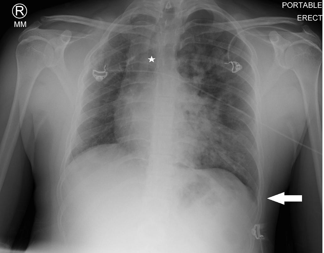
Article in Press 333 Clinical Practice and Cases in Emergency Medicine
Emergency
Images in
Medicine
Intermountain Healthcare, Department of Emergency Medicine, Park City, Utah
Image 1. An anteroposterior plain film of the chest revealing a deviated trachea (star) and a deep sulcus sign (arrow), in addition to fluffy infiltrates in the left lung fields.
supplementary oxygen. The final diagnosis was highaltitude pulmonary edema (HAPE) and absent right pulmonary artery.
DISCUSSION
Absent pulmonary artery is a rare condition, occurring in approximately 1 in 200,000 adults1 Usually it is accompanied by other cardiovascular abnormalities, but it can be isolated.2 Due to the nature of the embryologic development, it most commonly occurs on the right side.3 Presentation and diagnosis vary but is typically found while evaluating for
CPC-EM Capsule
What do we already know about this clinical entity?
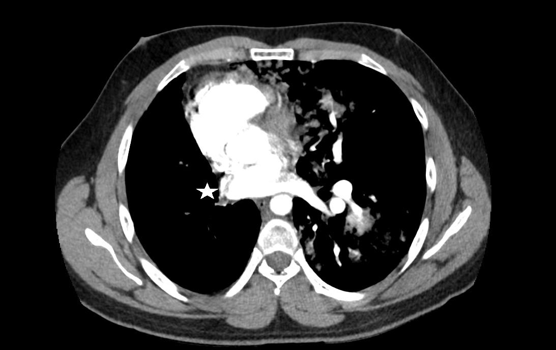
Absent pulmonary artery is a rare condition frequently diagnosed when evaluating for causes of dyspnea. Patients with this condition are particularly susceptible to high altitude pulmonary edema.
What is the major impact of the image(s)? It is important for clinicians to ensure that a radiologic findings are consistent with clinical conditions; if not, more investigation should be sought.
How might this improve emergency medicine practice?
This case is an excellent example of the importance of clinical correlation and not anchoring on a diagnosis. Had the clinician relied upon the radiologic interpretation, this patient could have had a catastrophic outcome.
pulmonary hypertension, suspected structural lung disease, or cardiac evaluations. Interestingly, HAPE is seen in approximately 10% of patients with an absent pulmonary artery.3-6 This is thought to be caused by development of pulmonary hypertension secondary to altitude and exercise at higher elevation.5
The authors attest that their institution requires neither Institutional Review Board approval, nor patient consent for publication of this case report. Documentation on file.
Address for Correspondence: Austin Smith, MD, Intermountain Healthcare, Department of Emergency Medicine, 900 Round Valley Drive, Park City, Utah, 84060. Email: austinsmith@utexas.edu.
Conflicts of Interest: By the CPC-EM article submission agreement, all authors are required to disclose all affiliations, funding sources and financial or management relationships that could be perceived as potential sources of bias. The authors disclosed none.
Copyright: © 2022 Dillon et al. This is an open access article distributed in accordance with the terms of the Creative Commons Attribution (CC BY 4.0) License. See: http://creativecommons.org/ licenses/by/4.0
Clinical Practice and Cases in Emergency Medicine 334 Volume 6, no. 4: November 2022
Dillon et al.
Absent Pulmonary Artery Presenting as High-altitude Pulmonary Edema
Image 2. A coronal view of a contrast-enhanced computed tomography angiography revealing an absent right pulmonary artery (star) and hypoplastic right lung.
Image 3. An axial view of a contrast-enhanced computed tomography angiography revealing an absent right pulmonary artery (star).
REFERENCES
1. Bouros D, Pare P, Panagou P, et al. The varied manifestation of pulmonary artery agenesis in adulthood. Chest. 1995;108(3):670–6.
2. Presbitero P, Bull C, Haworth SG, et al. Absent or occult pulmonary artery. Br Heart J. 1984;52(2):178–85.
3. Ten Harkel AD, Blom NA, Ottenkamp J. Isolated unilateral absence of a pulmonary artery: a case report and review of the literature. Chest 2002;122(4):1471-7.
4. Rios B, Driscoll DJ, McNamara DG. High-altitude pulmonary edema with absent right pulmonary artery. Pediatrics. 1985;75(2):314-7.
5. Hackett PH, Creagh CE, Grover RF, et al. High-altitude pulmonary edema in persons without the right pulmonary artery. N Engl J Med 1980;302(19):1070-3.
6. Fiorenzano G, Rastelli V, Greco V, et al. Unilateral high-altitude pulmonary edema in a subject with right pulmonary artery hypoplasia. Respiration. 1994;61(1):51-4.
Volume 6, no. 4: November 2022 335 Clinical Practice and Cases in Emergency Medicine
Dillon et al. Absent Pulmonary Artery Presenting as High-altitude Pulmonary Edema
Championing individual physician rights and workplace fairness JOIN CAL/AAEM! BENEFITS - Western Journal of Emergency Medicine Subscription - CAL/AAEM News Service email updates - Discounted AAEM pre-conference fees - And more! CAL/AAEM NEWS SERVICE - Healthcare industry news - Public policy - Government issues - Legal cases and court decisions In collaboration with our official journal FACEBOOK.COM/CALAAEM FOLLOW US @CALAAEM HTTP://WWW.CALAAEM.ORG
CALIFORNIA ACEP ANNIVERSARY
50


