
VOLUME 99, NUMBER 4 | OCTOBER, NOVEMBER & DECEMBER 2022 DR. CYNTHIA SOUTHERN MEET YOUR NEW VDA PRESIDENT >> PAGE 36 PEER REVIEWED PERIODONTAL MEASUREMENT TERMINOLOGY REVISITED >> PAGE 13 VADENTAL.ORG


Address: 1054 31st Street NW, Suite 540 Washington, D C 20007 Phone: (202) 735 5382 Medical Arts Real Estate Services & Advisory Email: information@thegenaugroup.com G E T I N T O U C H “ W E A R E T H E O N L Y D E N T A L T E N A N T B R O K E R A G E I N V I R G I N I A ” THE GENAU GROUP: MARESA
IN THIS ISSUE
99,
COLUMNS
3 STRENGTH IN NUMBERS
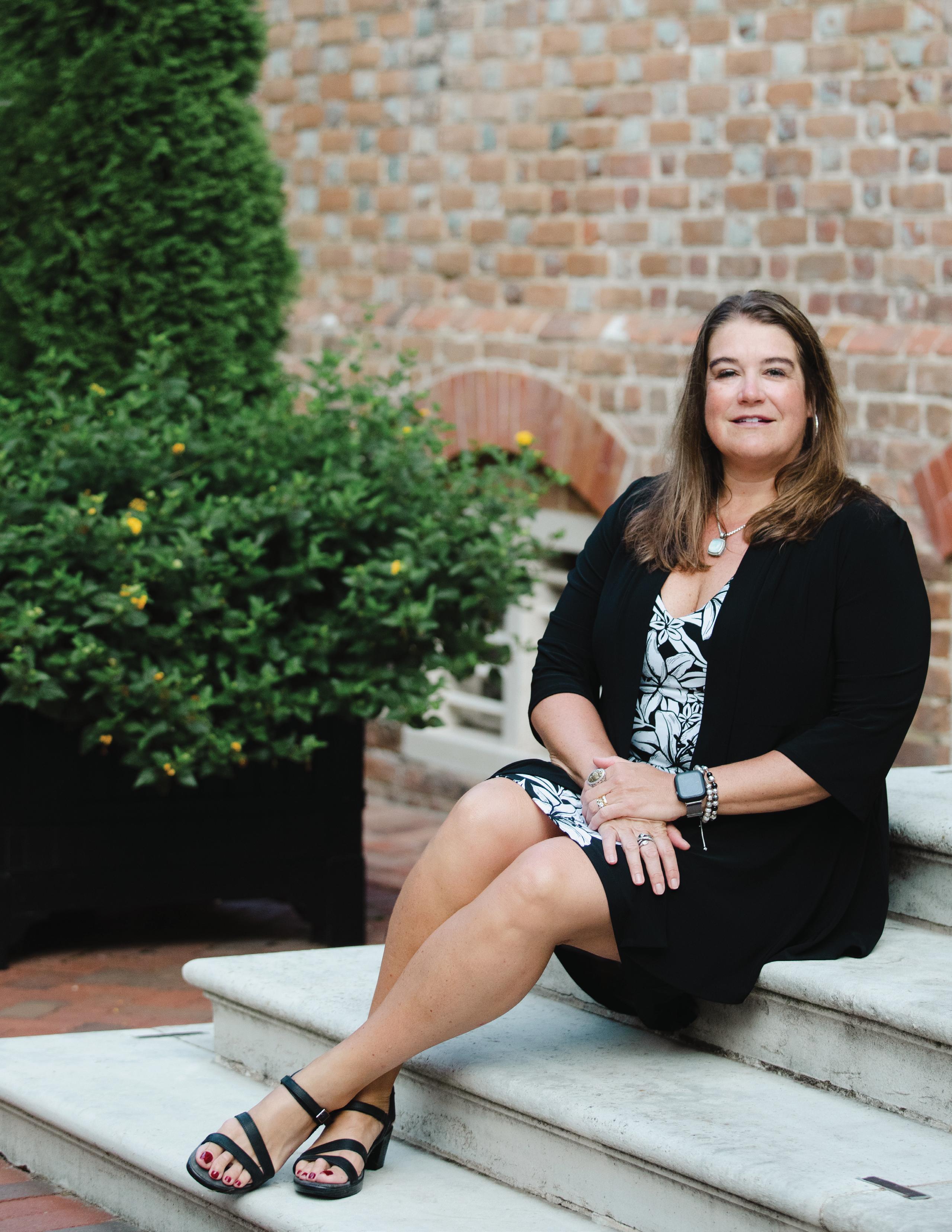
Dr. Cynthia Southern
5 NOT FADE AWAY Dr. Richard F. Roadcap
7 WHERE IS DENTISTRY HEADING? WHAT IS THE ADA DOING ABOUT IT? Dr. Gary D. Oyster
SCIENTIFIC
13 PERIODONTAL MEASUREMENT TERMINOLOGY REVISITED Drs. Weinberg, Segelnick, Fine, and Palomo
THROUGH THE LOOKING GLASS THE FANTASTICAL WORLD OF ORAL PATHOLOGY Dr. Sarah Glass
ORAL SURGERY ABSTRACTS
ADVOCACY
21 THE VDA POLITICAL ACTION COMMITTEE NEEDS YOUR HELP! Dr. Bruce Hutchison
RECENT VDA TOOTH PAC EVENTS Laura Givens
PUTTING INSURANCE DOLLARS TO WORK FOR PATIENTS Ryan L. Dunn
FEATURES
36 MEET YOUR NEW VDA PRESIDENT AN INTERVIEW WITH DR. CYNTHIA SOUTHERN
RESOURCES
45 TAKE ACTION: INFLATION IS HIDING IN YOUR INSURANCE POLICIES TOO E. Andrew Gerner
47 VIRGINIA BOARD OF DENTISTRY NOTES Dr. Ursula Klostermyer


49 DID YOU KNOW? A SERIES FROM THE VIRGINIA BOARD OF DENTISTRY
51 PEER REVIEW: A THIRTY-YEAR PERSPECTIVE Neil A. Landy, DMD, MSD
52 WHAT ACCOUNTS SHOULD YOU DRAW FROM FIRST IN RETIREMENT? Jimmy Pickert
54 PROMOTING YOUR PRACTICE’S VOLUNTEERISM IN THE COMMUNITY Michaela Mishoe
57 DENTAL DETECTIVE SERIES WORD SCRAMBLE Dr. Zaneta Hamlin
72 SCHOOL-BASED ORAL HEALTH PROGRAMS OFFER EFFECTIVE PLACE-BASED CARE Sarah Bedard Holland
UNIVERSITY CONNECTIONS
ACADEMIC YEAR, NEW ACADEMIC CHALLENGES Lyda Sypawka
WINNING PUBLICATION
OF
1
VOLUME
NUMBER 4 • OCTOBER, NOVEMBER & DECEMBER 2022
16
27
22
24
61 NEW
MEMBERSHIP 62 AWARDS & RECOGNITION 67 NEW MEMBERS AWARD
WINNER
THE2020 SILVERSCROLLAWARD
VA DENTAL
JOURNAL
EDITOR-IN-CHIEF Richard F. Roadcap, D.D.S., C.D.E. BUSINESS MANAGER Ryan L. Dunn MANAGING EDITOR Shannon Jacobs
EDITORIAL BOARD Drs. Ralph L. Anderson, Scott Berman, Carl M. Block, Gilbert L. Button, B. Ellen Byrne, Craig Dietrich, William V. Dougherty, III, Jeffrey L. Hudgins, Wallace L. Huff, Rod Klima, Thomas E. Koertge, James R. Lance, Karen S. McAndrew, Travis T. Patterson III, W. Baxter Perkinson, Jr., David Sarrett, Harlan A. Schufeldt, James L. Slagle, Jr., Neil J. Small, John A. Svirsky, Ronald L. Tankersley, Roger E. Wood
VDA COMPONENT ASSOCIATE EDITORS Dr. Zane Berry, Dr. Michael Hanley, Dr. Frank Iuorno, Dr. Stephanie Vlahos, Dr. Sarah Friend, Dr. Jared C. Kleine, Dr. Chris Spagna, Lyda Sypawka (VCU Class of 2024)
BOARD OF DIRECTORS
PRESIDENT Dr. Cynthia Southern, Pulaski
PRESIDENT ELECT Dr. Dustin Reynolds, Lynchburg
IMMEDIATE PAST PRESIDENT Dr. Scott Berman, Falls Church SECRETARY-TREASURER Dr. Zaneta Hamlin, Virginia Beach CEO Ryan L. Dunn, Goochland
SPEAKER OF THE HOUSE Dr. Abby Halpern, Arlington
COMPONENT 1 Dr. David Marshall
COMPONENT 2 Dr. Sayward Duggan COMPONENT 3 Dr. Samuel Galstan COMPONENT 4 Dr. Marcel Lambrechts
COMPONENT 5 Dr. David Stafford
COMPONENT 6 Dr. Marlon A. Goad
COMPONENT 7 Dr. Caitlin S. Batchelor
COMPONENT 8 Dr. Justin Norbo
ADVISORY Dr. Lyndon Cooper ADVISORY Dr. Ralph L. Howell
EDITOR Dr. Richard F. Roadcap
VCU STUDENT Brett Siegel, VCU Class of 2023
VOLUME 99, NUMBER 4 • OCTOBER, NOVEMBER & DECEMBER 2022
VIRGINIA DENTAL JOURNAL (Periodical Permit #660-300, ISSN 0049 6472) is published quarterly (January-March, April-June, July-September, October-December) by the Virginia Dental Association, 3460 Mayland Ct, Ste 110, Richmond, VA 23233, Phone (804)288-5750.

SUBSCRIPTION RATES Members $6.00 included in your annual membership dues.
Members – Additional Copy: $3.00 Non-Members- Single Copy: $6.00 Non-Member outside the US: $12.00 Annual Subscriptions in the US: $24.00 outside the US: $48.00
POSTMASTER Second class postage paid at Richmond, Virginia.
©Copyright Virginia Dental Association 2022
MANUSCRIPT, COMMUNICATION & ADVERTISING Send address changes to: Virginia Dental Journal, 3460 Mayland Ct, Ste 110, Richmond, VA 23233.
Managing Editor, Shannon Jacobs 804-523-2186 or jacobs@vadental.org
2
STRENGTH IN NUMBERS
Dr. Cynthia Southern

Let me start by saying I am honored to speak to you as your next President. I have big shoes to fill following Dr. Berman. He has been a wonderful leader of our Association this past year; it has been a pleasure working with him. I can assure you I’m not near as funny as Scott. Hopefully, you will be entertained by my Southern accent. Many of you who know me realize that speaking to a group is my least favorite part of being in a leadership position.
I hope everyone enjoyed the Virginia Dental Showcase in Williamsburg. Williamsburg is the home of my alma mater, The College of William & Mary, not to mention all of the ties to our state and nation’s history. I love coming back to my old stomping ground.
I have spent time reflecting on all the years I’ve been a member of this Association. There are so many wonderful memories: CE, Golf tournaments, and President’s parties to name a few. One thing that kept coming to my mind was the wonderful people I’ve met. I have had so many mentors: Dr. Terry Dickinson, Dr. Ted Sherwin (who kept adding to my list of obligations), Dr. Anne Adams, Dr. Benita Miller, and so many more. There is one who had the most significant influence, my father. My dad was a dentist; many of you knew him. He believed in organized dentistry and gave me no option but to be involved. He served on the Council on Finance. When he passed, my component asked me to take over his position. I agreed, and honestly, it has been an incredible journey for me. I have learned so much about our Association. I have also made so many friends across the state.
While reflecting, I remembered how much my dad loved dentistry. He was rarely
upset, but he told me his frustrations about being a practicing dentist. The two he always mentioned were governmental regulation imposing on his practice and dealing with third-party payers (occasionally, he complained about a contentious VDA House of Delegates). I’m pretty sure he lost his hair complaining about third-party payers. He kept a copy of the dental practice act in his desk drawer. He believed in protecting this profession and cautioned me about supporting regulations that would make changes to this act.
battled third-party payers in the past. Remember assignment of benefits, the de minimis clause, and tele-dentistry. I am grateful to our past leaders who promoted legislation to protect our profession and kept the dental practice act intact.
And this year, finally, we have a Medicaid reimbursement increase after 20 years of the same fee schedule. And let’s not forget COVID sent us into chaos. I never imagined we would have a pandemic that would force us to close our offices. Our Association provided us guidance through the COVID pandemic, and thankfully we are now considered essential. We were one of the first professions to recover. This would not have happened without the leadership of Dr. Elizabeth Reynolds, Dr. Frank Iuorno, the Back-to-Work Task Force, our amazing CEO Ryan Dunn, and all of the wonderful members of our VDA team.
Our Association has so many arms. PAC needs our contributions. VDSC needs our help. Increasing non-dues revenue benefits the entire Association. The VDA Foundation needs our support. MOM projects are finally up and running again. There are so many people in the Commonwealth who still do not have access to a dentist. Our Fellows, Committees, Councils, and Task Forces continue their hard work.
When I talk to dentists around the state, they tell me the biggest problems in their practice are advocacy, third-party payers, and workforce shortages. Imagine that 20 years later, we are dealing with some of the same problems. These issues affect all dentists, solo practitioners, corporate dentists, and associate dentists, maybe in different ways, but they affect us all. I can’t say that we can solve these problems this year. What I do believe is that our Association is strong. We have
I recall what our President said in his address last year. “Every time people feel dentistry is facing too many challenges to remain viable, it usually leads to the next Golden Age of Dentistry.” Post-COVID, is that the Golden age? Maybe it’s the fact that we are still here and are as strong, if not stronger than pre-COVID. We are facing challenges, the threat of declining market share, declining contributions to our PAC, and members struggling to keep
3 MESSAGE FROM THE PRESIDENT
>> CONTINUED ON PAGE 27
“We need to continue to increase membership and retain the members we have... Asking our specialty organizations to join us so we are all working together as a team, specialist or not; we are all dentists.”
Private Banking Solutions for Dental Practices & Practitioners

4 TowneBank.com/DentalBanking | Member FDIC | NMLS#512138 | Equal Housing Lender | * Credit approval required
• Dedicated Local Bankers • Business & Private Banking • Practice Acquisition/Buy-out Options Michelle S. Butler | ExEcutivE vicE PrEsidEnt | dirEctor of PrivatE Banking t 757-638-7521 | MichEllE.ButlEr@townEBank nEt Our consultative Private Bankers are always available to help. • Loans & Lines of Credit* • Custom Mortgage Solutions*
NOT FADE AWAY
Elected representatives serve their constituents. “Constituents” are those individuals who reside in that political jurisdiction and may or may not be registered to vote. Even if the constituent did not vote in favor of a particular solon, or vote at all, they are nonetheless entitled to representation.
Both the VDA and the VCU School of Dentistry, although not elected to public office, have separate communities of interest, populated by their constituents. Contemporary jargon would label them as “stakeholders”, a term frequently used in grant proposals, press releases, and public relations forays. It might be easier for each party to identify their communities as people we care about and dispense with the terminology.
And both the VDA and VCU have similar but somehow different, problems in representing their constituents. VCU is the only dental school in the state, but barely one-third of dentists licensed in Virginia are alumni. According to the Virginia Department of Health Professions, a 2021 survey (in which 87% of dentists participated) revealed that 36% of dentists in Virginia received their professional (i.e., dental) degree in Virginia.1 Furthermore, among those licensed within the last five years, only 23% received their professional degree in the state.2 It is curious to note that, based on DHP’s survey, 31% of these most recent licensees received their high school diploma in Virginia, indicating a trend for state high school grads to look elsewhere for a dental education.
Another way to measure this demographic can be found in the pages of the Journal. Considering the previous four issues, Volume 98, #4, through Volume 99, #3, 63 of 245 new VDA members,
or about 25%, were graduates of VCU School of Dentistry. Is this scientific? Perhaps it is not. The numbers could be skewed in either direction by dentists practicing part-time (or not at all), the 10% or more who didn’t answer the survey or a host of other factors. However, the Journal numbers are based on one year’s new members, nearly all of whom are actively practicing in the state at the time of their membership application.
licensing examinations by a large number of state boards of dentistry has done much to increase the mobility of recent graduates. For example, the ADEX dental licensing examination, which is scheduled to be held at VCU in April 2023, is accepted by 48 of 50 states.4
Given that the percentage of VCU alumni in the 2021 dentist workforce is greater than those licensed in the past five years3, or the past one year, it would seem that the number of VCU alumni as a percentage of the workforce is declining. It’s not that VCU grads are stricken with wanderlust, or the state of Virginia is seen as a mecca for dentists from yon regions. In years past, it was very difficult if not impossible, for dentists to practice in another state, due to board licensing requirements and the need to recruit patients for board exams. The SRTA exam I took in 1977 allowed me to apply for licensure in three states outside of Virginia. Thus, older practitioners, now leaving the workforce in large numbers due to retirement, were more likely to be graduates of a school within the state borders. Reciprocity, licensure by credentials, and the acceptance of certain
Also, Virginia has only one school of dentistry (VCU) and is contiguous or in close approximation with Maryland/DC (two schools), North Carolina (two, soon to be three), Tennessee (soon to be two schools), Kentucky (two schools), and Pennsylvania (three). Not for one moment am I suggesting that Virginia needs another dental school. Despite being the 12th largest state in terms of population5, the dentist-population ratio is above the national average. The ADA estimates that the number of working dentists per 100,000 population in Virginia is 63.4, against a national average of 60.8.6 Yet, as anyone who has participated in a MOM Project will attest, many areas of the state still lack access to care due to a shortage of doctors in those regions.
The VDA, in some ways like VCU, faces a shrinking constituency, at least at home in Virginia. In 2007 I was asked to speak to an association meeting attended by many regulators. One of the first questions I was asked was “What percent of Virginia dentists are VDA members?” I answered, with great pride, 72%. Ah, the good old days! In 2022, that number hovers around 60%. Nearly every state dental association struggles to maintain market share. The number of members creeps up, but the percentage of active dentists who join continues to decline. The reasons are many and can’t be explored in these pages.
Both the VDA and VCU are anxious to engage the dental community in

5
Dr. Richard F. Roadcap
MESSAGE FROM THE EDITOR
“The VDA, in some ways like VCU, faces a shrinking constituency, at least at home in Virginia.”
>> CONTINUED ON PAGE 6
Virginia. The VDA communicates with its members, and VCU with its alumni (and students, of course) but those shares seem to be on a downward trajectory. I know the VDA has strategies to communicate with non-members (having engaged in some of those tactics) and I suspect VCU attempts outreach to alumni of dental schools near and far, who appear now to comprise 75% of Virginia’s dental workforce of the future. Dispensing with mission statements, the VDA represents the profession of dentistry, and VCU is the standard bearer for dental education in Virginia. The problem for both lies in communicating with parties outside their orbits.
Both the VDA and VCU stand to gain by sharing information. As Edward Bear, better known to us as Winnie, said to his friends “You can’t stay in your corner of the Forest waiting for others to come to you. You have to go to them sometimes.”7
Of course, there are risks involved. The greater risk would be for the footprint of both institutions to fade or wither away from doing nothing.
References
1. https://www.dhp.virginia.gov/ media/dhpweb/docs/hwdc/ dentistry/0401Dentist2021.pdf
2. https://www.dhp.virginia.gov/ media/dhpweb/docs/hwdc/ dentistry/0401Dentist2021.pdf
3. Ibid.
4. https://www.cdcaexams.org/ adex-acceptance-map/
5. https://www.census.gov/ quickfacts/geo/chart/ID/ PST045221
6. https://www.ada.org/ resources/research/healthpolicy-institute/dentistworkforce
7. A.A. Milne. Winnie-the-Pooh Methuen; London, 1926
While no one likes to think about it, things do happen and it’s always important to be prepared. Knowing your practice’s value can make the difference between selling your practice or having it become unsellable. That is why practice owners should have an up-to-date practice valuation.
A Henry Schein Dental Practice Transitions valuation considers both tangible and intangible assets of the practice and can provide the many key factors which influence the practice’s value


Contact us today to schedule a complimentary, confidential consultation!


Bobby Anderson Eileen Anderson, CPA, CVA Transition Sales Consultants
C: (804) 334-0872 or (804) 334-0871
Robert.Andersoniii@henryschein.com


Eileen.Anderson@henryschein.com


PRACTICE SALES
VALUATIONS
TRANSITION PLANNING

6 © 2022 Henry Schein, Inc. No copying without permission. Not responsible for typographical errors.
Your practice is one of your most important assets. DO YOU KNOW WHAT IT’S WORTH?
www.henryscheindpt.com •
•
•
21PT4334_plans options worth_7.5x4.5_.indd 3 1/25/21 6:51 PM
MESSAGE FROM THE EDITOR
>> CONTINUED FROM PAGE 5
WHERE IS DENTISTRY HEADING?
WHAT IS THE ADA DOING ABOUT IT?
Gary D. Oyster, DDS; ADA Trustee, 16th District

As the Baby Boomers retire and the millennials and Gen-Xers take over what will our profession look like? The ADA and the 16th District are trying to work with this diverse group of new dentists to make them feel included. Only by having a strong state and national organization can we have any influence on the direction of the profession. The ADA and most societies in the tripartite are not keeping pace with the growth in the dental profession. The ADA is on course to dip below 50 percent market share. Many state societies are on the same path. Regardless of your workplace model there are benefits to being a member of the tripartite organization.
To counter this loss of market share the ADA is proposing a change in its governance structure to be more inclusive and open to younger and more recent graduates. Strategic Forecasting rather than Strategic Planning will make the ADA more responsive to member, state, and local societies. The new governance structure will keep the Councils, Commissions and Board of Trustees as they are. Data would suggest that the clear pathway to growth is in welcoming dentists into membership regardless of practice modality, public, private-solo, or group, and providing greater value to these members at every career stage and keeping them engaged. The Strategic Forecasting model will help engage younger dentists.
There will be a Strategic Forecasting Committee that will oversee several subcommittees and work groups. The four work groups will focus on member issues, tripartite issues, administrative issues, and public issues. Issues that arise during the year between sessions of the House of Delegates will be sent to the work group for a quick response.
The work groups will be made up of House members and if needed, consultants. To help make this new governance arrangement successful several strategies including the new ADA Member App are designed to put the ADA value into the hands of all dentists regardless of practice modalities. In addition, the cumbersome ADA Connect will be replaced by an advanced Microsoft 365 to allow dentists to communicate in real time with each other and ADA staff and leadership. None of us will miss ADA Connect!
New dentists, in particular, across all practice modalities have emphasized that they need to see more value from the ADA around financial issues, wellness, and careers. This is true of dentists pursuing practice ownership, employment, research and education, or public service. The Mobile App will give state and local societies the ability to stay connected with new dentists as they progress from place to place throughout their careers. In addition a new ADA podcast created by and for dentists, with a personal document vault for saving professional documents in one place on their phone, a personalized content feed where members select topics they are most interested in to deliver content to match their preferences within the app’s feed, and a career pathways content experience to enable dental students to learn more about career options for early career dentists. The new reality is that our newer colleagues are going to be more mobile as licensure restrictions are relaxed and the demographics of our profession are rapidly evolving.
I hope that many of you will attend the ADA SmileCon meeting in Houston in early October. A marketing launch plan to drive downloads has been created to
promote and highlight the new features at SmileCon and through the fourth quarter of 2022. The plan includes paid media, search ads, social media promotion, influencer outreach and utilization of the ADA’s communication channels to increase downloads of the app.
The ADA’s State Government Affairs team continues to address medical necessity issues, along with other Medicaid and Medicare related topics. Advocacy continues on in the areas of McCarranFerguson reform, military dental care, military spouse licensing relief act, general access to care issues and student reform, especially with regard to the Resident Education Deferred Interest (REDI) Act. Section 1557 of the Affordable Care Act, which addresses discrimination and was minimized under President Trump, is potentially coming back to full force under President Biden. The ADA will monitor activity on this issue.
New Initiatives also aim to reduce the administrative complexity of joining. We hope to make it easy to join for the individual dentist paying their own dues or for the employees paying for multiple memberships, emphasizing value and values.
Organized dentistry welcomes all dentists. We are all in this together. Insurance intervention, government mandates, public opinion, and social media influence all of us to some degree and we need to stand together to have a profession and not a trade. I have enjoyed being the ADA Trustee from the 16th District and welcome any ideas that any of you have how as to how the ADA and I can improve.
7 TRUSTEE’S CORNER
“Thanks for an amazing VDA! LOVED the Pediatric Dentistry CE courses! I hope you all will continue to do it in the future!”


photos.


8
“This year’s Virginia Dental Association meeting was awesome. Great to connect with our team and doctors from across virginia and talk about dentistry!”
Please visit us on Facebook to see more
“Loved the pairing of dental students with the doctors. Courses offered were relevant. Enjoyed the ability to get together in person again!”
“the Meeting was well organized. It had the best setup for that site, and I have been attending for many years. Support staff gets 10+ for friendly help.”


Please visit us on Facebook to see more photos.


9
“We all had a GREAT time. It was a first-class event, for sure. Fun for the whole team! You and your team exceeded our expectations. Definitely going to return next year!”


“Beautiful historic setting, well organized and a chance to meet up with fellow Hygienists.”


Please visit us on Facebook to see more photos.


10 SCIENTIFIC


11 2023 Hilton – Norfolk The Main September 21-24, 2023 SAVE THE DATE Virginia Academy of Pediatric DentistryJOINT MEETING WITH:
ADA MEMBER BENEFIT
Tackle Insurance Issues with the ADA Third Party Payer Concierge™

12 Do you have a dental insurance question or concern? If so, the ADA Third Party Payer Concierge is here to help! This is a free, personalized service for VDA and ADA members. Get assistance from ADA insurance experts on: • Nuances of doing business with dental insurance companies • Coordination of benefits • Claim denials and assistance with the appeals process • Understanding confusing explanation of benefits language • And more available at ada.org/dentalinsurance! This is just one way the ADA and VDA empower our members to make the best decisions for their patients and practice.
Call or email the Concierge with your issue from 9:30 a.m. to 6:00 p.m. EST Monday through Friday. Phone: 800-621-8099 dentalbenefits@ada.org
PERIODONTAL MEASUREMENT TERMINOLOGY REVISITED
Mea A. Weinberg, DMD, MSD, RPh; Stuart L. Segelnick, DDS, MS; James Burke Fine, DMD; Leena Palomo, DDS, MSD
Many periodontal terms are used in literature but are not completely defined or understood and there may be some confusion about whether some of these terms can be used synonymously. Understanding these terms is crucial when reading and interpreting research articles and in treatment and evaluating outcomes of clinical therapy.1 This article will explore different periodontal measurement terminology including periodontal probing, critical probing depth, clinical attachment loss, clinical attachment level, relative attachment level, and gain of attachment.
The term “pocket depth” is usually used inappropriately when taking probing depth measurements. The word “pocket” means the probing site is not healthy. A sulcular depth ranging between 1-3 mm is generally considered health. Probing depth is the distance in millimeters from the gingival margin to the deepest point the probe reaches.2 Limitations of periodontal probing include probing technique (i.e., improper angle of the probe, amount of pressure applied to the probe), size of the probe, and severity of tissue inflammation.
In health the probe penetrated 0.70 ± 0.56 mm apical to the coronal end of the JE and in the presence of inflammation the probe will usually stop at the level of the dentogingival complex.3,4 The ideal probing forces are established to be between 20 and 25 g (0.20-0.25 N).3,5,6 The probing tip diameter varies from different probes. Garnick and Silverstein reported that probe tips need to have a diameter of 0.6 mm for approximate probing depths.7 Additionally, varying forces and diameter tips gave different histological measurements in healthy and inflamed tissue.7 The larger the tip diameter, the less the probe could reach
the inflamed connective tissue. The probing pressure was found to be directly related to the force of the probe whereas inversely proportionate to the probe tip diameter. On interproximal posterior teeth, the periodontal probe needs to be angled so the tip of the probe reaches the col area which is directly under the contact point of the tooth.8 This angulation can differ depending on tooth position and location of tooth contact. The col is nonkeratinized tissue that connects the buccal and lingual papilla and is an initial site for the start of inflammation.
There has been much confusion regarding the terms clinical attachment loss and clinical attachment level. Not only is there a continuous debate about whether these terms are synonymous, but also there is the misperception of the definition of each term.9 Additionally, many clinicians use the abbreviation “CAL”. What does CAL stand for? This abbreviation is seen in different articles as either clinical attachment level or clinical attachment loss or both ways.10 Is the term clinical attachment loss (CALoss), also referred to as loss of clinical attachment, loss of attachment, connective tissue attachment loss, or attachment loss, synonymous with clinical attachment level? To answer this question, the clinical and histological features of the term will be reviewed.
Clinical attachment level (CAL) is a clinical measurement or value approximation of the level of the epithelial attachment to the tooth surface and can change over time. Thus, clinical attachment levels are indicators of past destruction of periodontal attachment and can be used to monitor the progress of periodontitis. It is an assessment of all the supracrestal soft tissue components and reflects the histormorphological aspect of the periodontium.
Clinical attachment level is measured from the distance of the cementoenamel junction to the base of the probeable crevice or the tip of the periodontal probe.9,11 It is a measurement that is performed preoperatively (surgical or nonsurgical) and the difference between the two measurements will determine the loss or gain of attachment after treatment. Since the level of attachment can be different around the circumference of a tooth, it is important to “walk” the probe in millimeter increments rather than taking the probe out at each measured site. On the other hand, probing depth is measured from the free gingival margin to the apical location of the tip of the periodontal probe. Since the position of the gingival margin is not static and migrates, a more accurate measurement is the clinical attachment level which uses the CEJ as a static reference point rather than the free gingival margin. The term relative attachment level uses a fixed reference point, such as a crown margin, cusp tip or a stent as the static reference point when the CEJ is not visible.12
Histologically, clinical attachment loss refers to the pathological destruction of the dentogingival complex allowing for the apical and lateral migration of the junctional epithelium (epithelial attachment) onto the root.13 Clinical attachment loss only refers to soft tissue destruction or the extent of detachment of the periodontal tissues from the root surface in periodontitis.13 It is the actual distance the dentogingival complex migrates apically. Radiographic alveolar bone loss will usually be undetected initially but can occur as a subsequent pathological event to the loss of connective tissue attachment and usually occurs by 6-8 months after attachment loss.14
13 SCIENTIFIC
>> CONTINUED ON PAGE 14
Clinical attachment loss is determined by measuring the clinical attachment level, which can exist as pocketing with or without marginal tissue recession, or recession with no pocketing (Figure 1), or both inflamed pocketing and marginal tissue recession, as long as it goes below the CEJ.15,16 Marginal tissue recession can be seen in a non-diseased mouth due to anatomic, traumatic or mechanical factors as well as in patients with inflammatory periodontal disease.13,17 If a tooth is displaced off of the basal bone then the resulting gingival recession, if it results in clinical attachment loss may or may not be caused by periodontitis or mechanical trauma.
An example for determining the clinical attachment level is: if periodontal probing was 3 mm on the mesial of a tooth and there was no marginal tissue recession or enlargement and the gingival margin is located at the CEJ or 0.5 mm coronal to the CEJ as is in health then, the clinical attachment level is 3 mm (probing depth

of 3 mm and 0 mm of recession) which means that there is no clinical attachment loss.18 Marginal tissue recession which is the apical displacement of the gingival margin onto the root surface, is an indication that attachment loss has occurred.3 Another example: 3 mm probing depth and 2 mm of recession. There is 5 mm of attachment loss (because of the recession) and the clinical attachment level is 5 mm which means that the junctional epithelium (epithelium attachment) is attached more apically onto the root due to the recession. Thus, if the clinical attachment level is 5 mm apical to the CEJ, it is understood there is 5 mm of loss of clinical attachment.
The next term is clinical gain of attachment. The most important outcome of periodontal therapy is to achieve a gain of clinical attachment which indicates the efficiency of the periodontal therapy. A gain of clinical attachment indicates a better healing result compared to the reduction of the
probing depth.19 Attachment gain denotes a coronal migration of the periodontal supporting structures which refers to a to a decreased penetration of the probe at the base of the pocket due to increased proportion of collagen fibers with the gingival connective tissue (after inflammation is reduced), which combined with formation of a long junctional epithelium will produce increased resistance of penetration of the probe tip. During the healing of non-regenerative therapy, a long junctional epithelium forms after the connective tissue is destroyed as a result of periodontal disease and the JE moves apically until it reaches intact connective tissue at which point it stops migrating.20
In conclusion, attention to the use of the appropriate terminology presented in this article will help the clinician to better understand the reasoning for the proper use of probing to diagnosis a periodontal case as well as the outcomes of periodontal therapy.
The authors propose the terms clinical attachment loss and clinical attachment level not be used interchangeably. The abbreviation for clinical attachment level should be “CAL” and the abbreviation for clinical attachment loss or attachment loss should be “CALoss”. It is called clinical attachment loss because it is measured histologically. Clinical attachment level (CAL) is the clinical approximation of the level of attachment of the epithelial attachment to the tooth surface; it is a measurement of probe tip penetration relative to the CEJ. CALoss is exclusively used to refer to the loss of clinical attachment level which occurs following the destruction of gingival connective tissue attachment with apical migration of the epithelial attachment creating a periodontal pocket with or without marginal tissue recession. Early CALoss is not evident on radiographs since there is no alveolar bone loss at this time; it occurs subsequently to the loss of attachment.
14 SCIENTIFIC
>> CONTINUED FROM PAGE 13
Figure 1
References
1. Needleman I, Garcia R, Gkranias N et al., Mean annual attachment, bone level, and tooth loss: A systematic review. J Periodontol 2018; 89(S1): S120-S139.
2. Schwartz M, Lamster I, Fine JB. Clinical Guide to Periodontics. 1st ed. 1994. Elsevier. Netherlands.
3. Polson AM, Caton IB, Yeaple RN, Zander HA. Histological determination of probe tip penetration into gingival sulcus of humans using an electronic pressure sensitive probe. I Clin Periodontol 1980; 7:479-488.
4. Listgarten MA. Periodontal probing: What does it mean? J Clin Periodontol 1980; 7:165176.
5. Garnick JJ, Keagle J, Searle J, Thompson W. Gingival resistance to probing forces (ii). Gingival resistance to probing forces (II). The effect of inflammation and pressure on probe displacement in beagle dog gingivitis. J Periodontol 1989; 60:498-505.
6. Armitage GC, Svanberg GK, Loe H. Microscopic evaluation of clinical measurements of connective tissue attachment levels. J Clin Periodontol 1977; 4:173-190.
7. Garnick JJ, Silverstein L. Periodontal probing: probe tip diameter. J Periodontol. 2000 Jan;71(1):96-103.
8. Hefti AF. Periodontal probing. Crit Rev Oral Biol Med. 1997;8(3):336-356.
9. Fritz PC. Clinical attachment level – how to calculate and interpret this important measurement. Oral Health. October 1, 2013.
10. Barbosa VL, Angst PDM, Stadler AF, et al., Clinical attachment loss: estimation by direct and indirect methods. Int Dent J 2016;66(3):144-149.
11. Karmakar S, Prakash S. Clinical attachment level: an unsung
hero in periodontal diagnosis. IJAR 2019; 7(4):106-111.
12. American Academy of Periodontology. Glossary of Terms. 2001. 4th ed. The American Academy of Periodontology, Chicago, IL.
13. Armitage G. Clinical evaluation of periodontal diseases. Periodontol 2000; 1995(7):39-53.
14. Goodson JM, Haffajee AD, Socransky SS. The relationship between attachment level loss and alveolar bone loss. J Clin Periodontol. 1984 May;11(5):348-359.
15. Ryan ME. Clinical attachment level change as an outcome measure for therapies that slow the progression of periodontal disease. J Int Acad Periodontol 2005 Oct;7(4 Suppl):162-71; discussion 172-174.
16. Beck JD, Koch GG. Characteristics of older adults experiencing periodontal attachment loss as gingival recession or probing depth. J Periodontal Res. 1994 Jul;29(4):290-298.
17. Chrysanthakopoulos NA. Gingival recession: Prevalence and risk indicators among young Greek adults. J Clin Exp Dent 2014; 6(3):e243-249.
18. Vandana KL, Haneet RK. Cementoenamel junction: An insight. J Indian Soc Periodontol 2014 Sep;18(5):549-554.
19. Kotsilkov K, Popova C. Effectiveness of target antimicrobial therapy of severe chronic periodontitis part I: reduction of gingival inflammation and active periodontal disease sites. JofIMAB 2010;16(4):24-26.
20. Wazen R, Nanci A. 2016. Chapter 15. Repair and regeneration of oral tissues. In: Nanci A. (Ed.) Ten Cate’s Oral Histology. 9th ed. pp. 729-766. Netherlands: Elsevier Publishing.
Authors: Mea A. Weinberg, DMD, MSD, RPh
Clinical Professor Department of Periodontology and Implant DentistryNew York University College of Dentistry
Email: maw2@nyu.edu
Stuart L. Segelnick, DDS, MS
Adjunct Clinical Professor Department of Periodontology and Implant DentistryNew York University College of Dentistry
Email: eperiodr@aol.com
James Burke Fine, DMD
Senior Associate Dean for Postdoctoral Academic and Student Affairs, Professor of Dental Medicine (in Periodontics), Attending Dental Surgeon on the Presbyterian Hospital Dental Service, Chief Practice Director of Faculty Practices at the College of Dental Medicine, Columbia University College of Dental Medicine
Email: jbf1@cumc.columbian.edu
Leena Palomo, DDS, MSD
Professor & Chair, Ashman Department of Periodontology and Implant DentistryNew York University, College of Dentistry Email: lp2706@nyu.edu
15 SCIENTIFIC
LOOKING GLASS THROUGH THE
WITH DR. SARAH GLASS
Cases are presented by Michaela Banks, a dental student at the Virginia Commonwealth University School of Dentistry.
EXPLORE THE FANTASTICAL WORLD OF ORAL PATHOLOGY

Editor’s Note: Dr. Sarah Glass is a board certified Oral and Maxillofacial Pathologist. She works as an assistant professor at VCU School of Dentistry, and her job responsibilities include teaching, working in the biopsy service, and seeing oral medicine patients.

A 53-year-old male presents to the clinic for a periodic exam. Upon the intraoral exam, there is a red and white lesion on the ventral surface on the tongue. The lesion is not able to be wiped off and there is no obvious cause of irritation. The patient reports noticing it about six months ago and reports no pain associated with the area. What is your suspected diagnosis?


16
A 58-year-old female presents to the clinic with blue, non-painful, swelling on the left mandibular premolar gingiva. Patient has not been to a dentist for many years and does not recall when the swelling started. What is your suspected diagnosis?


A 16-year-old male patient comes to the pediatric dental clinic for a screening appointment. Upon evaluating the radiographic image, you note a mixed density lesion on the right mandible between the incisors and premolars. The lesion seems to be impeding the eruption of #27 and has a radiolucent rim.

17 SCIENTIFIC
>> ANSWERS ON PAGE 18
1. Severe epithelial dysplasia causes atypical architecture and cytological features in the entire thickness of the stratified squamous epithelium. An immediate biopsy or referral to a specialist is recommended for a speckled leukoplakia/ erythroleukoplakia due to classification as an oral potentially malignant disorder.


2. The gingival cyst of the adult histologically shows a cyst lined with epithelium without any inflammation. The location of this lesion is often in the mandibular canine to premolar region. The cystic fluid of these lesions gives them a blue appearance.

3. A compound odontoma often presents with a radiolucent rim filled with multiple radiodensities that represents the organization of enamel, dentin, cementum, and pulp. Often, these lesions prevent eruption of teeth. They are typically found on routine panoramic images commonly around the age of 14.

>> THROUGH THE LOOKING GLASS ANSWERS CONTINUED FROM PAGE 17
Career Center connects employers with premier dental professionals
Job Seekers:

— CAREERS.VADENTAL.ORG — • EMAIL your job directly to job seeking professionals • PLACE your job in front of our highly qualified members • SEARCH our resume database of qualified candidates • MANAGE jobs and applicant activity right on our site • LIMIT applicants only to those who are qualified • FILL your jobs more quickly with great talent Employers: • POST multiple resumes and cover letters or choose an anonymous career profile that leads employers to you • SEARCH and apply to hundreds of fresh jobs on the spot with robust filters • SET UP efficient job alerts to deliver the latest jobs right to your inbox • ASK the experts advice, get resume writing tips, utilize career assessment test services, and more
VDA
19
2022 Cor e Sponsor s and Suppor ter s


The generosity of our financial supporters helps ensure that we not only improve the oral health of our neighbors in need, but it also helps make the VDAF stronger and more sustainable.


We TRULY APPRECIATE the companies, foundations, and individuals who fully embrace our mission to provide access to dental care for underserved Virginians.


20
Richard & Caroline T. Gwathmey Memorial Trust, Bank of America, N.A., Trustee
THE VDA POLITICAL ACTION COMMITTEE NEEDS YOUR HELP!
Bruce Hutchison, DDS; VDA PAC Chair

With your help, the Virginia Dental Association (VDA) led the successful fight for a 30% increase in dental Medicaid reimbursement rates in Virginia’s state budget. Not only is this the largest new investment of state funding in oral healthcare in the history of Virginia, it’s also the largest percent increase in dental Medicaid rates of any state in the country this year. This new state and federal funding will help address the gap between reimbursement rates through the Medicaid program and private insurance, which has been growing since the last increase in 2005. We are thankful for the hundreds of VDA members, students and partners who advocated for this necessary increase, and for the state’s lawmakers and Governor Youngkin for supporting this vital investment.
This win for oral healthcare attests to our incredibly strong profession and organization, however; we all know that reimbursement rates are just one challenge we face from third-party payers. Among other things, an economic downturn, workforce challenges and unfair and burdensome practices from insurance companies impact our ability to serve the most vulnerable patients. It is for that reason that we must remain vigilant in protecting patients and our profession. We were able to secure an impactful increase in dental Medicaid rates - with further effort and elevated support, we are certainly capable of also working towards an increase with private insurers in the future.
The cost to remain at the table has sharply increased: Nine of the ten most expensive House races in Virginia’s history have come in the last three years. And if you’re not at the table, you’re on the menu.

We need your financial support for the VDA PAC now. I strongly believe in its importance and have increased my donation this year. I’m personally asking you to contribute at least $100 more this year to support the next phase of critical work for the VDA PAC. You can easily donate at the level you wish at www.vadental.org/vda-pac.
The VDA has historically been a model for legislative success, but we must continue to adapt to represent our profession. Our successes show exactly what can happen when we join together in action. Thank you in advance for your contribution – When We Give Together, We Have a Stronger Voice. If you have questions, please contact Laura Givens, givens@vadental.org or 804-523-2185.
21 ADVOCACY
RECENT VDA TOOTH PAC EVENTS
Laura Givens, Director of Legislative and Public Policy

VDA Hosts an Advocacy Day for VCU Dental Students: Students Recognized for their Efforts this Year and Encouraged to Participate in Future VDA Activities

The VDA hosted a two-part advocacy event for VCU dental students on August 10th. The first part of the program was a lunch and learn with special guest Delegate Carrie Coyner. She talked to nearly 50 students about the General Assembly and the importance of their advocacy as legislators tackle legislation impacting dentistry. Delegate Coyner expressed why it was imperative to engage with Virginia House and Senate members and shared how best to go about communicating with them.
Part two of the program was a reception at the Graduate Hotel where nearly 50 students celebrated the 30% increase in dental Medicaid reimbursement rates – an effort that was led by the VDA and many VCU students were involved in advocating for this vital increase. Several VDA dentists were a part of this
event where they met and mingled with students who were eager to learn more about advocacy and the opportunities and resources that the VDA has to offer.

22 ADVOCACY
VDA PAC Fundraisers for Delegate Knight and Senators Barker and Saslaw
The VDA continues to ambitiously support campaigns for legislators and this year we had an exceptionally successful event for Delegate Barry Knight.

On August 23rd, over 40 dentists and guests gathered at the Princess Anne Country Club in Virginia Beach to show support for Delegate Knight. Drs. Michael Morgan and Dag Zapatero chaired the event with help from steering committee members Drs. Zaneta Hamlin, Dani Howell and Anthony Peluso. Delegate Knight represents the 81st District in the Virginia General Assembly and serves as Chairman of the Appropriations Committee.

Dr. Bruce Hutchison chaired a fundraising event on September 28th for Senators George Barker and Dick Saslaw in Tysons Corner with help from steering committee members Drs. Scott Berman, Rod Klima, Melanie Love and Nate Schoenly. Senator Barker (39th district) co-chairs the Senate Finance and Appropriations Committee while Senator Saslaw (35th district) serves as Chairman of the Senate Commerce and Labor Committee. Both Senators also serve on the Senate Education and Health Committee.
The VDA is most appreciative to all who made generous contributions to Delegate Knight and to everyone who attended the event.
We encourage members to contribute and attend VDA PAC Fundraisers as they are a wonderful opportunity to gather socially with your friends and colleagues and meet with legislators in an intimate setting. The VDA PAC appreciates VDA member involvement in steering committees to make these fundraising events successful.
23 ADVOCACY
Component % of 2022 Members Contributing to Date 2022 VDA PAC Goal Amount Contributed to Date Per Capita Contribution % of Goal Achieved 1 (Tidewater) 33% $45,500 $62,602 $304 138% 2 (Peninsula) 37% $27,500 $19,750 $330 67% 3 (Southside) 33% $14,000 $14,000 $287 100% 4 (Richmond) 21% $67,750 $40,354 $313 58% 5 (Piedmont) 30% $30,000 $19,225 $291 63% 6 (Southwest VA) 42% $25,250 $13,800 $314 55% 7 (Shenandoah Valley) 26% $30,000 $22,015 $345 72% 8 (Northern VA) 24% $135,000 $75,150 $295 56% TOTAL 31% $375,000 $266,896 $310 71% TOTAL CONTRIBUTIONS: $266,896 MUST RAISE $108,104 TO REACH GOAL 2022 GOAL: $375,000
PUTTING INSURANCE DOLLARS TO WORK FOR PATIENTS
Ryan Dunn, CEO

Those of you who have been VDA members for a long time know the importance of being engaged with dental policy at the state level. The work the ADA does in Washington is important. But things also tend to move slowly in Washington, if at all.
In Virginia, our citizen legislators meet for a couple of months out of the year and return to their day jobs in law firms, small businesses, or even two who work in dental offices. Change can happen in the blink of an eye, and that’s why the relationships and trust built up by the VDA and its members over the years are so important.
What we don’t talk about as often, is the importance of what happens in other states. I’m going to use my column this month to highlight one such issue that should be important to every VDA member and every practicing dentist in Virginia.
Massachusetts voters will decide on a statewide ballot initiative this November that would require dental third-party payers to spend at least 83% of premiums on member dental expenses and quality improvements instead of administrative expenses. Carriers that do not meet this minimum standard would be required to refund the difference to covered individuals and groups.
This is a consumer protection bill. If a patient or their employer is purchasing dental insurance, there should be transparency and assurances that the money is going to its intended purpose of providing dental care.
But we’re in Virginia, home to the first Thanksgiving. Why should we care what happens in the Commonwealth to the north?
Insurance companies are predictably using scare tactics and spending huge sums of money in opposition to this ballot initiative. They want to defeat the initiative and send a message to all of us in doing so.
“Make no mistake that a victory in Massachusetts will open the door wider for every other state to hold third-party payers accountable and increase transparency. It’s in the best interest of dentists and our patients.”
accountable will take a united effort of dentists across the country.
Our VDA Board and our Third-Party Payer Task Force members are following this issue closely and will remain engaged.
I encourage you to learn more about the initiative in Massachusetts at voteyeson2fordental.com
If you have family or friends in Massachusetts, talk to them about why this is important.
And if you haven’t already, make plans to join us in Richmond from January 26-28 for Dental Days at the Capitol.
Massachusetts voters will decide their initiative in November and our Legislative Reception, Lobby Day, and House of Delegates meetings are where we chart the course for dental policy in Virginia.
Make no mistake that a victory on the ballot measure in Massachusetts will open the door wider for every other state to hold third-party payers accountable and increase transparency. It’s in the best interest of dentists and our patients. That’s why the ADA has committed $5 million to support the ballot question in Massachusetts.
At the Virginia Dental Showcase in Williamsburg, our 16th District ADA Trustee Dr. Gary Oyster shared that he has personally donated $100,000 to support this initiative. It’s important.
We had a successful 2022 Virginia General Assembly session in which we fought for and won a 30 percent increase in reimbursements in the dental Medicaid program. To hold other third-party payers
24 ADVOCACY
Today!
VDA Legislative Reception
p.m.


25
Thursday, January 26, 2023 6:00
- 8:30 p.m. | Omni Richmond Sample hors d’oeuvres and cocktails while you advocate for your profession and patients! Register by September 19th to receive $5 off. VDA Lobby Day Friday, January 27, 2023 Omni Richmond 7:00 a.m. - 11:00 a.m. Buffet Breakfast & Visit with Legistators 11:00 a.m. - 12:30 p.m. VDA House of Delegates (2nd Session on Saturday, January 28) 12:30 p.m. Lunch and Presentation SAVE THE DATE Visit vadental.org/dental-days for more information! Register
Dentistry is about people.
Selling your practice is not just a transaction, it’s a transition. Find the perfect match for those who rely on you while getting the best value for your practice.
DDS match.com
Hi, we’re Todd and Sheryl. We help doctors to successfully transition their practice through our foundational pillars of expertise, professionalism, transparency and confidentiality.

It all starts with a conversation. Begin your transition today.

26
Serving MD, D.C. and VA | (443) 422–9509 | sgarfinkel@ddsmatch.com
AUTOFLUORESCENCE-GUIDED SURGERY FOR THE TREATMENT OF MEDICATION-RELATED OSTEONECROSIS OF THE JAW (MRONJ): A RETROSPECTIVE SINGLE-CENTER STUDY
Otto S., et al. Oral Surg Oral Med Oral Path Oral Radiol. 2021;
Medication-related osteonecrosis of the jaw (MRONJ) is a devastating condition that is known to be caused by antiresorptive or antiangiogenic medications. These medications are prescribed to patients with various conditions including metastatic bone lesions, multiple myeloma, and osteoporosis. MRONJ is commonly characterized by an exposed bone in the oral cavity for more than 8 weeks that is associated with gingival ulceration in patients with no history of radiation therapy. The management of MRONJ is still controversial and varies between conservative approach and surgical resection with safety margins. Fluorescence-guided surgery is one surgical way to manage MRONJ, in which ultraviolet or blue light is used to differentiate between necrotic (pale or no fluorescence) and vital (bright green light) bone during the surgery. This retrospective, single-center, cohort study aimed to evaluate the efficacy of
autofluorescence-guided bone resection as a treatment option for MRONJ patients.
The study included 75 patients diagnosed with 82 MRONJ lesions (52 in the mandible and 31 in the maxilla) according to AAOMS guidelines and treated at the Department of Oral and Maxillofacial Surgery and Facial Plastic Surgery, Ludwig-MaximiliansUniversity, Munich, Germany, between 2008 and 2018. MRONJ stages were identified as stage 0 (3 lesions 3.7%), stage 1 (3 lesions, 3.7%), stage 2 (62 lesions, 75.6%), and stage 3 (14 lesions, 17%). Autofluorescence-guided bone resection was performed under general anesthesia using VELscope system with approximately emission light between 400-440 nm wavelength. All patients received antibiotics preoperatively and postoperatively with a minimum follow-up of 3 months.
The study found that 81.7% of the lesions showed complete mucosal healing in the absence of inflammation and pain after first surgery, while it was 90.2% after revision surgery, when wound dehiscence occurred. Of note, four lesions showed complete mucosal healing but with persisting oroantral communication, three lesions with persisting hypoesthesia in lower lip, and three lesions with protection plate. Overall, the study showed that using autofluorescence-guided surgery is a safe and successful treatment option in managing MRONJ in all stages as a promising minimally invasive tool. Due to the recent use of autofluorescence-guided surgery in MRONJ management further prospective studies are needed.
their teams full. Do we continue to sit back while the Board of Dentistry makes changes that complicate our delivery of care? Could our assistants be trained to scale coronally? This could help offices with the hygiene shortage. Wouldn’t this also help with access to care? How can dentists provide superior dental care with a partial team when reimbursement rates are stagnant, and inflation is rising rapidly? We can’t solve this by working faster. We can’t wait around to fill our teams while more hygienists, assistants, and office managers are educated.
What is the solution? I honestly don’t know, but I do believe our strength is in our numbers. We need to continue to increase membership and retain the members we have. We need to keep our specialty organizations informed of our efforts. Asking our specialty organizations to join us so we are all working together as a team, specialist or not; we are all dentists. Getting our Vision and Mission out to all dentists and showing the value of membership. People don’t know what they don’t know. Our advocacy is strong; we need to let others know.
Thank you for the opportunity to serve you. I look forward to this upcoming year. I know that one year is not enough time to make drastic changes, but I hope that in a year, our VDA will be stronger than today. In my office, we have a saying posted, “we may not have it all together, but together we have it all.” Let’s work together and help our VDA thrive today and continue positive momentum into the future.
27 ORAL SURGERY ABSTRACTS
Ahmad M. AlAli, BDS MSc; Intern, Oral and Maxillofacial Surgery, VCU Medical Center
131(5): 519-526
>> CONTINUED FROM PAGE 3
ORAL AND MAXILLOFACIAL SURGEONS’ OPPORTUNITY TO IDENTIFY PATIENTS AT HEIGHTENED RISK OF A FIRST MYOCARDIAL INFARCTION
Friedlander AH, Lee UK, Chang TI, Bostrom KI. J Oral Maxillofac Surg. 2018: 76 (10): 2041-2043
Coronary disease is a major health issue in the United States, with approximately 700,000 individuals being hospitalized each year for their first heart attack. For most Americans, the first occurrence of a myocardial infarction (MI) occurs during the mid-6th to early 7th decade of life. In addition to these 700,000 individuals, there are an additional 165,000 individuals who experience “silent” myocardial infarcations, and 450,000 individuals who die from coronary heart disease each year.
As oral health care providers, we have the unique experience in being able to identify some of these “at risk” patients on a daily basis, using technology and skills we already possess. By looking at panoramic images, we are able to assess and look for atherosclerotic plaques located in a patient’s coronary arteries. In 1997, Dr. Daniel Laskin discussed and “implored” oral and maxillofacial surgeons to look for these plaques when assessing patient’s panoramic images during pre-surgical planning. He explained that the plaques were radiopaque and are usually found “2.5 cm posterior and/ or inferior to C2 to C4.” He also stated, “when in the bifurcation, the plaques often have a lobular appearance and
when in the internal carotid artery, a linear configuration.” Dr. Laskin emphasized that these plaques were not only risk factors for thrombus/embolus formation, but also served as risk factors for stroke, and “risk indicators of concomitant CHD” which is the leading cause of death in the US. Studies conducted in Sweden supported Dr. Laskin’s statements, and showed that the presence of a carotid artery plaque on a panoramic image was significantly associated with an individual’s first MI. In this study, researchers looked at panoramic images of individuals 75 and younger who had the panoramic image taken 6-10 weeks before having an MI. This study primarily looked at Caucasian individuals, but the findings were extremely similar to the multiethnic Atherosclerosis Risk in Communities (ARIC) study. This study looked at several different things, but of note looked at a “calcium score,” based on imaging studies which showed calcium hydroxyapatite deposits coexisted with coronary artery calcification. The score correlated with the severity of stenosis and was found to be a significant and valid risk factor for asymptomatic patients having incidental CHD events.
In short, this study re-emphasizes the importance of taking a more global approach when treating our patients and prioritizing their overall health while not becoming hyper-focused on a single issue (e.g., only teeth). As clinicians, we ultimately are responsible for everything that is present in the radiographs we take. Regardless of specialty, it is our responsibility to be aware of these findings and the potential risk factors associated with them, so we can provide our patients with the appropriate care and medical consultations.
28 ORAL SURGERY ABSTRACTS
Dr. Nikki Youd; Resident, Oral and Maxillofacial Surgery, VCU Medical Center
EVALUATION OF BACTERIAL COLONIZATION AND CLINICAL PROPERTIES OF DIFFERENT SUTURE MATERIALS IN DENTOALVEOLAR SURGERY
Proper closure of wound edges during dentoalveolar surgical procedure is crucial to the success and healing of the procedure. There are many different types of dental suture materials that can be used, and many properties of each suture material have been researched, with tensile strength being the most explored property. The ideal suture material would have high resistance to traction, sufficient flexibility to minimize damage to oral mucosa, and also good dimensional stability. Additionally, an important consideration is different postoperative microbial colonization properties of various suture types.
Yaman et al. aimed to compare different suture materials and their postoperative microbial colonization. This randomized prospective study included 43 patients (32 females and 11 males) who were undergoing full bony impacted mandibular third molar extractions. The inclusion criteria included having impacted mandibular third molars which were planned for surgical extractions. The exclusion criteria included pregnant or breastfeeding patients, smokers, patients on anticoagulants, patients with current systemic viral, bacterial, or fungal infections, and smokers. All patients underwent surgical extraction of mandibular third molars, with special attention paid to controlling all preoperative, peri-operative, and postoperative factors. Ten different suture
materials were used for mucoperiosteal flap closure: poly-glycolide-colactide (PLGA), fast absorbable PLGA (FAPLGA), poly-glycolic acid-cocaprolactone (PGCL), polydioxanone (PDO), silk, polypropylene (PP), polyvinylidene difluoride (PVDF), polyamide (PA), polyester, and polytetrafluoroethylene (PTFE). Afterwards, patients were recalled on day 7 for suture removal and these sutures were submitted for microorganism quantification using PCR methods. Additionally, different clinical parameters were studied such as ease of handling, ease of suture removal, suture slack, and wound healing (using Landry Index). A total of 172 samples were included in the study, with at least 15 samples of each suture type.
It was shown that bacterial colonization occurred in all suture samples with the multifilament sutures showing a greater microbial load (P<0.001) compared to monofilament sutures. Of the 172 suture samples, PA had the lowest microbial load, with the highest bacterial load observed was PLGA. It was also found that polyester sutures were more difficult to manipulate than the other sutures, with no statistical difference between ease of handling for the other sutures. Additionally, monofilament sutures were associated with better wound healing than multifilament sutures (P=0.019). In terms of dental plaque accumulation, it was shown that the
entire suture of the polyester type was covered with a thick layer of dental plaque, while the only sutures without dental plaque accumulation were PP sutures. Furthermore, nonabsorbable sutures showed significant better wound epithelization than absorbable sutures (P<.001).
Overall, the current study suggests that due to the increase in bacterial colonization, multifilament sutures should not be applied for prolonged periods due to higher risk of infection. Additionally, it is suggested that PP sutures be used for trauma wounds because of the lack of dental plaque accumulation and bacterial shelter. This study also suggested that tissue reaction to absorbable sutures may adversely affect wound healing. Nevertheless, the type of suture material a provider uses should be patient-specific and depend on many unique factors, such as type of surgery performed, the tissue quality of the patient, and the cooperativeness of the patient. Using all these unique factors and understanding all features of different suture types is crucial for the post-operative success of the patient’s wound.
29 ORAL SURGERY ABSTRACTS
Talal Beidas, DDS; Resident, Oral and Maxillofacial Surgery, VCU Medical Center
Yaman D, Paksoy T, Ustaoglu G, Demirci M. J Oral MaxillofacSurg. 2022; 80(2):313-326
RADIOTHERAPY AND THE SURVIVAL OF DENTAL IMPLANTS: A SYSTEMATIC REVIEW
Shokouhi B, Cerajewska T. Brit J Oral Maxillofac Surg. 2022;
In patients undergoing radiotherapy for head and neck cancer, the intraoral effects can be detrimental to oral function. Damage to the mucosa, salivary glands, and other structures may lead to loss of dentition and salivary hypofunction, causing xerostomia or mucositis. These effects can complicate restorative options such as removable prostheses for edentulous areas, and implants may be indicated. The literature regarding implant success in irradiated patients has been controversial as definitions of success may vary regarding bone loss and acceptable implant function. To better understand the implant success in irradiated patients, a systematic review was performed to evaluate survival of implants in patients who previously underwent radiotherapy for a primary head & neck tumor in native bone.
A focused query was used through databases including Medline, CENTRAL, and Web of Science in which seven studies were identified investigating implant survival in affected areas of irradiation. All studies measured survival rate of implant placement on patients with a history of radiotherapy affecting the site of implant surgery, with a follow-up period of 12 months or more and without prior use of bone grafts or hyperbaric oxygen therapy. No restriction was placed on the
year of publication.
Across seven studies, 441 patients and 1,502 implants were evaluated. Implant survival in the mandible was significantly higher than in the maxilla (p = 0.04) over mean follow-up periods of 1 to 14 years (OR 5.03). Implant survival rate was highest in the first 5 years following placement, with reduction after 5 years. With regard to radiation doses, results suggested doses over 50Gy to be associated with lower survival rates. The fixed effect model was used to compare survival rate with non-irradiated patients, in which significantly increased survival was seen in implants placed in nonirradiated bone (p < 0.001) over a mean follow-up period of 1 to 3.8 years (odds ratio (OR) 4.77).
Implant success and survival are imperative factors that drive our ability to recommend treatment to these populations. Determining implant success is variable as values for acceptable function, bone loss, and patient satisfaction are not consistent across the literature. However, implant survival is a marker for success that is replicable across varying methodologies. Strong associations of survival were associated with radiation dose and timing of surgery. Recent theories suggest a process of
radiation-induced fibrosis (RIF) and atrophy, which is progressive and occurs over several years. This then becomes a chronic pathological process and may be a plausible explanation for the decreasing survival rate of implants over time. Existing literature has suggested decision-making guidelines based on radiation dosage, reporting exposures over 65Gy to be considered relatively high risk for implant success without adjacent therapy such as HBO. Based on the results of this systematic review, implant rehabilitation should not be deemed a contraindication for head and neck cancer patients with a history of radiotherapy. Considerations such as dosage of radiation and timing of implant placement prior to or following radiotherapy should be communicated with patients as they can alter treatment outcomes. However, an accurate analysis of survival rate against time was not possible due to the wide variation in reporting. Further high-quality research and randomized controlled trials are required for further conclusions regarding survival rate against time.
30 ORAL SURGERY ABSTRACTS
Breanna Irizarry, DMD; Resident, Oral and Maxillofacial Surgery, VCU Medical Center
60(4):422-429
Roanoke Valley Collec�ng $400K per year. Mainly FFS pa�ent base. 4 ops with room to add more. Seller is re�ring but would like to stay on for a transi�on. Building for lease and possible future sale.
Loudoun County The prac�ce generates over $500K per year in revenue. The cash flow is strong and pa�ent base is 100% FFS. There are 4 ops, digital x-ray, and a strong staff in place. Real estate is for sale which includes a nice apartment above the dental prac�ce that buyer can occupy or rent out.
Alexandria Grossing $900K per year. 6 treatment rooms. PPO & FFS pa�ent base. Highly desirable area. Seller re�ring.
Richmond Located in high-traffic, high-visi bility area. Stand alone building with 3 ops. Windows in each room. Collec�ng $200K. Lots of poten�al to do more!
Norfolk Consistently genera�ng over $800K per year. 7 operatories with room for expansion. Office is paperless with digital x-ray. Seller is re�ring.
Southwest Virginia Collec�ng $400K per
year. 100% FFS pa�ent base. Free standing building for sale or lease. Seller flexible on transi�on. Lots of room for growth.
Hampton Roads Collec�ng $1M per year. 7 operatories. Located on high traffic street with lots of visibility. FFS/PPO pa�ent base. Refers out endo, perio and oral surgery.

Southwest Virginia Long-established general den�stry prac�ce available in a charming southwest Virginia town with the real estate available.This prac�ce has 5 operatories and consistently generates over $300,000 per year with 95% FFS pa�ent base. This is a fantas�c growth opportunity. The seller is re�ring but is flexible on �meline with the transi�on.
Newport News Grossing around $800K per year. Currently has 7 operatories with room to grow in a 2500+ square feet space. The office is paperless and fully digital.
Hampton Roads Collec�ng $400K per year. Mainly PPO pa�ent base. 4 ops with room to expand. Seller wishes to stay on. Great satellite opportunity.
Complimentary practice appraisals. Complimentary consultations.


31 800-516-4640 | www.bridgewaytransi�ons.com | info@bridgewaytransi�ons.com
Fairfax County Collec�ng over $1M per year. PPO/FFS pa�ent base. Highly desirable area near retail and restaurants. Seller felxible on transi�on �meline.
Prac�ces for Sale
Dr. James Willis has acquired the prac�ce of Dr. Douglas Phillips Charlo�esville, Virginia
DOES GRAFTING THE JUMP GAP IN IMMEDIATELY PLACED ANTERIOR IMPLANTS USING VESTIBULAR SOCKET THERAPY INFLUENCE THE LABIAL BONE THICKNESS?
Immediate implant placement after extraction of anterior teeth demands a higher technical skill level. However, this procedure offers patients an overall decreased treatment time from extraction to restoration delivery. This contrasts with conventional implant placement where the Oral and Maxillofacial Surgeon extracts a tooth, grafts the socket, and waits 3-4 months prior to placing an implant. Overall, the conventional route ranges from seven to twelve months, compared to the potential for three to six months with immediate implant placement. One complicating factor which requires attention is the “jumping gap,” which is defined as the gap formed between the inner surface of the labial plate of bone and the implant surface. Neglecting the jumping gap can result in suboptimal outcomes including early implant failure, exposed implant threads with gingival show-through and darkening, and an overall decreased patient satisfaction.
This double-blind randomized controlled clinical trial from a private practice
included twenty-two patients from November to December 2019 who underwent extraction and immediate implant placement. These patients were assigned to either group 1 where the jumping gap was grafted, and group 2 where the jumping gap was not grafted. In group 1, the jumping gap was grafted with a mixture of autogenous bone chips and deproteinized bone mineral at a ratio of 3:1. CBCT measurements were taken at baseline, prior to tooth extraction and were compared to CBCTs obtained 12 months postoperatively, after implant placement. These images were superimposed to compare buccal plate thickness, which was defined as the distance between the implant surface and the outer surface of the labial bone.
The Mann Whitney U test was utilized for comparing between groups, while the Wilcoxon signed-rank test was utilized for within-group comparisons.
This study found that grafting the jumping gap with the aforementioned material did, in fact, enhance the thickness of the
labial bone plate when implementing the vestibular socket therapy in the esthetic zone. Among the study population, 8 men and 11 women with a mean age of 45 years were included. The mean overall bone thickness was 1.45mm preoperatively and 2.95mm post-operatively in group 1 (the grafted group). The mean overall bone thickness was 0.79mm preoperatively and 1.98mm postoperatively in group 2 (the non-grafted group). This study, based on the clinical improvement in labial bone thickness, recommended grafting the jumping gap of the labial bone plate during immediate implant placement in the esthetic zone to obtain enhanced cosmesis.
32
Christopher Loschiavo, DMD; Resident, Oral and Maxillofacial Surgery, VCU Medical Center
Elaskary A. et al. J Oral Maxillofac Surg.
2022; 80(8): 1398-1407 ORAL SURGERY ABSTRACTS
PREVENTION OF LINGUAL NERVE INJURY IN THIRD MOLAR SURGERY: LITERATURE REVIEW
Lingual nerve anatomy is highly variable and an important consideration during dentoalveolar surgery, particularly lower third molar removal. Given the common nature of the procedure by both general practitioners and oral and maxillofacial surgeons, it is necessary to understand the risk of lingual nerve damage and techniques to reduce the overall incidence of lingual nerve complications. The overall incidence of lingual nerve damage in dentoalveolar surgery is quite low, but the consequences are significant for the patient. This literature review aimed to assess the location of the lingual nerve in the third molar region and compared the incidence of both temporary and permanent lingual nerve damage following third molar removal using different techniques.
Given the heterogeneity of studies evaluating lingual nerve location and damage following third molar extraction, the data is limited in terms of significant correlations associated with the procedure. Variables such as surgeon experience, anatomic location of the nerve and third molar, local anesthesia used, and patient specific factors such as age and bone resorption are all important considerations that can impact postsurgical outcomes.
In terms of topographical evaluation of the lingual nerve, the included studies noted a large variability in lingual nerve location in the posterior mandible. Statistical analysis was difficult due to the heterogeneity in landmarks used for measurement of the lingual nerve. With these factors in mind, the authors report that the lingual nerve is in close proximity to the lingual alveolar crest in up to 62% of cases, and can travel at the level, or above the superior aspect of the alveolar ridge in up to 17.6% of cases. The average distance between the mandibular third molar lingual alveolar wall and the lingual nerve was 3.05 mm, ranging from 0.57 to 9.3 mm. The average
vertical distance between the lingual nerve and the superior aspect of the alveolar ridge was cited as 7.24 mm, ranging from 2.28 to 16.8 mm, emphasizing the wide variability in lingual nerve position. Other factors such as alveolar bone loss may also have an association with nerve position, and some studies suggest that alveolar bone loss can lead to a more superior position of the lingual nerve.
Based on the studies evaluated in this review, the authors compared three different surgical techniques to extract mandibular third molars and assessed the incidence of temporary or permanent lingual nerve damage following surgery. Only studies that followed the patient for a minimum of six months were used. The three techniques compared were the lingual split technique, the buccal approach, and the buccal approach plus lingual flap retraction. Overall, the results were similar for each surgical approach and did not show a significant correlation between a specific technique and incidence of permanent nerve damage. However, in terms of temporary nerve damage, there was a significant association between lingual flap retraction and temporary alterations in sensation.
Aside from incision location and approach to accessing the lower third molar, this review also compared outcomes when performing buccal sectioning of the tooth. The authors report that judicious use of the handpiece when sectioning the tooth can improve outcomes and reduce risk of lingual nerve injury, so long as the lingual plate is not encroached with the bur. It is recommended to section the tooth in an incomplete fashion in order to ensure the lingual wall is not damaged. In reference to periradicular bone removal, or ostectomy as it is stated in the article, there is a significant association with lingual nerve injury when bone is removed in the distal and distolingual sites.
Aside from the surgical instrumentation, it is important to consider local anesthetic and suture technique in relation to lingual nerve outcomes. This review suggests avoiding higher concentration anesthetics such as articaine and prilocaine in this region, as it has been cited that the lingual nerve is more commonly affected by chemical injury than the inferior alveolar nerve. Also, although rare, needle trauma to the lingual nerve is a possibility, but typically presents as a temporary nerve damage rather than permanent. When closing at the end of the case, the authors suggest passing suture as close to the ridge as feasible, as suturing more apical can increase the risk of either temporary or permanent lingual nerve damage.
Overall, this review evaluated general considerations when extracting lower third molars and suggestions for possibly reducing lingual nerve injury. The lingual nerve location is highly variable among different patients and even within the same patient. There are many factors to consider when determining the cause of a lingual nerve injury; however the techniques presented in this review are an excellent way to attempt to avoid lingual nerve complications. Although no approach presented had a significant correlation with permanent nerve damage compared to the others, the authors of this review recommend to avoid lingual flap retraction if possible given that temporary lingual nerve damage incidence is increased when retracting lingual tissues during surgery. As always, each practitioner should use their best clinical judgement to determine the appropriate approach for each third molar surgery.
Dr. Hunter Watson; Resident, Oral and Maxillofacial Surgery, VCU Medical Center
33 ORAL SURGERY ABSTRACTS
Pippi R, Spota A, Santoro M. J Oral Maxillofac Surg. 2017;75(5):
890-900
PREDICTION OF RESIDUAL ALVEOLAR BONE HEIGHT IN THE POSTERIOR MAXILLA AFTER DENTAL EXTRACTIONS
The replacement of missing dentition in the posterior maxilla with dental implants is a common procedure that can often be complicated by lack of bone height or width. There are a number of factors that influence bone volume in the posterior maxilla including age, gender, sinus pneumatization, history of trauma, extractions resulting in significant removal of adjacent bone. Extraction of bone in the posterior maxilla usually results in decreased volume. Insufficient bone for placement of dental implants may necessitate addition procedures such as sinus lift via lateral approach, sinus intrusion or crestal approach at the time of extractions or afterwards. The ability to predict remaining bone volume after extraction would ideally allow the surgeon the ability to predict need for grafting or not.
To this date, a method to predict the amount of bone volume remaining after extraction of maxillary molars has not been demonstrated. The purpose of this study was to develop a method for predicting the post extraction alveolar bone height in the posterior maxilla. The authors conducted a retrospective cohort study in which patients who were treated for replacement of a missing tooth or teeth with dental implants from January 1, 2008 to January 31, 2019. Overall,
63 patients were included in the study after all inclusion and exclusion criteria were assessed. All patients had maxillary first molar extractions performed by one author and all patients had socket preservation with allograft cancellous bone with placement of collagen resorbable membrane. Sites were left to heal for minimum of 12 weeks prior to dental implant placement. Potential predictor variables included age, gender, medical comorbidities and time between extraction and imaging. The primary outcome variable was having more than 6mm of bone at the center of the alveolar on post extraction CBCT measured from alveolar crest to sinus floor. There were 3 measurements done in this study. Distance from furcation to sinus floor, from alveolar crest to sinus floor and from palatal root tip to sinus floor.
The results of the study mostly looked at the height from furcation to sinus floor. Based on their findings, if there was less than 6.7mm of bone height from furcation to sinus floor prior to extraction, then post extraction there was only a 7.1% chance that more than 6mm of bone height would be present post extraction/ healing. However, if there was greater than 6.7mm of bone height from furcation to sinus floor prior to extraction, the post extraction there was a 61.9% change of
having greater than 6mm of bone height post extraction/healing. They suggested that if less than 6.7mm of bone was present from furcation to sinus floor prior to extraction than addition surgery to increase height would be required such as sinus augmentation.
In my opinion, there were a few study limitations and factors that need to be addressed. The sample size was small for such a study. This study also did not mention how long they followed the patients post extraction before determining the final bone height. They also did socket preservation on every single patient and many studies have demonstrated that socket preservation is not necessary for posterior extraction sites as long as it is a four-wall defect. Personally, this was a good study to make you think whether or not you should have the discussion with patient regarding postsurgical interventions needed such as sinus lift as this is an additional cost to the patient and could result in patient being upset if they did not know they would have to undergo further procedures.
34 ORAL SURGERY ABSTRACTS
Soheil Rostami, DDS; Resident, Oral and Maxillofacial Surgery, VCU Medical Center
Clarot S, Christensen B, Chapple A, Block M. J Oral Maxillofac Surg. 2022; 80(3): 517-524
AMERICAN ASSOCIATION OF ORAL AND MAXILLOFACIAL SURGEONS’ POSITION PAPER ON MEDICATION-RELATED OSTEONECROSIS OF THE JAWS - 2022 UPDATE
Ruggiero SL et al. J Oral Maxillofac Surg. 2022; (80)5:
Dentoalveolar surgery has been identified as one of the most common predisposing factors (62-82 percent) for development of MRONJ. As new data emerges it is important to update treatment guidelines regarding MRONJ. This review from AAOMS looks comprehensively at the most up to date research on the topic.
This paper focused on clinical guidelines for dental treatment in patients on the anti-resorptive medications: bisphosphonates (BPs) and denosumab (DMB). DMB is an antibody against RANK-L, inhibiting osteoclast function and bone resorption. BPs (Fosamax, etc.) attach to hydroxyapatite binding sites on bone, especially surfaces undergoing active remodeling. Anti- resorptive therapy results in dysfunction of osteoclasts leading to poor bone remodeling which increases risk of MRONJ.
Anti-resorptive therapy varies by condition; the condition being treated will have an impact on medication dose and route of administration. These factors influence risk of developing MRONJ. Risk of developing MRONJ after anti-resorptive therapy is higher in patients with bone malignancy when compared to treatment of patients with osteoporosis.
Patients treated with antiresorptives (BPs and DMB) for osteoporosis that undergo dentoalveolar surgery have
a risk of developing MRONJ that is between 0 percent to 0.15 percent for BPs, and 1 percent for DMB. Cancer patients treated with antiresorptives that undergo dentoalveolar surgery have a risk between 1 percent and 5 percent. There is a 0.5 percent risk of developing MRONJ after implant placement in patients treated with DMB. At present there is insufficient data on other antiresorptives to generate astute risk association. Studies showed MRONJ occurrence in the mandible (75 percent), maxilla (25 percent), and in both jaws (4.5 percent).
Prophylactic dental treatment is essential in reducing the number of dentoalveolar surgeries in the anti-resorptive patient population; inflammatory dental disease increases risk for MRONJ development. Strategies for prevention include: smoking cessation, diabetes optimization, primary closure of extraction sites, pre and postoperative antibiotic usage, antimicrobial mouth rinses, and good oral hygiene. In cancer patients using dentures, there was an increase in association with MRONJ, possibly related to ill fit and gingival irritation.
There is inconclusive evidence on the efficacy of a drug holiday in reducing the incidence of MRONJ. In fact, studies show patients treated with DMB that take a drug holiday can have rebound increases in bone resorption. If a drug
holiday is recommended it is best to plan treatment to limit time off of medications. Anti-resorptive therapy is crucial for reducing the morbidity and mortality of fragility fractures. The risk of developing MRONJ should not be prioritized over the reduction of these fractures.
In low stage cases, first line treatments typically include non-operative therapies (antibiotics and chlorhexidine rinse); if disease is refractory, operative (debridement) therapy becomes the treatment of choice. Hyperbaric oxygen and ozone therapies have not been proven efficacious in the treatment of MRONJ.
As more data is generated, treatment recommendations will continue to evolve. Keeping up with evidence based treatment will allow for safe and effective management of patients at risk for development of MRONJ.
35 ORAL SURGERY ABSTRACTS
Dr. Mel Savarese; Resident, Oral and Maxillofacial Surgery, VCU Medical Center
920-943
MEET YOUR NEW VDA PRESIDENT
AN INTERVIEW WITH DR. CYNTHIA SOUTHERN

I have asked every incoming VDA President the same question: Why do you want to be VDA President?

Okay; I can honestly say I would rather work behind the scenes to get things done. But I can say I just felt honored to serve the members as VDA President.
What challenges do you expect to face this year? Will there be any surprises?
Our biggest challenge going forward is the workforce shortage. Also, there are going to be issues revolving around licensure that will come before the state board. And there may be circumstances involving “at home” internet-based dental care that will need to be resolved. The VDA Board needs to decide what our policy on licensure is, and not let outside groups like Americans for Prosperity push us in one direction.
Your family has a dental “legacy.” Tell us more about it.
My dad graduated in ‘73 from MCV and spent two years in the Air Force in
Wichita, Kansas. He opened his practice in Pulaski in ‘75. It was while I was at William & Mary that I decided to pursue a career in dentistry. My daughter Emily is now a D-1 student at VCU and my son Paul will be taking his DAT soon and applying next year. I started a scholarship at VCU in memory of my dad for students from southwest Virginia and adjacent areas, who intend to practice in that area of the state.
What led you to choose a career in dentistry?
It’s interesting. I’m sure that’s not what I wanted to do at first. Growing up I worked in my dad’s office, and I didn’t want any part of it. But one day I was out jogging on Duke of Gloucester Street, and it suddenly came to me that I wanted to pursue a career in dentistry. I called my dad and told him he needed to sit down. When I told him on the phone all I heard at first was silence. The rest is history. I found a place to take the DAT and managed to schedule an interview. My first interview was with (Dr.) John Svirsky,
who just happened to be a classmate of my dad’s.
We know your daughter is entering dental school. Do you have any advice for current students?
That’s a hard one. I’d tell them to study hard and ask for help. That is, ask for help from an upperclassman. Also, join ASDA so that you will get to meet the students ahead of you. They need to look for contacts outside the school and in the community. And, of course, attend all VDA-sponsored events.
Staff shortages are handcuffing the delivery of dental care. What can the VDA, and organized dentistry at large, do to mitigate the problem?
One thing we need to look at is allowing assistants to do coronal scaling, that would be a big help. Also, we need to look at large practices that are doing well in hiring staff and find out what models are
36
FEATURE
working. We could establish hiring pools at the component level so that members could find new employees. VDA webinars on hiring and retaining staff would be a good idea. Unfortunately, there are no quick fixes to this workforce problem.
I ask this question of every President: Why should a new graduate join and remain a member of the VDA?
I can think of two right away: advocacy and mentorship. A new member or new graduate really doesn’t appreciate or understand advocacy. What is more important is mentorship. We don’t spend enough time developing mentorship. At times there has been a disconnect with the dental school. We need to find a way to put more part-time and adjunct faculty in the school so that students have these contacts. I understand that the school has academic and accreditation concerns from having too many adjunct faculty on the staff.
Who are some of your mentors in dentistry? Outside the profession, do you have any heroes?
Of course, my dad was the greatest. I can also say (Drs.) Terry Dickinson, Ted Sherwin (he never let me say no), Anne Adams, and Benita Miller. My hero, outside the profession, is one of my patients. She lost her 13-year-old daughter to cancer. Later, she became septic from a kidney infection and lost four fingers and both of her legs. She spent three months in ICU. Her hobby was horseback riding, but now she has taken it up again, and she has continued to work at her previous job. She has been a great inspiration to me.

Do you have an avocation or hobby? Tell us about it.
I do have a hobby: I love bodybuilding! I built a gym next to my office. I gave it up for a while when the kids were small, but now I compete in bench press and pushpull competitions. I consider vacations one of my hobbies too!

Finally, and most important, what do plan to be doing five years from now?
In five years, I want to be on vacation... and watching my daughter and son practice dentistry once they’re out of school. But I’ll still be involved in organized dentistry in some way.
37 FEATURE
WHAT FACTORS ARE ASSOCIATED WITH IMPLANT FAILURE?
As endosseous dental implants become more accessible, it becomes increasingly important to recognize patient-specific risk factors that lead to implant failure. Clinicians should assess the health issues that can have an adverse effect on implant success to improve patient selection and management. Failure of dental implants can be divided into two categories, early or late, depending on the treatment stage. Early failures are a function of limited osteointegration and occur within one year before the implant is restored. Implants removed within one year are more likely to be removed for infection. Late failures occur after the implant has been loaded for a period of time. Late failures include malposition or bone loss which are significantly more likely to occur after the one-year mark. Implant failures are multifactorial and risk factors include smoking, diabetes, bruxism, penicillin allergy, alcohol use, osteoporosis, bisphosphonates, opioid use, depression, peripheral vascular disease and many others. Appropriate risk discussion with the patient is recommended as they are often modifiable with either medications or behavior change.
This retrospective case-controlled study aimed to identify risk factors associated with implant failure and to determine
how these factors can be used to predict failure over time. This study enrolled patients who had one or more implants removed from December 1, 2007 to February 29, 2020. During the study period, the investigators included 224 subjects, with 82 (36.6%) subjects experiencing an implant failure. This study included the following inclusion criteria as follows: (1) all patients without exception who had at least one implant removed by the senior author from December 7, 2007 to February 29, 2020, (2) only the first implant to fail was included for each patient with a failure, (3) Patients who had at least 1 implant placed by the senior author in 2012 without implant removal with follow-up were included in the control group. The primary outcome variable was whether the patient’s implant failed. Control patients were those without implant failure. The time periods examined for implant failure were T1 (0.1 to 1.0 years), T2 (1.1 to 4.0 years), and T3 (4.1 to 29 years).
Significant risk factors for early implant failure (T1) were osteoporosis, alcohol use, and penicillin allergy. Risk factors for implant failures within 1 to 4 years (T2) were osteoporosis, smoking status, and depression. Risk factors for implant failures after 4 years (T3) were alcohol use, smoking status, and osteoporosis.
Given that osteoporosis had an increased risk across all time periods, extra consideration should be given towards monitoring progression of this disease.
Alcohol use, smoking, depression, and penicillin allergy were all associated with an increased probability of failure within 1 or more of the periods considered.
Limiting factors of this study include a sample size too small to appropriately measure comorbidities. Also, there is a lack of information concerning the experience level of the outside clinicians who treated 40% of the patients. Despite these limitations, the study shows all clinicians should be familiar with the health issues described and understand their adverse effect on implant success. Patient risk assessment will help decrease implant failure and inform patients of the potential long-term effects they will experience over time.
38
Dr. Moe Fawaz; Resident, Oral and Maxillofacial Surgery, VCU Medical Center
Block MS, Christensen BJ, Mercante DE, Chapple AG. J Oral Maxillofac Surg.
2021;79(1):91-97. ORAL SURGERY ABSTRACTS
CHARACTERISTICS AND MANAGEMENT OF DENTAL IMPLANTS DISPLACED INTO THE MAXILLARY SINUS: A SYSTEMATIC REVIEW
Int J Oral Maxillofac Surg.
As oral implantology continues to expand, so do complications. Displacement of dental implants into the maxillary sinus represents a rare but potentially dangerous complication that warrants appropriate recognition, response, and prevention. Common complications include oroantral fistula and maxillary sinusitis but can progress to pansinusitis, orbital infection, secondary migration of the implant, or intracranial infection in extreme cases. The purpose of this article is to describe characteristics, management, and temporal evolution of this increasingly common complication.
A literature search was conducted from the first reported case in 1990 through December 2021. Search included full displacement of dental implants into the maxillary sinuses in humans, articles in English or French. Two authors reviewed the articles, assessing quality of evidence and risk of bias. A total of 450 articles in PubMed and Sopus were initially identified with 73 studies ultimately included in the review: 14 case series and 59 case reports. Given the heterogeneity of case reports, they were naturally less amenable to meta-analysis. As such the authors completed a descriptive analysis, assessing quantitative variables and examining trends within each of the decades of the search range. The studies described 299 dental implant displacements in 285 patients.
Over half, 55%, of the patients were male, with a mean age of 51.9 (range 19-88). Pre-implant surgery (e.g. sinus lift) was not described in any of
the cases. Strangely, the site was not specified in 55.2% of the cases but the most commonly reported implant displacement site was maxillary first molar (23.7%). In cases reporting the timeframe of displacement, postoperative displacement was six times more likely than intraoperative -- typically during functional loading in the first six months following placement. In these cases, 56.2% of patients were symptomatic with maxillary sinusitis, oroantral communication, and/or pain. Management of the displaced implant included surgical removal in 93.4% of cases with lateral approach, Caldwell-Luc, or endoscopic nasal surgery the most commonly used approaches. Twelve cases were observed without treatment and eight cases reported spontaneous expulsion into the nasal cavity (including two cases of ingestion).
The literature has reflected an increase in reported displacement of dental implants from 1990-2021 including a greater than threefold increase in the last decade. The lateral approach has been the most common retrieval method over the past two decades, but the advent of endoscopic nasal surgery has made it increasingly popular in the last decade.
The authors speculate that the increased prevalence of maxillary sinus displacement of dental implants may be due in part each to the increase in dental implant placement (reportedly up 14% per year from 1999-2016), increased reporting, and the changing experience level of dentists placing implants.
Demographic risk factors such as sex or age did not seem to influence likelihood of implant displacement. Reported risk factors - and thus warning signs for the cautious implant surgeon - included location of implant (upper first molar), pneumatization of the maxillary sinus, type IV bone, lack of primary stability, and significant bone defect (less than 4 mm bone height). Importantly, implant displacement usually occurred postoperatively. This revealed a lack of osseointegration, but the authors also suggest the risk of blindly uncovering a buried posterior implant.
Mitigation of implant displacement includes thorough pre-operative analysis (including three-dimensional imaging), placement by an experienced practitioner, and removal of posterior maxillary implants without sufficient primary stability. Suspected displacement should be quickly followed by appropriate imaging and surgical removal, though only 15.4% of displaced implants were close to the sinus floor. Caldwell-Luc approach thus remains the gold standard for this type of sinus surgery, with care taken to avoid injury to adjacent neurovasculature. Excellent patient care warrants a practitioner aware of these complications and early surgical management to prevent excessive patient morbidity.
Peter Arvanitis, DDS; Resident, Oral and Maxillofacial Surgery, VCU Medical Center
39 ORAL SURGERY ABSTRACTS
Seigneur
M, Hascoët E, Chaux AG et
al.
https://doi.org/10.1016/ j.ijom.2022.06.009i
ANTIBIOTICS IN DENTOALVEOLAR SURGERY, A CLOSER LOOK AT INFECTION, ALVEOLAR OSTEITIS AND ADVERSE DRUG REACTION
Azher S, Patel A. J Oral Maxillofac Surg.
Antibiotics are commonly used prophylactically and therapeutically in the dental practice. Most are prescribed during established infections, which accounts for 10% of all antibiotic prescriptions. Symptoms of infections include pain, purulent discharge, trismus, swelling, temperature, increased c-reactive protein levels, and salivary neutrophil counts. Commonly misdiagnosed as an infection is alveolar osteitis; a common complication following extractions that usually presents 3 to 7 days postoperatively due to early loss of the blood clot resulting in alveolar exposure. Pain, foul taste, halitosis, activity reduction, and multiple return visits are the sequalae. Risk factors include smoking, oral contraceptives, menstruation cycle, surgery length, surgeon experience, surgical trauma, and active irrigation. This literature review discusses effects of antibiotic treatment on infection, alveolar osteitis, and adverse reaction rates in the context of third molar extractions, dental implants, infective endocarditis, medication-induced osteonecrosis of the jaw (MRONJ), and osteoradionecrosis (ORN).
A systematic review found that antibiotics reduce risk of infection by 70% and alveolar osteitis by 38% in patients undergoing third molar surgery with mild adverse reactions seen for one of every 21 people treated. Additionally, it has been shown that regardless of type, frequency, dose or delivery, any antibiotic can reduce risk of inflammatory complications. Conversely, the use of antibiotics to promote success of dental implants demonstrates preoperative dosing
or extended doses post-operatively showed no significant difference in pain, purulence, swelling, and wound dehiscence. For infective endocarditis, which is a very rare complication with absolute risk of 1:14,000,000, antibiotics provide a 49% protective efficacy for first ever endocarditis occurring in only 13% of patients with a predisposing heart lesion; therefore, implying little benefit from prophylaxis in decreasing the total endocarditis burden. Furthermore, studies have shown that antibiotic therapy is beneficial in controlling infection with established MRONJ cases due to its main objective being to control infection, minimize necrosis, and support soft tissue healing in which antimicrobials show success. In comparison, prophylactic antibiotics in osteoradionecrosis has shown only a 6% reduction of incidence and is therefore insufficient for prevention of delayed healing complications.
The most commonly used antibiotics in the dental practice consist of Amoxicillin, Augmentin, and Clindamycin with side effects ranging from skin rashes to pseudomembranous colitis (often seen with Clindamycin) to anaphylaxis in rare scenarios. Less commonly used antimicrobials include Metronidazole, aminoglycosides, and tetracyclines which are used cautiously due to high renal toxicity and teratogenic risks. Overall, Amoxicillin has the highest safety level with the least adverse reactions whereas Clindamycin has the highest risk profile.
This article proposed recommendations to better standardize antimicrobial prescribing. It concluded routine antibiotic use is not recommended and should only
be prescribed for at-risk cases including immunosuppression, operative difficulty, or established infection. If selecting an antibiotic, Amoxicillin is the drug of choice for any dentoalveolar surgery due to its broad oral bacteria coverage and safer adverse risk profile. Clindamycin and amoxicillin-clavulanic acid have higher gastrointestinal adverse risk profiles compared with amoxicillin however it may be beneficial to prescribe concurrent probiotics in order to reduce diarrheal complications. Amoxicillin-clavulanic acid, metronidazole, and azithromycin may be used as alternatives when amoxicillin and clindamycin are unavailable. In conclusion, there is some evidence that systemic antibiotics decrease inflammatory complications however use should be limited to a single preoperative dose rather than prescribed over multiple days. Antibiotics are not recommended with dental implants to increase longterm success due to little evidence for the reduction of postoperative infection and the use of antibiotics to prevent MRONJ, ORN, and infective endocarditis is controversial due to limited clinical trials. Most of the time, postoperative antibiotics are given in long courses when preoperative antibiotics are indicated. This is likely due to timing, ease of administration, or lack of clinical knowledge regarding dental guidelines.
Peter Broccoli, DDS; Resident, Oral and Maxillofacial Surgery, VCU Medical Center
40
2021; 79(11): 2203-2214 ORAL SURGERY ABSTRACTS
SURGICAL TREATMENT OF IMPACTED MANDIBULAR SECOND MOLARS: A SYSTEMATIC REVIEW
Han T, Christensen B. J Oral Maxillofac Surg. 2022; 80(1): 29-36
The prevalence of impacted mandibular second molars is low but is a condition that will likely present from time to time over a dental career. Unlike most impacted third molars, there is typically room in the arch to incorporate the impacted second molar into functional occlusion. This can be accomplished in several different ways. The second molar may be exposed and bonded, but it is more challenging and inconsistent due to its distal location from the arch wire. It can also be extracted, in hopes that the third molar will then develop normally and drift mesially into its place. However, this method is also unpredictable and must occur at just the right time, before the third molar is fully developed.
Two other approaches include surgical exposure and surgical uprighting. Surgical exposure consists of removing bone and soft tissue over the impacted molar and allowing it to erupt spontaneously. Surgical uprighting consists of exposing the tooth and then elevating it above the height of contour, into the occlusal plane. There is some reluctance to accept surgical uprighting for fear of complications and there are also few studies comparing these different treatments.
The aim of this paper is to assess the efficacy of surgical exposure and uprighting in the management of
impacted second molars, by evaluating the frequency with which these methods result in proper positioning of the tooth and the frequency of complications. The authors conducted a systematic review, evaluating the outcomes of positioning of the tooth in the dental arch and complications such as pulpal obliteration or calcification, infection, root resorption and root fracture.
After surgical exposure they found that 22 of 27 (81.5%) impacted mandibular second molars were successfully positioned in the arch, with no reported complications. After surgical uprighting they found that 374 of 408 (91.7%) mandibular second molars were successfully positioned in the arch with rates of pulpal obliteration or calcification, infection, root resorption and root fracture reported at 27.1, 1.9, 14.9, and 1.0% respectively.
These findings show that successful positioning of the impacted second molars is possible via surgical exposure alone at 81.5% or via surgical uprighting at 91.7%. However, the evidence for surgical exposure is limited by the low number of studies (two cohort studies and one case series) and low number of total teeth evaluated (27). The evidence for surgical uprighting is much stronger with more studies (2 cohort studies and five case series) and more teeth (408).
Another limitation is the complete lack of randomized controlled trials published on the topic.
Although there were complications with surgical uprighting and surgical exposure had none, the complication rates were low, and the impact of the complications were minimal. Furthermore, the complications that occurred did not necessarily mean that the treatment was unsuccessful. For example, root resorption and pulpal obliteration may remain asymptomatic and unimpactful. Additionally, surgical uprighting has the advantage of significantly reducing overall treatment time since the tooth does not have to spontaneously erupt after exposure. Overall, surgical exposure and surgical uprighting are both viable options for treating an impacted second molar, with stronger evidence for uprighting; however, the topic should be studied further with long-term prospective studies to support the findings.
Ross Gemmill, DDS; Resident, Oral and Maxillofacial Surgery, VCU Medical Center
41 ORAL SURGERY ABSTRACTS
Lower third molar surgery is one of the most common procedures in oral surgery and comes with the risk of damage to the inferior alveolar nerve. Damage occurs in up to 13.2% of cases, though it is rarely permeant. Root proximity to the IAN is the most important factor when considering risk for nerve damage. Authors have found a positive correlation between various radiographic signs and IAN damage. This study attempted to evaluate which factors were associated with a greater probability of IAN damage during extraction of third molars. Awareness of risk factors can aid us in surgical planning, as well as allow us to provide the patient with clear expectations when explaining the risks of the procedure.
A prospective observational study was performed on 92 patients who underwent surgical extraction of a lower third molar that was radiographically overlapped with
the mandibular canal. Surgical difficulty was assessed using a scale that assigned 1-3 points to each of the following variables: tooth inclination (mesioangular/ vertical = 1, horizontal = 2, distoangular = 3), depth of impaction (modified Winter classification: A/B = 1; C1 = 2; C2 = 3), Pell and Gregory class (I = 1, II = 2, III = 3), root morphology (fused or slightly divergent = 1, strongly divergent = 2, apical anomalies = 3), proximity to the IAN (none = 1, contiguity = 2, embrication = 3), and maximum mouth opening (>4 cm = 1, 3-4 cm = 2, <3 cm = 3). Difficulty scores thus ranged from 6-18
Temporary IAN damage occurred in 10.9% of cases and lasted from 18180 days. IAN damage was more common in more difficult surgeries with longer duration of surgery. The number of radiographic risk markers and decreased maximum incisal opening
were significantly correlated to greater risk of IAN damage. Other positive, but non-significant factors found to be associated with greater risk of IAN damage were greater BMI, age, and number of roots. Factors that were not found to be correlated with IAN nerve damage were intro-operative IAN exposure, radiographic discontinuity of the mandibular nerve cortex, and whether the tooth as sectioned with a handpiece or not. These factors should be evaluated prior to surgery to allow for proper surgical assessment and to allow for appropriate discussion of surgical risks with the patient.
ANTIBIOTIC PROPHYLAXIS IN ORAL AND MAXILLOFACIAL SURGERY: A SYSTEMATIC REVIEW
Milic T, Raidoo P, Gebauer D. Brit J Oral Maxillofac Surg. 2020; 59(6): 633–642
Medication-related Surgical site infections (SSIs) are a common complication of oral and maxillofacial surgery that have the potential for significant morbidity and mortality. For this reason, preoperative, perioperative, and postoperative antibiotic prophylaxis is often considered and employed to reduce the incidence of SSIs. However, the risk of adverse events related to administration of these medications may outweigh the benefits of their use. One must consider the
possibilities of antibiotic resistance, adverse side effects, medication interactions, as well as additional financial cost to both to patient and institutions to name a few. The aim of this study was to review current literature to examine whether the use of antibiotic prophylaxis was supported in the pre, peri, and postoperative settings of specific procedures.
In this systematic review, a total of 531 papers were retrieved, including meta-
analyses, systematic reviews, and randomized control trials that compared different antibiotic protocols. Procedures reviewed included treatment of dental abscesses, dental extractions, implants, trauma, TMJ, orthognathic, malignant and benign tumor removal, and bone grafting. Of the papers reviewed, 98 were included in the final systematic review. The overall quality of evidence was assessed using the GRADE method. A strong recommendation was defined
42
ORAL SURGERY ABSTRACTS
Dr. Kane Louscher; Resident, Oral and Maxillofacial Surgery, VCU Medical Center
A PROSPECTIVE OBSERVATIONAL STUDY ON THE VARIABLES AFFECTING THE RISK OF INFERIOR ALVEOLAR NERVE DAMAGE DURING LOWER THIRD MOLAR SURGERY WITH NERVE/ROOT PROXIMITY
Pippi R, De Luca S, Pietrantoni A. J Oral Maxillofac Surg. 2022; 80(1): 13-21
>
MARGINAL BONE LOSS ONE YEAR AFTER IMPLANTATION: A SYSTEMATIC REVIEW OF DIFFERENT LOADING PROTOCOLS
The ability to restore areas of human tooth loss is one of the mainstays of dental treatment. There are many options in order to restore edentulous areas. The use of endosteal implants to restore edentulous spaces has become an increasingly popular treatment option. These can be placed as single tooth implants or can be used for retention of fixed and removable dentures. The marginal bone loss (MBL) is defined as the amount of bone loss immediately around the implant also known as periimplant bone loss. Prevention of MBL is one of the most important factors in the success of prosthetic rehabilitation with implants. Increased MBL can lead to poor aesthetic outcomes and patient dissatisfaction as well as overall implant failure. For this reason, it is important to look at the factors that influence MBL.
The study aimed to compare different loading protocols and their influence on MBL. The loading protocols included were immediate, immediate non-occlusal, early,
and conventional. The systematic review included 22 randomized control trials with inclusion criteria of follow-up at one year after implant placement. Marginal bone loss was measured at one-year followup with intraoral periapical radiographs. Immediate loading is defined as loading within 48 hours of implant placement. Early loading is defined as loading greater than 48 hours but earlier than 3 months after implant placement. Conventional loading is defined as implant loading any time after 3 months of implant placement. The study found an estimated MBL of 0.390 mm for immediate non-occlusal loading, 0.457 mm for immediate loading 0.488 for early loading, and 0.852 for conventional loading implant protocols. The conventional loading protocol showed significantly higher MBL than the other protocols. These results were deemed statistically significant. However, the values of MBL between conventional loading protocol and the other protocols are similar, thus it cannot be determined if the results are clinically relevant. There
was not enough data to assess whether there was a relationship between MBL and the success rates of the implant prostheses. The conclusion drawn from this systematic review and meta-analysis is that the immediate loading protocol is a reasonable alternative to the conventional loading protocol with regards to outcomes 1 year after implant placement. The study leaves many avenues for further research on this topic. There is a need for randomized controlled trials, which compare these loading protocols with long-term follow up. It would be beneficial for there to be studies that correlate the success and survival rates of implants and prostheses in order to come to more clinically relevant conclusions.
Jennifer Van Hook, DMD; Resident, Oral and Maxillofacial Surgery, VCU Medical Center
where the majority of the published literature of level II and above supported the recommendation.
Based on this systematic review of currently available evidence, Milic et. al published that prophylactic antibiotic use is recommended in surgical extraction of third molars, comminuted mandibular fractures, TMJ replacement, complex implants in which grafting or multiple implants are involved, and in clean
contaminated tumor removal procedures. These recommendations are further categorized in to pre, peri, and postoperative administration with specific recommendations on antimicrobial choice and dosage in table format, which can be found for review in the published paper.
These recommendations must be interpreted with broader factors influencing the risk/benefit decision in mind, including degree of contamination,
duration of operation, and likely pathogenic organism. It is also worth mention that clinical decisions regarding the use of prophylactic antibiotics is only one part of the strategy to reduce SSIs. A comprehensive approach is of utmost importance, including adequate debridement and good surgical technique.
43 ORAL SURGERY ABSTRACTS
Kipley J. Powell, DDS; Resident, Oral and Maxillofacial Surgery, VCU Medical Center
Sommer M, Zimmermann J, Grize L, Stubinger S. Int J Oral Maxillofac Surg. 2020; 49(1): 121-134


44
TAKE ACTION: INFLATION IS HIDING IN YOUR INSURANCE POLICIES TOO
You won’t receive a notice from your insurance company, nor did you or your agent request any changes, but due to inflation, the limits on your property policies are probably too low. Stated bluntly, if you experience a significant property loss, replacement-cost claim payments from your insurance policies may not be sufficient to replace your damaged or destroyed property. This is not only a business property concern but also a residential property concern, and inflation exacerbates the problem with each passing month.
Inflation is often referred to as “the silent thief” because of its ability to reduce the purchasing power of each dollar of your savings and income. The passive erosion of dollar value becomes active and acutely apparent for those who unexpectedly need to replace damaged or destroyed property using insurance proceeds. A house or office that could have been built for $400,000 two years
ago may easily cost $600,000 to rebuild today. Inventories of new and used automobiles are limited and sold at a premium. Costs of parts and repairs of existing automobiles are significantly higher too. The tables and graphs below provide a snapshot from the Consumer Price and Producer Price indices
of selected inputs to property claim adjustment:
Consumer Price Index (CPI-U)

Motor Fuel: 49.1%
New Vehicles: 12.6%
Used Cars and Trucks: 16.1%
Producer Price Index
Automotive Parts, Including Tires, retailing: 15.6%
Construction Machinery and Equipment: 11.5%
Commercial Furniture: 13.7%
Household Furniture: 12.8% Household Appliances: 15.5% Home Electronic Equipment: 12.1%
Floor and Floor Coverings retailing: 8.1% Textile House Furnishings: 7.2%
The higher cost of virtually everything required to fix or replace damaged property means you need higher replacement cost limits or a robust extra expense endorsement on property policies for your business and your personal property. Without adequate limits, the victim of a significant property loss is faced with an array of bad options to cover a shortfall from insurance proceeds:

1. Borrow money at higher interest rates than you have paid in decades
2. Actively reduce your savings (if sufficient)
3. Liquidate investments at a significant loss compared to the market valuations investors enjoyed over the last several years
45 RESOURCES
E. Andrew Gerner, CFP® ; President, R. K. Tongue Co., Inc.
>> CONTINUED ON PAGE 46 Table 1
Fortunately, insurance limits are usually increased easily and at a relatively low cost. VDA members are invited to contact your agent or Virginia Dental Services Corporation endorsed insurance broker, R.K. Tongue Co., Inc., for a consultation. Copies of your policy “declarations pages” and a ten-minute conversation with R.K. Tongue’s specialized agents and brokers is usually all that is required to assess the adequacy of your dental office and/or personal lines of insurance. Consultations to VDA members are offered with no cost or obligation.
For business insurance: Mike “Fitz” Fitzpatrick fmfitzpatrick@rktongue.com (410) 752-3154

Mike Urbanik, CRIS, CWCC murbanik@rktongue.com (410) 369-3957
For personal insurance: Brian Turek bturek@rktongue.com (410) 752-4778

Sources: Table 1. Consumer Price Index for All Urban Consumers (CPI-U): U.S. city average, by expenditure category, May 2022 https://www.bls.gov/news.release/cpi. t01.htm
Table 1. Producer price index percentage changes for selected commodity groupings by Final Demand-Intermediate Demand category, seasonally adjusted [May 2022] https://www.bls.gov/news.release/ppi. t02.htm
Dr. Adam Hogan
46 RESOURCES
>> CONTINUED FROM PAGE 45
Find us on social media757-404-9885 The Atlantic Implant Institute is located in Virginia Beach, Virginia. Dr. Adam Hogan provides customized one-on-one training and group courses for dentists interested in learning more about oral reconstruction with dental implants and the AOX procedure. E-mail or call for more information director@theatlanticimplantinstitute.com
VIRGINIA BOARD OF DENTISTRY NOTES
SEPTEMBER 9, 2022
Ursula Klostermyer, DDS, PhD

The Board of Dentistry has two new board members. They were welcomed by Board president Dr. Nathaniel Bryant. They are Dr. Bill Bigelow (absent) and Ms. Emelia McClennan (from the Virginia Beach area), a representative for dental hygienists.
Acceptance of Exams for Licensure
Public comments were reviewed regarding the Board’s decision to accept only one examination for licensure, which will become effective in January 2023. At the most recent Exam Committee meeting, a motion was made to recommend that the Board accept only the ADEX examination for licensure of dentists and dental hygienists in Virginia.
Dr. John Harris requested that the Board kindly reconsider the decision. During his 31-year licensure examinations, he noted that dental boards have become more user-friendly. In the past all dental boards were accepted in Virginia, making it easier for applicants to gain licensure. He feels that a competition of different dental board examinations stimulates innovation and improves the exams and their delivery. A monopoly of one examination agency might increase the price, with no benefit for the users. He encourages the Board to accept all exams and testing administration that fulfill the Board-required criteria.
Ms. Suzanne Porter, a SRTA administrator from the Virginia Beach office, asked the BOD to reconsider their decision. Ms. Richael Cobler, the Executive Director of the Central Regional Dental Testing Service, who had traveled from Kansas to speak at this BOD meeting, kindly asked the Board to reconsider their decision as well. Dr. Ed Mullins, a former BOD member, spoke on behalf of the negative impact of accepting only one test. He stated that this Board decision would be a disservice if only one
testing agency was accepted in Virginia. With several testing agencies accepted, it would be more user-friendly for the applicant and more competition would keep the pricing in balance as well.
The ADEX exam is accepted in 48 states. Only Delaware and New York do not accept it. During the Board discussion, members were asked if they wanted to discuss the public comments. Initially, there was no emphasis to discuss this matter further. DHP Director Dr. David Brown recommended follow-up with the individuals who presented their concerns at this meeting. Dr. Bryant appreciated this but stated that the BOD had extensively discussed the topic of only accepting the ADEX testing, and not accepting other examinations. The Virginia BOD will therefore eliminate compensatory exams and only accept conjunctive exams. To clarify the terminology: A ‘Compensatory Exam’: A single passing decision is made for the entire exam. Poor performance in one area can be compensated for by doing well in other areas. Whereas ‘Conjunctive Exam’: A candidate must receive passing scores in each individual content area to receive an overall passing decision. Poor performance in one area will result in an overall failing result. A motion was made to review and revisit this new examination requirement and the Board members unanimously voted against it, meaning that the new requirement for only accepting ADEX will not be revisited and that this will become effective in January 2023. It will be effective for all dentists and dental hygienists seeking licensure in Virginia, who have not held a license in another state. It was clarified that dentists and hygienists who are licensed in another state can apply for licensure in Virginia by credentials and,
if they meet all requirements, and are in good standing with the other state, they will be able to obtain a license.
Regulatory Reduction
Dr. David Brown, Director of the Department of Health Professions, reported that the Governor places great emphasis on reducing the regulatory items accumulating in his office. A new regulatory management office was formed and will take care of managing them.
Nominees Accepted
Dr. Hendricksen reported from the Nominating Committee and all the suggested new members for the coming year were accepted with all in favor.
CE Audit System
Ms. Barrett, DHP Policy Analyst, reported on the status of regulatory actions. Pending regulations, which are of most interest to the VDA and our members, are the Dental Assistant training in infection control, CE requirements for jurisprudence, and digital scan technicians. These are all at the Secretary’s desk.
A topic discussed was the CE audit procurement process. All Board members voted to agree to a specific vendor CE auditing company. The company, CE Broker, was selected to provide an easy way for all dentists and dental hygienists to track their mandatory CE credits. The BOD will have access to this information and will make licensure dependent on the fulfillment of the CE requirements.
This should be completely free of charge for the BOD, and users (dentists and hygienists). It was not mentioned who will cover the costs for this service.
Many states in the US have already implemented a CE audit system. In the beginning, participation was voluntary
47 RESOURCES
>> CONTINUED ON PAGE 50
Keep


48
more of what you earn... while streamlining your practice with Best Card Online and BCPay. Accept Website Payments Customized Reporting Customer Card Vault Text-to-Pay Automatic Posting of Payments BCPAY BENEFITS + FEATURES VDA members save an average $4,221 (28%) annually on credit card processing fees See what Best Card can save your practice! Email or fax a recent processing statement to Compare@BestCardTeam.com or 866-717-7247 for a no-obligation savings analysis and a $5 Amazon card. BestCardTeam.com/VA 877.739.3952
Regulations KNOWING IS HALF THE BATTLE
DID YOU KNOW?
A SERIES FROM THE VIRGINIA BOARD OF DENTISTRY
Radiation
Did you know it is unprofessional practice if a dentist knowingly or negligently violates any applicable statute or regulation governing ionizing radiation in the Commonwealth of Virginia, including but not limited to current regulations promulgated by the Virginia Department of Health?
18VAC60-21-70 (A) (2) of the Regulations Governing the Practice of Dentistry.
Prescription Monitoring Program
Did you know it is unprofessional practice if a dentist allows unauthorized use or disclosure of confidential information received from the Prescription Monitoring Program?
18VAC60-21-70 (A) (3) of the Regulations Governing the Practice of Dentistry.
Maintaining and Dispensing Scheduled Drugs
Did you know it is unprofessional practice if a dentist fails to maintain and dispense scheduled drugs as authorized by the Virginia Drug Control Act (Chapter 34 (§ 54.1-3400 et seq.) of Title 54.1 of the Code) and the regulations of the Board of Pharmacy?
18VAC60-21-70 (A) (4) of the Regulations Governing the Practice of Dentistry.
Sexual Conduct
Did you know it is unprofessional practice if a dentist has sexual conduct with a patient, employee, or student that is unwanted or nonconsensual or the sexual contact is a result of the exploitation of trust, knowledge, or influence derived from the professional relationship or if the contact has had or is likely to have an adverse effect on patient care?
18VAC60-21-70 (B) (1) (2) of the Regulations Governing the Practice of Dentistry.
49
RESOURCES
until it became mandatory. A similar evolution will likely transpire in Virginia.
Disciplinary Cases
Ms. Sacksteder reported on the disciplinary cases received and closed. From January to August 2022 there were 283 cases received: 243 cases were closed without violation, and 67 cases were closed with a violation. Most of these cases were patient care related. There were 40 cases where the standard of care (Diagnosis/treatment) was an issue.
There were 14 business practice issues, such as advertising, default on student loan, solicitation, records, inspections, audits, self-referral of patients, required to report not filed, prescription blanks, or disclosure.
Four individuals were practicing a profession without holding a valid license as required by statue or regulations. Four individuals were unable to safely practice as they were impaired due to the use of alcohol, illegal substances, prescription drugs or were incapacitated due to mental, physical or medical conditions.
Next Meeting
This BOD meeting was adjourned after only an hour and a half and the next Board Business meeting is scheduled for December 2, 2022 at 9:00 a.m.
Editor’s Note: Dr. Klostermyer, a VDA member, practices prosthodontics in Richmond. Information is presented here for the benefit of our readers, and is deemed reliable, but not guaranteed. All VDA members are advised to read and comprehend all Board of Dentistry regulations and policies.

50 RESOURCES
>> CONTINUED FROM PAGE 47 BE AWARE OF THE HEALTH OF YOUR DENTAL PRACTICES! D I D Y O U K N O W T H A T D E N T I S T S I N P R I V A T E P R A C T I C E S L O S E A B O U T 3 0 % O F T H E I R I N S U R A N C E P A Y M E N T S ? C o n t a c t u s T o d a y w e s l e y @ b r p s o l u t i o n s c o m w w w B R P s o l u t i o n s c o m ( 8 0 4 ) 8 3 5 0 7 6 2 B l u e R i d g e P r o v i d e r S o l u t i o n s 8 9 0 0 T h r e e C h o p t R d R i c h m o n d , V A 2 3 2 2 9
PEER REVIEW: A THIRTY-YEAR PERSPECTIVE
Neil A. Landy, DMD, MSD

Dentistry holds a special position of trust within society. This position is fostered by the profession’s commitment to high ethical standards of professional conduct, which have the benefit of the patient as their primary goal. Peer review serves the public and profession by providing an alternate means of achieving the resolution of disputes between patients and dentists.
A successful peer review process is efficient, expeditious, and is perceived to be unbiased and credible. Its success is never more undermined than through inconsistent, arbitrary procedures and policies. Consumer health care awareness, the dental benefit marketplace, and the dental practice environment have changed considerably since organized dentistry first recognized the need for, and benefits of, patient grievance committees. The ability of dental societies to recognize and respond to the varied demands imposed on individual dentists and the dental profession has evolved into a sophisticated and effective process.
Matters considered for peer review include the appropriateness of care, quality of care, and fees. With the review of fees, the committee must exercise great caution so as not to run afoul of state or federal statutes.
The Peer Review Committee is comprised of mainly generalists with a representation of each specialty. Each local component has a standing committee. Depending on the demographics of the community it could be one individual or several.
The goal of the committee is to provide mediation, not arbitration, for patientdoctor disputes. Mediation is generally non-binding, whereas the decision in arbitration is generally final and binding on the parties. Arbitration generally
proceeds under a set of formal rules and procedures that do not necessarily apply to mediation. The mediator should gather information from both parties and although the two versions of events may differ, try to find a middle ground for resolution. The mediator’s goal is to help the parties make a mutually agreeable decision. The mediator is not there to convince the parties to accept their decision or to convince them of their opinion on what is fair or best.
In my three decades of involvement with the peer-review process, most issues have been resolved mutually successfully without elevating conflicts to litigation or review by the state board of dentistry. The dentist is always well-served to keep this in mind when offered an off-ramp for an unhappy patient with a compromise that can be agreed upon. We have seen an increasing tendency for lawyers to encourage patients to seek the avenue of peer review for the purpose of gathering evidence that would help them in litigation. Another observation is a decrease in the total annual cases seen before the committee and the simultaneous increase in social media posts, such as Yelp® and Google® reviews. Finally, a number of conflicts have been observed when a doctor inadvertently (or purposefully) speaks ill of another doctor’s work. Mediation does not always succeed. Sometimes, the parties cannot or will not participate in mediation, either refusing at the outset or later derailing the process. In other cases, the parties may not be able to reach a mutually satisfying agreement, even though they have participated cooperatively. In such cases, the mediator may terminate the mediation and the parties are given the option of proceeding to an in-person clinical review. In rare cases, the mediator may become aware that the clinical care in the case
involves a serious question about the quality of care or a serious ethical issue. In those circumstances, the mediator may terminate the mediation and the case may be referred to an ethics committee or the state board of dentistry.
An important factor in the mediation process is the timeliness of the mediator’s follow up. It is imperative that complaints be acted upon promptly--preferably within 10 days and try to resolve the problem.
The role of peer review and mediation is a great service that organized dentistry offers its members. It is also an interesting learning experience as a committee member to see how the doctor-patient relationship has two points of view. Along with the facts of the dispute, the parties in a dispute bring their emotions, values, backgrounds, and perspectives. Through these human characteristics, most people can understand both sides of a dispute, have empathy for others as well as a sense of justice and fairness, and cooperate with others.
Reference: Peer Review in Focus, A.D.A. publication 11/21/2013.
Editor’s Note: Dr. Landy, a VDA member, practices periodontics in Virginia Beach
51 RESOURCES
WHAT ACCOUNTS SHOULD YOU DRAW FROM FIRST IN RETIREMENT?
Jimmy Pickert, CFA, CFP, CRPS; Portfolio Manager, ACG Wealth Management

It is common for retirees to have their investments spread across multiple types of accounts, and it can be confusing to know which account to draw from first in retirement. Making the wrong decision can lead to paying unnecessary taxes and ultimately not stretching your nest egg as far as it could go. Maybe you have a traditional IRA as well as some money in a Roth IRA. It might even be the case that a large chunk of your investment assets is in a normal, taxable brokerage account. Each of these accounts is different, and the order in which you draw them down over your retirement years can have a big impact on the growth and sustainability of your nest egg, not to mention how much you pay in taxes.
As with many other things when it comes to investing, there’s a general guideline but also a slew of caveats for which it might make sense to diverge from that guideline. The guideline suggests you withdraw money in the following order:
1. Income streams—pensions, social security, other passive income like rental properties, etc.
2. Taxable accounts
3. Traditional IRA/401(k)
4. Roth IRA/401(k)
The reasoning behind this guideline is to minimize and defer taxes wherever possible. First, any income streams you have should be considered when determining how much you take from your investment portfolio. This is money coming to you regardless of your actions, and you’ll likely be taxed on it, so you might as well use it as retirement income. It’s important to point out that this is not advisable to begin taking social security
right away—it may be that you’re better off if you delay taking social security because you’ll receive a higher benefit. But once you have determined to take it, it should be the first source of your retirement income. If there is a shortfall between your income streams and your retirement needs, you should then begin to take distributions from your taxable investment accounts. While you will likely need to realize capital gains in order to take these distributions, the tax rate that you’ll pay on long-term capital gains is less than the income tax rate you will pay on traditional IRA withdrawals. Again, caveats will be described below, but at this point, we are describing the default case.
Only after you’ve depleted your taxable accounts would you then begin to withdraw from your tax-deferred or “qualified” accounts. By delaying withdrawals from qualified accounts, you not only delayed the payment of income tax on distributions, but you have also given your investments more time to grow tax-free—without tax on capital gains or dividends—which can make a big difference.
After your taxable accounts, you should begin to take distributions from your traditional IRA. This is counterintuitive for those who have Roth IRAs, withdrawals from which aren’t taxed at all—you might ask: if the goal is to defer taxes, shouldn’t you start with the Roth and save the traditional IRA for last? What’s important to keep in mind here, though, is that by saving your Roth for last you are giving that account the most possible time to grow. There are no required minimum distributions for Roth IRAs, so you can delay drawing from this type of account for as long as you’d like. This means that you’ll have that much more that you can withdraw tax-free when the time does come.
The sequence defined above is the default order and it will be appropriate for many retirees. However, there are several important caveats to keep in mind.
IRA Required Minimum Distributions (RMDs)
Beginning in the year in which you turn 72 years old, you will be required to withdraw a certain amount from your traditional IRA or 401(k) each year. The exact amount depends on the account size and your life expectancy, and it injects two additional considerations that might cause you to diverge from the general guideline.
The first consideration is that if you need to take a required minimum distribution but also still have assets in other types of accounts, it’s likely advisable that you only take the minimum required from your traditional IRA and make up the shortfall with your taxable account.
The second consideration is for those whose traditional IRAs are of very high
52 RESOURCES
“It is common for retirees to have their investments spread across multiple types of accounts, and it can be confusing to know which account to draw from first in retirement.”
value. Imagine you are retired and in your sixties and that your IRA makes up a large portion of your liquid net worth. There’s a decent chance that by the time you’re forced to take an RMD, your RMD will be so big that it will push you into a higher tax bracket. For example, let’s imagine you are 65 right now with a traditional IRA worth $4,000,000, but your lifestyle only costs you $80,000 per year. Even if your IRA didn’t grow at all between now and your RMD age of 72, your first year’s RMD would be almost $146,000—significantly more than you need to live, even after accounting for taxes. There are a number of assumptions and tradeoffs involved with this consideration, but it may make sense to take a moderate amount from your IRA while you’re in your sixties so that by the time you turn 72 years old and start taking RMDs, your RMDs won’t be large enough to create an avoidable tax burden.
Highly Appreciated Investments
There’s also a scenario in which you may be better off not depleting your taxable investment accounts before withdrawing from your IRA assets. This scenario arises when you have one or more highly appreciated investments. There are a couple of reasons you may want to avoid selling such an investment. First, if the unrealized gain is high enough, you could potentially be paying more in taxes from realizing a full or partial gain than if you withdrew a comparable amount of money from your IRA. Second, if you don’t anticipate needing to sell this investment at any point for retirement income, you might as well leave it alone because your beneficiaries will see the cost basis step up to the investment’s value on the day you die, effectively eliminating the tax bill for them.
Other Considerations
While RMDs and highly appreciated investments are some of the more common reasons why your strategy might diverge from the standard order, there are a number of other factors that may come into play for you. You may have a complex estate structure that dictates how you generate your retirement income. You may have an annuity or the cash value of an unneeded life insurance policy. You may be in anticipation of an inheritance or some other future source of income that will change your calculus. The reality is that over the course of one’s life, investors tend to accumulate many things in their “financial attic.”
The information in this article applies to every investor approaching retirement, but how it impacts you specifically is unique. Rather than considering this article as individual investment advice, treat it as a guide through which you can identify key areas that require your attention for retirement success. Working with a professional can help you sort out the complexity in your portfolio to guide you toward the most efficient system for living off your investments in retirement.
See Important Disclosure Info: https://acgwealthmanagement.com/ important-disclosure-information/
53 RESOURCES
PROMOTING YOUR PRACTICE’S VOLUNTEERISM IN THE COMMUNITY
 Michaela Mishoe
From candy buy-back programs for Halloween to participating in free dental clinics, we know that Virginia dentists are incredibly generous with their time when it comes to supporting the community.

Have you marketed or shared your practice’s philanthropic efforts lately? If not, you should consider it: Promoting your charitable efforts can help attract patients, improve employee retention and raise awareness about nonprofits in your community.
According to polling, 85% percent of consumers have a more positive image of
a company that gives to charity, and 90% of consumers want to know how companies are supporting charitable causes. Other research shows that 93% of employees who volunteer with their company said they are happy with their employer and feel they become closer to colleagues.
In addition to bolstering your company’s image and supporting employee morale, promoting your commitment to a nonprofit can introduce a new cause to your patients. It’s a win-win.
Here are opportunities to promote your practice’s volunteerism.
ON SOCIAL MEDIA
Share photos on your social media accounts from events you and your employees attended such as food drives, fundraisers, or any other volunteer opportunities off-site or in your office. The content you share doesn’t necessarily have to be brand new. Repurpose photos from a year or two before to celebrate or remind followers of an upcoming annual event.
Spotlight a member of your team who went above and beyond as a volunteer, whether it was part of an office event or an experience that was during their personal time.
Go “live” during events. If you are participating in a volunteer event, consider going live on Instagram or Facebook. You also can use this opportunity to showcase a “day in the life” in your office and offer dental hygiene tips from your staff. Remember to follow HIPAA guidelines – while keeping an eye on the comments – as viewers may ask questions and send reactions. These “live” videos can be saved and shared on social pages later.
Partner with local influencers. There are family bloggers with a significant following on social media throughout Virginia. These influencers are everyday people who have an authentic following and can help amplify your practice’s volunteerism. You can partner to help spread the word for fundraisers you’re supporting or contests you’re holding. Note, there often will be a fee when working with influencers.
54 RESOURCES
Volunteers providing free dental cleaning and exams at a Mission of Mercy event at the University of Richmond.
IN YOUR OFFICE
Share information or pamphlets on nonprofits you’re supporting in the patient waiting area, whether it’s just for the month or throughout the year. The information can educate patients about a new cause they may be interested in supporting in the future.
Run a contest if you are participating in a candy buy-back program or collecting items for a charity. You also can share it on social media to encourage others to come by the office or participate online.
TRADITIONAL COMMUNICATIONS AND MARKETING
Include in patient newsletters. Along with general updates and important dates, include a volunteer section in your newsletter where you can encourage patients to become involved in upcoming events, reshare the photos from social media or include your employee spotlight.
Reach out to your local paper. Outside of events calendars, local newspapers love to highlight what’s going on in the community, especially if local nonprofits and individuals are directly impacted.
Contact the VDA’s communications team: The VDA’s communications and marketing teams are available to help you raise awareness about your nonprofit event or give back program. Send them an email or call for opportunities to partner on promotions.
However, if you decide to promote your practice’s volunteer efforts, make it fun and authentic. And don’t lose sight of the cause you’re supporting.

55 RESOURCES
Editor’s Note: Michaela Mishoe is an Account Coordinator at The Hodges Partnership
The Williamsburg Center for Dental Health collected over 250 pounds of school supplies for the Grove Christian Outreach Center.
It takes two steps to help patients get care


56
You proactively recommend the CareCredit credit card to patients. Have patients scan your custom link QR code where they can privately see if they prequalify, apply and pay with CareCredit. #1: #2: It’s financing simplified. To learn more scan this QR code. If you’ve yet to add the CareCredit credit card as a financing solution call 800-300-3046 (option 5). VDAQ422DA ®
DENTAL DETECTIVE SERIES
WORD SCRAMBLE Dr. Zaneta Hamlin

DENTAL DETECTIVE SERIES (DDS): WORD SCRAMBLE
LIANTPM
ALWMILGSUBRI CSOWES AH ORNTEISTRAO GNEBIN SMDOUR ELESWLSN CNIITIROFVAE
EMYPOOXNK
TTANSXE
AINCIOLED URDNRVTOEEE MCEPYIOOACT OOTR
RITIRSNOCPOEI IMCCSEOT OXOTB RUOTS ISEAML XAONSISIYL
57 RESOURCES
>> ANSWERS ON PAGE 59 Build your own custom worksheet at education.com/worksheet-generator © 2007 - 2022 Education.com
VIRGINIA
OF LABOR AND INDUSTRY

SERVICES
What is VOSH Consulta�on Services?


· We help small employers be�er understand and voluntarily comply with the Virginia Occupa�onal Safety and Health (VOSH) standards

· We offer FREE On Site Consulta�on services.


· We help employers iden�fy and correct poten�al safety and health hazards
· Priority is given to high hazard workplaces with 250 or fewer employees


Benefits of Working with VOSH
What do we offer?
Request Our Services


58
DEPARTMENT
CONSULTATION
· We can help develop your safety and health programs · Reduce worker injury and illness rates · Decrease workers compensa�on and other accident costs · Conduct a hazard assessment of your workplace · We come to you · No Penalty or Cost to you! · All our services are free
www.doli.virginia.gov/vosh programs/consulta�on consulta�on@doli.virginia.gov
DENTAL DETECTIVE SERIES (DDS): WORD SCRAMBLE
LIANTPM ALWMILGSUBRI CSOWES AH ORNTEISTRAO GNEBIN SMDOUR
ELESWLSN CNIITIROFVAE EMYPOOXNK TTANSXE AINCIOLED URDNRVTOEEE MCEPYIOOACT
OOTR RITIRSNCPOEI
IMCCSEOT OXOTB RUOTS ISEAML XAONSISIYL
IMPL ANT WILLIAMSBURG SHOWCASE RESTORATION BENIGN DORSUM WELLNESS VERIFICATION MONKEYPOX SEXTANT LIDOCAINE OVERDENTURE APICOECTOMY ROOT PERICORONITIS COSMETIC BOTOX
TORUS MESIAL ANXIOLYSIS
59 RESOURCES Build your own custom worksheet at education.com/worksheet-generator © 2007 - 2022 Education.com
>> WORD SCRAMBLE ANSWERS CONTINUED FROM PAGE 57

60 www.acgwealthmanagement.com
NEW ACADEMIC YEAR, NEW ACADEMIC CHALLENGES
Lyda Sypawka, Associate Editor; Class of 2024, VCU School of Dentistry

Entering clinic is one of the most exciting and nerve-wracking parts in the journey through dental school. There is so much to learn from every aspect of clinic, it almost seems impossible to retain every new thing you encounter in a day. With requirements increasing and protocols changing, the only thing the students can do is adapt and stay alert.
VCU uses axiUm®, https://www. exansoftware.com/axium/,which poses one of the greater obstacles to navigate as an entering D3. Thanks to the American Association of Women Dentists organization at VCU there is an Axium Clinic Survival Guide that students can purchase to make the computer side of dentistry a little easier to learn. Students also sell a Clinic Manual that is filled with step-bystep guides to every procedure done at the school along with instruments and materials needed for each appointment.
VCU students are firm believers of “teamwork makes the dream work”, which becomes very apparent once starting clinic. Didactic courses are less frequent during the last two years of dental school, meaning most knowledge and skills are attained through individual experience on a dayto-day basis. Sharing experience and lessons learned with other classmates is what makes VCU’s education strong and diversified.
Students and faculty were asked questions about the clinical side of dentistry. Here are their answers.
What is one of the most important things you should know when entering clinic?
“A couple important things to know when entering clinic is realizing the importance of connection and adaptation. Being able to connect with your patients in a brief amount of time pre-treatment will not only set the tone for that appointment but also subsequent ones. Being able to adapt during treatment is also a very important factor when entering clinic. During pre-clinical training, we are often given the perfect or ideal prep outline to practice. Often, when working on a real patient, the perfect outline doesn’t allow complete excavation of decay. Being able to conceptualize and prepare the best possible outline for a specific tooth is vital for practicing minimally invasive dentistry” – Carter Wright, D4 VCU Dental Student
What would you like to learn more about before entering clinic next year?
“As a D2, something I would like to learn more about is how to provide the best care for patients with disabilities. We often discuss how our patients will seek different needs and how important it is that we care for each patient individually. I want to learn more about what I can do to provide the best care for patients with disabilities as a student at the VCU dental clinic and as a dental provider in my community.” – Sanam Zadeh, D2 VCU Dental Student
What is your favorite part about being in clinic? What is your least favorite?
“I love interacting with my patients and providing a service. The first two years
of dental school were very self-focused so it feels great to finally be giving back and investing in someone’s health. One of the most challenging parts of the clinic is being able to quickly adjust to changes. My very first appointment was supposed to be a cleaning, but the patient came in with severe pain and needed an extraction. It was hard to completely change course when I had mentally and physically prepared for a certain appointment. You always have to expect the unexpected and try your best to stay cool, calm, and collected.”
– Anna Grace Patrick, D3 VCU Dental Student
What do you think students struggle with the most when entering clinic during their D3 year?
“1-The human factor- I feel the students have been working on the manikin for so long that they completely forget what a patient would be.
2- Patient management- This is a skill that they learn very slowly. For some of us it comes naturally, but for the most part it needs to be developed.
3-Awareness of the pulp and other soft tissues- Since all their work experience is on typodont teeth with no pulp the D3s sometimes are not able to differentiate between caries and an actual pulp exposure. One thing that I would say is that the dental students need to be aware of the pulp at all times during the caries management.
4-Workflow efficiency-This is very important on the clinic floor and the sooner the students understand and appreciate it, the more skilled they become in their clinical work.”
– Dr. Jitendra Jethwani, VCU Dental Faculty
61 RESOURCES
UNIVERSITY CONNECTIONS
AWARDS & RECOGNITION
DR. RALPH HOWELL, JR.
Leadership Award
Virginia Dental Association
DR. KATELYN LINDBERG New Dentist Award

Virginia Dental Association
DR. FRANK IUORNO, JR.

Presidential Citation Award

Virginia Dental Association
Dr. Jeena Devasia, Dr. Evan Garrison, Dr. David Stafford, Dr. Holly Lewis, Dr. Gloria Ward, Dr. Thomas Glazier, Dr. Daniel Stockburger, Dr. Zaneta Hamlin, Dr. Christine “Dani” Howell
VDA FELLOWS

Virginia Dental Association

DR. VANESSA N. STURZ

Presidential Citation Award Virginia Dental Association
DR. WILLIAM BENNETT
Presidential Citation Award Virginia Dental Association

MEMBERSHIP
62
DR. ABBY HALPERN Presidential Citation Award
Virginia Dental Association
DR. ROBERT LEVINE Presidential Citation Award Virginia Dental Association

SHANNON JACOBS Presidential Citation Award

Virginia Dental Association
NORTHERN VIRGINIA DENTAL CLINIC Presidential Citation Award

Virginia Dental Association


63 MEMBERSHIP >> CONTINUED ON PAGE 65
AWARDS & RECOGNITION (CONTINUED)
CATHY GRIFFANTI Presidential Citation Award

Virginia Dental Association
JACQUELINE TEPALE Dental Team Member Award Virginia Dental Association


CRISTIE ARAGON Dental Team Member Award
Virginia Dental Association
ABIGAIL OLVERA-LEON Dental Team Member Award
Virginia Dental Association

LATASHA RENEE’ HARDY Dental Team Member Award
Virginia Dental Association
DR. JEFFREY SOTACK
Special Service Award
Virginia Dental Association
MEMBERSHIP
DR. RODNEY KLIMA Emanuel W. Michaels Distinguished Dentist Award

Virginia Dental Association
VIRGINIA DENTAL JOURNAL
VOL 98 #1 –OUTSTANDING USE OF GRAPHICS Platinum Pencil Division 2 Award International College of Dentists
DR. N. RAY LEE Challenge Coin Recipient AAOMS

DR. SCOTT BERMAN

President’s Award
Virginia Dental Association

VIRGINIA DENTAL JOURNAL

VOL 98 #3 – HUMAN TRAFFICKING IN VIRGINIA BY VANESSA STURZ

Golden Pen Division 1 – Honorable Mention Award
International College of Dentists
MEMBERSHIP


66 Help new patients find you with a comprehensive digital marketing plan by ProSites. Request a demo today. Call (888) 932-3644 to learn more about dental marketing that works for your practice. BONUS: VDA members get a discount! prosites.com/vda In the Digital World, You Need a Digital Partner ®
WELCOME NEW MEMBERS THROUGH SEPTEMBER 1, 2022
Dr. Richard Greene – Richmond – East Carolina University School of Dental Medicine 2018
Dr. Joseph Abiad – Norfolk – OHMetrohealth Medical Center 2005
Dr. Ashwini Bichu – Virginia Beach – New York University College of Dentistry 2010
Dr. Natalie Datien – Norfolk - University of Detroit Mercy School of Dentistry 2013
Dr. Aldo Guevara – Virginia Beach –University of Illinois at Chicago College of Dentistry 2020
Dr. Nicholas Kovacevic – Norfolk –University of Pittsburgh School of Dental Medicine 2019
Dr. Alyson Kelly – Suffolk – Boston University Goldman School of Dental Medicine 2009
Dr. Kevin Rodriguez-Lichtenberg – Accomac – Virginia Commonwealth University School of Dentistry 2020
Dr. Hassiba Abdi – Glen Allen – Virginia Commonwealth of Virginia School of Dentistry 2022
Dr. Abdullatif Aldousari – RichmondVirginia Commonwealth University School of Dentistry 2022
Dr. Julie Anderson – Richmond – University of Kentucky College of Dentistry 2013
Dr. Gurvina Atwal – Richmond - Virginia Commonwealth University School of Dentistry 2022
Dr. Clairise Cash – Richmond - Virginia Commonwealth University School of Dentistry 2022
Dr. Rebecca Cerva – Richmond - Virginia Commonwealth University School of Dentistry 2022
Dr. Lyndon Cooper – Richmond – New York University College of Dentistry 1983
Dr. Julia Cove – Richmond - Virginia Commonwealth University School of Dentistry 2022
Dr. David Han – Richmond - Virginia Commonwealth University School of Dentistry 2022
Dr. Caitlin Harrah – Richmond - Virginia Commonwealth University School of Dentistry 2022
Dr. Alexandra Howell – Richmond – Augusta University College of Dentistry 2019
Dr. Jonathan Journett – Richmond –University of Maryland Dental School, Baltimore College of Dental Surgery 2022
Dr. Brandon Khor – Richmond – Oregon Health Science University School of Dentistry 2022
Dr. Bethany King – Richmond – West Virginia University School of Dentistry 2022
Dr. Elizabeth Kleefisch – Henrico –Louisiana State University School of Dentistry 2022
Dr. Abdulaziz Mallik – Richmond – Virginia Commonwealth University School of Dentistry 2017
Dr. Najjiyya Davenport – Prince George –Virginia Commonwealth University School of Dentistry 2017
Dr. Nickolas Felten – Midlothian –Virginia Commonwealth University School of Dentistry 2021
Dr. Ethan Jang – Chesterfield – Virginia Commonwealth University School of Dentistry 2022
Dr. Mansi Mehta – Chester – Missouri School of Dentistry and Oral Health 2022
Dr. Harley Page – Colonial Heights - Virginia Commonwealth University School of Dentistry 2022
Dr. Paul DelDonna – Richmond - Virginia Commonwealth University School of Dentistry 2022
Dr. Danielle DiGuardia – Richmond - Virginia Commonwealth University School of Dentistry 2022
Dr. James Earl – Richmond - Virginia Commonwealth University School of Dentistry 2022
Dr. Devyn Fleischhacker – Richmond –Stony Brook University School of Dental Medicine 2022
Dr. Christine Gauss – Richmond – University of Michigan School of Dentistry 2022
Dr. Andrea Marquez Saturno – RichmondVirginia Commonwealth University School of Dentistry 2022
Dr. Aidan McCormick – Richmond - Virginia Commonwealth University School of Dentistry 2022
Dr. Danielle Miller – Richmond – Virginia Commonwealth University School of Dentistry 2018
Dr. Jacob Mirpanah – Richmond - Virginia Commonwealth University School of Dentistry 2022
67 MEMBERSHIP
>> CONTINUED ON PAGE 69
Keep ‘em smiling!

As a
professional, you’ve got a growing practice to run and don’t have a lot of the time to deal with
Yet taking care of your
is critical to business success.
Partner with

the
with
to
the
the
dental
human resources.
people
Dominion Payroll,
VDA Member Perks* endorsed vendor, to provide your office
exceptional payroll and HR services freeing you
provide
best care for your patients. Just scan
QR code below to learn more and receive a quote. * VDA Member Perks include 15% off list price “Dominion Payroll has made payroll and reporting almost effortless for me. I also appreciate their SBA-specific reports when applying for SBA and PPP loans.” Dr. David W. Merrell, West End Pediatric Dentistry, Richmond, VA Learn how Dominion Payroll can help your practice today. Scan the QR code or go to empower.dominionpayroll.com/vda
Dr. Zachary Morrison – Richmond - Virginia Commonwealth University School of Dentistry 2022
Dr. Gonzalo Olaverria Salavaggione –Richmond – University of California at Los Angeles School of Dentistry 2021
Dr. Brittney Owen – Ruther Glen - Virginia Commonwealth University School of Dentistry 2022
Dr. Logan Passey – Ashland – University of Utah School of Dentistry 2022
Dr. Nital Patel – Glen Allen - Virginia Commonwealth University School of Dentistry 2022
Dr. Reema Rawal – Richmond - Virginia Commonwealth University School of Dentistry 2022
Dr. Yotom Rabinowitz – Henrico – State University of New York at Buffalo School of Dental Medicine 2019
Dr. Melchior Savarese – Richmond –Virginia Commonwealth University School of Dentistry 2022
Dr. Zachary Shapiro – Richmond - Virginia Commonwealth University School of Dentistry 2022
Dr. Grant Shaw – Richmond - Virginia Commonwealth University School of Dentistry 2022
Dr. Vanessa Sturz – Richmond - Virginia Commonwealth University School of Dentistry 2022
Dr. Sonia Talegaonkar – Richmond - Virginia Commonwealth University School of Dentistry 2022
Dr. Tanner Townsend – Henrico - Virginia Commonwealth University School of Dentistry 2022
Dr. Laney Vaughan – Richmond - Virginia Commonwealth University School of Dentistry 2022
Dr. Shilpa Vijaya – Glen Allen - Virginia Commonwealth University School of Dentistry 2022
Dr. Annelise Westermeier – RichmondState University of New York at Buffalo School of Dental Medicine 2022
Dr. Thomas Winkler – Richmond - Virginia Commonwealth University School of Dentistry 2022
Dr. Nicole Youd – Richmond – University of Iowa College of Dentistry 2022
Dr. Dustin Murray – Bristol – University of Minnesota School of Dentistry 2022
Dr. Ewing Threet – Martinsville – University of New England College of Dental Medicine 2017
Dr. Benjamin Curling – Forest - Virginia Commonwealth University School of Dentistry 2022
Dr. Charleton Goodman – Roanoke –Boston University Goldman School of Dental Medicine 2022
Dr. Landon Holley – Danville - Virginia Commonwealth University School of Dentistry 2022
Dr. Joshua Irby – Halifax – Virginia Commonwealth University School of Dentistry 2022
Dr. Ali MacDonald – Lynchburg – Augusta University College of Dental Medicine 2022
Dr. Raymond Marion – Lynchburg –Virginia Commonwealth University School of Dentistry 2022
Dr. Stephanie Till – Roanoke – Virginia Commonwealth University School of Dentistry 2019
Dr. Aniqa Zaheer – Lynchburg – Louisiana State University School of Dentistry 2020
Dr. Shreya Gill – Stephenson – Rutgers School of Dental Medicine 2022
Dr. Akriti Gupta – Charlottesville –Massachusetts Berkshire Medical Center 2022
Dr. Emily Heffner – Keswick – Temple University The Maurice H Kornberg School of Dentistry 2022
Dr. Dushyanthan Nithiyananthasothy –Rockbridge – New York University College of Dentistry 2022
Dr. Bryan Scalf – Charlottesville – Virginia Commonwealth University School of Dentistry 2022
Dr. Alexandra Smith – Winchester –University of Louisville School of Dentistry 2013
Dr. Hong-Anh Tran – Charlottesville – Case Western Reserve University School of Dental Medicine 2021
69 MEMBERSHIP
>> CONTINUED ON PAGE 70
>> CONTINUED FROM PAGE 67
Dr. Hussein Alhasan – Fairfax – NYEastman Dental Center (University of Rochester) 2021
Dr. Hashim Alhassany – Falls Church –Columbia University College of Dental Medicine 2016
Dr. Rafil Ali – Burke – Howard University College of Dentistry 2019
Dr. Tamanah Anwari – Ashburn - Virginia Commonwealth University School of Dentistry 2022
Dr. Mina Armanious – Ashburn – University of Minnesota School of Dentistry 2019
Dr. Megan Borak – Arlington – West Virginia University School of Dentistry 2017
Dr. Sooji Choi – Fairfax – University of Pennsylvania School of Dental Medicine 2020
Dr. Daniel Chun – McLean - Virginia Commonwealth University School of Dentistry 2022
Dr. Marc Eun – Fairfax – Case Western Reserve University School of Dental Medicine 2017
Dr. Anisa Fanaeian – Loudoun – Tufts University School of Dental Medicine 2022
Dr. Dina Ghaly-Habib – Arlington – Columbia University College of Dental Medicine 2015
Dr. Jessica Gill – Fairfax - Virginia Commonwealth University School of Dentistry 2022
Dr. Brielle Gough – Alexandria – Augusta University College of Dental Medicine 2021
Dr. Nada Hammood – Reston – University of Pennsylvania School of Dental Medicine 2022
Dr. Sung Yong Han – Arlington - University of Detroit-Mercy School of Dentistry 2016
Dr. Farah Khan – Woodbridge – Howard University College of Dentistry 2021
Dr. Dae Young Kim – Fairfax – Indiana University School of Dentistry 2022
Dr. Jin Young Kim – Fairfax – Western University of Health Sciences College of Dental Medicine 2022
Dr. Steven Kim – Fairfax – University of Illinois at Chicago College of Dentistry 2017
Dr. Kapil Kotadia – Ashburn – Virginia Commonwealth University School of Dentistry 2021
Dr. Bao-Tan Le – Sterling – Midwestern University College of Dental Medicine-Illinois 2022
Dr. Mujahida Mahmoodi – Fairfax – Howard University College of Dentistry 2021
Dr. Ifrah Malik – Loudoun – University of Pennsylvania School of Dental Medicine 2022
Dr. Niklas Malmstrom – Fairfax – University of Maryland Dental School, Baltimore College of Dental Surgery 2020
Dr. Lily Marucci – Fairfax – Midwestern University College of Dental MedicineArizona 2017
Dr. Dong Hun Moon – Aldie - Virginia Commonwealth University School of Dentistry 2022
Dr. Tehrim Naseer – Alexandria – University of Mississippi School of Dentistry 2021
Dr. Youn Seon Park – Fairfax - Virginia Commonwealth University School of Dentistry 2022
Dr. Naghmeh Pashmini – Fairfax – Case Western Reserve University School of Dental Medicine 2014
Dr. Nidhi Patel – Woodbridge – Virginia Commonwealth University School of Dentistry 2022
Dr. Ryan Peters – Arlington – University of Pennsylvania School of Dental Medicine 2014
Dr. Sharmin Rahman – Alexandria –Midwestern University College of Dental Medicine-Illinois 2022
Dr. Ahmed Saadoon – Fairfax – Loma Linda University School of Dentistry 2021
Dr. Saleha Saiyed – Aldie – University of Pennsylvania School of Dental Medicine 2022
Dr. Jenny Shulsky – Prince William –LECOM College of Dental Medicine 2018
Dr. Khushbinder Sidhu – Bristow – Howard University College of Dentistry 2022
Dr. Aadil Toor – Woodbridge – University of Nebraska Medical Center College of Dentistry 2022
70 MEMBERSHIP >> CONTINUED FROM PAGE 69
IN MEMORY OF:
Name City Date Age
Dr. Mark A. Hammock Waynesboro
July 7, 2022 67
Dr. Fred Anthony Knaysi Richmond July 7, 2022 71
Dr. Dwight W. Newman Durham, NC August 19, 2022 100
Dr. Claude D. Richardson Richmond March 4, 2022 97
Dr. L. Warren West Fredericksburg July 21, 2022 71
71 MEMBERSHIP
SCHOOL-BASED ORAL HEALTH PROGRAMS OFFER EFFECTIVE PLACE-BASED CARE
Sarah Bedard Holland; CEO, Virginia Health Catalyst
School-based oral health programs (SBOHPs) are an important and effective public health approach to providing place-based care in communities. During a short visit to a mobile unit on school grounds or at a portable dental chair set up in the school gym, students can access preventive and restorative dental services, like dental sealants, fluoride varnish, fillings, oral health education, and referrals. Children can also establish a dental home at the local dental clinic to receive comprehensive, coordinated care moving forward.
Healthcare providers who deliver care to students through school-based health programs address multiple access barriers, like transportation and navigating the demanding schedules of working parents. These programs also support equitable health outcomes since they predominantly serve students from lowincome families who may be uninsured or underinsured. The pandemic has further exacerbated these issues, resulting in an overwhelming need for dental care among children.
Dental safety net clinic staff across Virginia are learning how to implement successful SBOHPs in their area thanks to a learning collaborative convened by Virginia Health Catalyst, the Virginia Department of Health, the Department of Education, and the Delta Dental of Virginia Foundation. In this year-long program, clinic staff were partnered with local school nurses to learn best practices in all aspects of program implementation, from understanding the necessary equipment to communicating with school boards and parents. In the first cohort, seven clinic teams were able to serve over 700 students in 25 schools from January-June 2022, despite the challenges of COVID-19 restrictions in schools. Cohorts two and three are currently underway.
You don’t have to work at a federally qualified health center to offer placebased care! Private practice dentists and their teams often visit long-term care facilities and early education programs like Head Start to provide services and connect people with a dental home. By
bringing dental care to people where they are, these programs can directly address acute oral health concerns and ensure continued access to care in the long term.
Learn more about SBOHPs and placebased oral health care:
• School-based oral health: https://vahealthcatalyst.org/ school-based-oral-health/ (Virginia Health Catalyst)
• Dental sealant programs: https://www.cdc.gov/oralhealth/ dental_sealant_program/schoolsealant-programs.htm (CDC)
• Mobile and portable dental programs and examples: https://www. ruralhealthinfo.org/toolkits/oralhealth/2/mobile-dental-servicesmodel (Rural Health Information Hub)
If you have questions about starting a SBOHP at your dental clinic, contact Ericca Facetti, VP of Clinical and Community Care, at efacetti@vaheathcatalyst.org
 USPS REQUIRED POSTINGS – STATEMENT OF OWNERSHIP


72 RESOURCES


Association 3460 Mayland Ct., Ste. 110 Richmond, VA
Abyde abyde.com | 800-594-0883 x1
ACG Wealth Management acgworldwide.com/vda | 800-231-6409
ADA Visa Credit Card from U.S. Bank adavisa.com/36991 888-327-2265 ext 36991
Bank of America Practice Solutions bankofamerica.com | 800-497-6067
Best Card BestCardTeam.com | 877-739-3952
CareCredit carecredit.com/dental | 866-246-9227
Dominion Payroll empower.dominionpayroll.com/vda 804-355-3430 ext. 118


iCoreConnect land.iCoreConnect.com/VA15 888-810-7706
Professional Protector Plan (PPP) protectorplan.com | 800-683-6353
ProSites prosites.com/vda | 888-932-3644

RK Tongue, Co., Inc. rktongue.com | 800-683-6353
The Dentists Supply Company tdsc.com/virginia | 888-253-1223
TSI tsico.com/virginia-dental | 703-556-3424

VDA Member Perks is a service mark of the Virginia Dental Association. VDA Member Perks is a program brought to you by the Virginia Dental Services Corporation, a for-profit subsidiary of the Virginia Dental Association. Peer Reviewed Members-Only Benefits ENDORSED PRODUCTS & SERVICES Credit Card Processing Virginia Dental
23233
