TDA
Texas Dental Journal

AUGUST/SEPTEMBER 2023
400 AN OPEN LETTER TO ADA MEMBERS THE VALUE OF A UNITED TRIPARTITE AND THE ADA
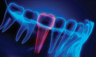
416 IMPACT OF MAXILLOFACIAL GROWTH ON IMPLANTS PLACED IN ADULTS: A NARRATIVE REVIEW
Dimokritos Papalexopoulos, DDS
Theodora-Kalliopi Samartzi, DDS Panagiotis Tsirogiannis, DDS Nikitas Sykaras, DDS, MSc, PhD
Aspasia Sarafianou, DDS, MSc, PhD
Stefanos Kourtis, DDS, PhD Aikaterini Mikeli, DDS
Originally printed in the Journal of Esthetic and Restorative Dentistry. Reprinted with permission.
436 PARTNER FOR SUCCESS: THE DYNAMICS AND ADVANTAGES OF STARTING A DENTAL PRACTICE WITH A PARTNER
Katie E. Stuchlik, DDS, FAGD COVER
The visitor center in San Angelo is a serene structure with stone and rock materials, surrounded by trees and gardens.


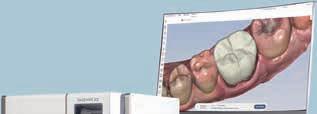
394 Texas Dental Journal | Vol 140 | No. 7 tdaperks.com Financial & Real Estate (iCoreConnect) iCoreHuddle instantly reveals revenue potential for each patient. 888-810-7706 Your staff no longer has to run dozens and dozens of reports. It’s all there. For every patient on the schedule. TDA members receive a 25% discount. Mention “TDA Perks.” Learn more or book a demo. Visit tdaperks.com (Financial & Real Estate) or scan the QR code below. Practice analytics at your fingertips Instantly see all revenue generating opportunities like: • Patient recall monitoring • Production numbers • New-patient metrics Have everything needed to help you: • Increase collections • Reduce time spent on insurance verifications • Understand case acceptance in real time
Spore Check System is accurate, easy to use, and inexpensive.
Week by week, you’ll know whether or not your sterilizer is working properly for just $159/sterilizer/year.
• An in-house lab, full-time microbiologist and years of experience mean accurate results delivered quickly, and that you’ll have help tracking down problems.
• After processing a test strip, just insert it into the pre-addressed envelope, and initial the receipt in the binder.
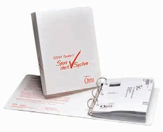
• If your test is positive, you’ll be called immediately.
800-555-6248
• If you forget to test for more than two weeks, you’ll be sent a reminder.
• You’ll receive free problem-solving consultations with OSHA Review’s microbiologist and and automatic reports after every ninth test.
• Access your test results anytime online, easily and privately.
1Since Texas regulations recognize ADA recommendations as State guidelines, Texas State Board Rule 108.24 states that “sterilization equipment and its adequacy shall be tested and verified in accord with the American Dental Association recommendations.” ADA and CDC recommendations include weekly spore testing.

www.tda.org | August/September 2023 395 To learn more,
tdaperks.com (Compliance & Supplies)
scan the QR code or visit
Spore testing is not optional.1
FEATURES
400 AN OPEN LETTER TO ADA MEMBERS
THE VALUE OF A UNITED TRIPARTITE AND THE ADA
405 ADDITIONAL ENTRIES: MEMBERSHIP AWARDS RECOGNITION
416 IMPACT OF MAXILLOFACIAL GROWTH ON IMPLANTS PLACED IN ADULTS: A NARRATIVE REVIEW
Dimokritos Papalexopoulos, DDS
Theodora-Kalliopi Samartzi, DDS
Panagiotis Tsirogiannis, DDS
Nikitas Sykaras, DDS, MSc, PhD
Aspasia Sarafianou, DDS, MSc, PhD
Stefanos Kourtis, DDS, PhD
Aikaterini Mikeli, DDS
This article was originally published August 5, 2022, in the Journal of Esthetic and Restorative Dentistry, Volume 35, Issue 3. Reprinted with permission.
436 PARTNER FOR SUCCESS: THE DYNAMICS AND ADVANTAGES OF STARTING A DENTAL PRACTICE WITH A PARTNER
Katie E. Stuchlik, DDS, FAGD
Editorial Staff
Jacqueline M. Plemons, DDS, MS, Editor
Juliana Robledo, DDS, Associate Editor
Nicole Scott, Managing Editor
Barbara Donovan, Art Director
Lee Ann Johnson, CAE, Director of Member Services
Editorial Advisory Board
Ronald C. Auvenshine, DDS, PhD
Barry K. Bartee, DDS, MD
Patricia L. Blanton, DDS, PhD
William C. Bone, DDS
Phillip M. Campbell, DDS, MSD
Michaell A. Huber, DDS
Arthur H. Jeske, DMD, PhD
Larry D. Jones, DDS
Paul A. Kennedy, Jr., DDS, MS
Scott R. Makins, DDS, MS
Daniel Perez, DDS
William F. Wathen, DMD
Robert C. White, DDS
Leighton A. Wier, DDS
Douglas B. Willingham, DDS
The Texas Dental Journal is a peer-reviewed publication. Established February 1883 • Vol 140 | No. 7
Texas Dental Association
1946 S IH-35 Ste 400, Austin, TX 78704-3698
Phone: 512-443-3675 • FAX: 512-443-3031
Email: tda@tda.org • Website: www.tda.org
Texas Dental Journal (ISSN 0040-4284) is published monthly except January-February and August-September, which are combined issues, by the Texas Dental Association, 1946 S IH-35, Austin, TX, 78704-3698, 512-443-3675. PeriodicalsPostage Paid at Austin, Texas and at additional mailing offices. POSTMASTER: Send address changes to TEXAS DENTAL JOURNAL, 1946 S IH 35 Ste 400, Austin, TX 78704. Copyright 2023 Texas Dental Association. All rights reserved.
Annual subscriptions: Texas Dental Association members $17. Instate ADA Affiliated $49.50 + tax, Out-of-state ADA Affiliated $49.50. In-state Non-ADA Affiliated $82.50 + tax, Out-of-state Non-ADA Affiliated $82.50. Single issue price: $6 ADA Affiliated, $17 Non-ADA Affiliated. For in-state orders, add 8.25% sales tax.
TMOM 2023 Events
TMOM, Edinburg: September 15-16, 2023
THE TEXAS DENTAL JOURNAL’S CALENDAR will include only meetings, symposia, etc., of statewide, national, and international interest to Texas dentists. Because of space limitations, individual continuing education courses will not be listed. Readers are directed to the monthly advertisements of courses that appear elsewhere in the Journal
Contributions: Manuscripts and news items of interest to the membership of the society are solicited. Electronic submissions are required. Manuscripts should be typewritten, double spaced, and the original copy should be submitted. For more information, please refer to the Instructions for Contributors statement included in the online September Annual Membership Directory or on the TDA website: tda.org. All statements of opinion and of supposed facts are published on authority of the writer under whose name they appear and are not to be regarded as the views of the Texas Dental Association, unless such statements have been adopted by the Association. Articles are accepted with the understanding that they have not been published previously. Authors must disclose any financial or other interests they may have in products or services described in their articles.
Advertisements: Publication of advertisements in this journal does not constitute a guarantee or endorsement by the Association of the quality of value of such product or of the claims made.
396 Texas Dental Journal | Vol 140 | No. 7
contents
HIGHLIGHTS
Calendar of Events 431 In Memoriam 436 Value for Your Profession: Expert Tips for Minimizing Credit Card Fees 438 Classifieds 446 Index to Advertisers calendar
396


www.tda.org | August/September 2023 397 Learn more at TXHealthSteps.com Join 225,000+ medical professionals who get free CE with Texas Health Steps Online Provider Education. Choose from a wide range of courses developed by experts, for dental experts like you. Courses on topics such as caries risk assessment and dental quality measures are available 24/7. Content on the Texas Health Steps Online Provider Education website has been accredited by the UTHSCSA Dental School Office of Continuing Dental Education, Texas Medical Association, American Nurses Credentialing Center, National Commission for Health Education Credentialing, Texas State Board of Social Worker Examiners, Accreditation Council for Pharmacy Education, Texas Academy of Nutrition and Dietetics, Texas Academy of Audiology, and the International Board of Lactation Consultant Examiners. Continuing Education for multiple disciplines will be provided for some online content. Texas Health Steps is health care for children from birth through age 20 who have Medicaid. Dental CE courses you can put into practice.
JKJ Pathology
Oral Pathology Laboratory
John E Kacher, DDS
¥ Available for consultation by phone or email
¥ Color histology images on all reports

¥ Expedited specimen shipping with tracking numbers
¥ Reports available online through secure web interface
Professional, reliable service with hightechnology solutions so that you can better serve your patients. Call or email for free kits or consultation.
jkjpathology.com

281-292-7954 (T)
281-292-7372 (F)
johnkacher@jkjpathology.com
Protecting your patients, limiting your liability
Board of Directors Texas Dental Association
PRESIDENT Cody C. Graves, DDS 325-648-2251, drc@centex.net
PRESIDENT-ELECT
Georganne P. McCandless, DDS 281-516-2700, gmccandl@yahoo.com
PAST PRESIDENT Duc “Duke” M. Ho, DDS 281-395-2112, ducmho@sbcglobal.net
VICE PRESIDENT, SOUTHWEST Richard M. Potter, DDS 210-673-9051, rnpotter@att.net
VICE PRESIDENT, NORTHWEST Summer Ketron Roark, DDS 806-793-3556, summerketron@gmail.com
VICE PRESIDENT, NORTHEAST Jodi D. Danna, DDS 972-377-7800, jodidds1@gmail.com
VICE PRESIDENT, SOUTHEAST Shailee J. Gupta, DDS 512-879-6225, sgupta@stdavidsfoundation.org
SENIOR DIRECTOR, SOUTHWEST Krystelle Anaya, DDS 915-855-1000, krystelle.barrera@gmail.com
SENIOR DIRECTOR, NORTHWEST
Stephen A. Sperry, DDS 806-794-8124, stephenasperry@gmail.com
SENIOR DIRECTOR, NORTHEAST
Mark A. Camp, DDS 903-757-8890, macamp1970@yahoo.com
SENIOR DIRECTOR, SOUTHEAST Laji J. James, DDS 281-870-9270, lajijames@yahoo.com
DIRECTOR, SOUTHWEST Melissa Uriegas, DDS 956-369-9235, meluriegas@gmail.com
DIRECTOR, NORTHWEST
Adam S. Awtrey, DDS 314-503-4457, awtrey.adam@gmail.com
DIRECTOR, NORTHEAST
Drew M. Vanderbrook, DDS 214-821-5200, vanderbrookdds@gmail.com
DIRECTOR, SOUTHEAST
Matthew J. Heck, DDS 210-393-6606, matthewjheckdds@gmail.com
SECRETARY-TREASURER*
Carmen P. Smith, DDS 214-503-6776, drprincele@gmail.com
SPEAKER OF THE HOUSE*
John W. Baucum III, DDS 361-855-3900, jbaucum3@gmail.com
PARLIAMENTARIAN**
Glen D. Hall, DDS 325-698-7560, abdent78@gmail.com
EDITOR**
Jacqueline M. Plemons, DDS, MS 214-369-8585, drplemons@yahoo.com
LEGAL COUNSEL
Carl R. Galant
*Non-voting member
**Non-voting
398 Texas Dental Journal | Vol 140 | No. 7
The glidewell.io™ system is much easier to use than my prior system. It couldn’t mill a BruxZir® crown. It didn’t have a lab for me to contact like Glidewell does. With the glidewell.io system, that’s what you’re paying to have — at a fraction of the cost. And it’s wonderful.


www.tda.org | August/September 2023 399
INTRODUCTION
Having been in use for more than 35 years in the field of dentistry, dental implants are a well-established treatment choice for the restoration of edentulous areas, yielding high survival rates.1 Many studies have focused on certain aspects of implant therapy, such as surgical trauma, periimplantitis, occlusal overload, the relationship between implant platform and bone margin, inter-implant distance, and continuous area disturbance, which may lead to technical and biological complications.2-8 However, the relationship between implant placement and adult facial growth has not been thoroughly investigated.9
Implants do not follow the course of jaw growth.10 The previous study using animal models was the first to demonstrate that implants do not adapt to dimensional changes, and this assumption was later confirmed in another study.10,11 Teeth are surrounded by the periodontal ligament that allows them to adapt to the developmental changes by continuous eruption and passive movement. The absence of this feature around implants, which are anchored in the alveolar bone, leads to the implants behaving similar to ankylosed teeth.12–14 This is the main reason why their use in children and adolescents is usually contraindicated.15,16 In this age group, implants are used with extreme caution, mainly in cases where anodontia or oligodontia is present.15,17 Growth complications are almost absent in these patients even after many years of observation. Implant placement in these cases is necessary to restore function and esthetics and enhance patient psychology.18
Facial growth is first completed in the transversal plane, followed by the sagittal and, later, the vertical plane.19
Studies with implants placed as reference points have further clarified jaw development.20–23 Maxillary growth is closely related to the growth of cranial structures, while mandibular growth is related to stature development.
Complications of implants placed before growth cessation include diastema formation, midline shift, asymmetries, palatal “displacement,” lack of the labial bone, anatomic cavity penetration (e.g., sinus), infraocclusion, and obstruction of the physiologic process of mesial drift.19
Some researchers have proposed the use of mini-implants to temporarily restore edentulous areas since it is considered that their small diameter (ca. 2 mm) does not affect jaw growth. However, the literature provides controversial findings regarding this approach.23,24
As a general rule, girls undergo a growth spurt around the age of 12 years (9–14 years), while in boys, this happens around the age of 14 years (11–17 years).19 However, chronological age does not always relate to developmental age. For the latter to be accurately defined, different methods have been proposed, such as wrist X-ray analysis and superimposed cephalometric radiography.19
Interestingly, changes in the maxillofacial complex continue to take place in adulthood as well. Until recently, this fact was rarely mentioned in the literature, mainly because of the short observation periods.25 However, these changes might be the cause of biological, technical, and esthetic complications that might compromise the success of implant therapy.
According to the recent systematic review, every fifth and every second implant placed is at risk of considerable infraposition or interproximal contact loss, respectively, at some point in the future.9 Nevertheless, no exclusion criteria were set regarding age.
The present narrative review aimed to report the impact of continuous craniofacial changes on implants and implant-supported rehabilitations placed in adult patients, describe the causes for alterations that occur even after active growth cessation, and provide clinicians with an array of prognostic factors as well as possible solutions.
MATERIALS AND METHODS
PubMed, Cochrane, Scopus, and Ovid databases were searched for relevant studies, namely evidence-based research articles regarding the effects of continuous maxillomandibular changes on implants placed in adults, published from January 1990 until July 31, 2021.
An initial study sample with key papers in the field was consolidated in order to aid the development and sensitivity verification of the search strategy. After an initial screening of the available literature infraocclusion and interproximal contact loss were highlighted as the two major complications arising from the bidirectional relationship between implants and maxillomandibular changes. Consequently, the key search terms were defined accordingly and the appropriate MeSH terms were grouped as follows: “Implants AND maxillofacial growth AND adults,” “implants AND continuous craniofacial
418 Texas Dental Journal | Vol 140 | No. 7
growth,” “implants AND infraocclusion,” “implants AND infraposition,” “implants AND interproximal contacts,” “implants AND proximal contacts,” and “implants AND open contacts”. A supplementary manual search was also conducted.
Peer-reviewed articles on the relationship between lifelong changes and implants placed in adults and clinical studies in adult patients were set as the inclusion criteria. Articles not written in English, duplicate articles, and articles not referring to implants placed in adult patients or not allowing for the extraction of data referring to adult patients were excluded from further evaluation. Contact with the authors of articles not reporting crucial information was attempted. If no answer was received within 2 months of the request, then the study was not included in the present review.
Two reviewers (D.P. and T.K.S.) evaluated all articles for appropriateness after screening the titles, abstracts, and full texts. The reference lists of the included articles were screened to identify any relevant studies, applying the same inclusion and exclusion criteria. Discrepancies between the two reviewers were discussed until both reached an agreement. The sensitivity of the search strategy was verified by both reviewers since all articles from the initial study sample were identified among the included studies.
RESULTS
The search strategy identified 889 records. After excluding irrelevant articles and thoroughly investigating the references of the articles that met the inclusion parameters, 36 studies were
included in the review (Figure 1). Among them, 9 clinical studies reported on implant infraocclusion, 9 clinical studies reported on proximal contacts loss, one clinical study investigated displacement of teeth adjacent to implants, one retrospective study evaluated implant submersion rates, and there were 8 case reports and eight reviews.
Synthesis of the studies included certified implant infraocclusion and proximal contact loss of implant restorations as the two major clinical problems arising from the interaction with continuous maxillomandibular changes. Consequently, this section was constructed in order to critically evaluate and present the results and clinical considerations regarding these two complications, after a thorough exposition of the maxillofacial growth mechanisms.
www.tda.org | August/September 2023 419
Databases searched: • Pubmed • Cochrane • Scopus • Ovid Initial records: 889 48 abstracts screened 32 full texts assessed for eligibility 36 studies included in the review: • 5 prospective studies • 15 retrospective studies • 8 reviews • 8 case reports 14 records identified through reference screening 841 records excluded: • Duplicate records • Irrelevant data 16 records excluded: • Irrelevant data • Studies not referring to adults 10 records excluded: • Unavailable data • Studies not referring to adults or not possible to derive data for adults
FIGURE 1. Flowchart of the article selection process
Lifelong maxillofacial growth
Developmental changes during the active growth period are obvious; however, growth is continuous during adulthood, albeit to a lesser extent.25 A retrospective study showed that the relative changes between implants and teeth occurred in patients in the active growth period as well as in adulthood.26
Lifelong changes of the maxillofacial complex are attributed to various mechanisms. The maxilla and mandible grow away from the cranial base, moving downward and forward.27 A 1.6-mm increase in face height, 80% of which was associated with the lower third, throughout a period of 20 years was measured in a previous study.28
According to the results of a clinical study on women aged 20–50 years, the anterior face height increased by 3–3.5 mm throughout the study period. This was attributed to the rotation of the mandible in a backward direction. However, in men, a forward rotation tendency was reported.29,30
Continuous bone remodeling takes place until late adulthood.28 An increase in the dentoalveolar height has also been reported.29 These findings are further substantiated by a study that also found an increase in the vertical overbite.31,32
Moreover, tooth eruption can be observed at any age, and there is a continuous tendency for uprighting (2–3).27 The average eruption speed of the maxillary incisors is 1.5 mm/ year during growth and 0.1–0.2 mm afterward, even after the age of 18.33 For the maxillary central incisor, eruption between the ages of 9 and 25 reaches 6 mm, with a simultaneous buccal displacement of 2.5 mm. Regarding the first maxillary molars, the corresponding values are 8 mm and 3
mm.21 According to the previous study, the mean eruption of teeth adjacent to implants ranged from 0.13 to 1.75 mm over a 3 year observation period.34 Another study on Swedish women reported negligible changes in the molars but distinct changes in incisors and canines, reaching values of 1.2 mm within 10 years.35 Moreover, the clinical crown length tends to increase through a combination of gingival recession (mainly in molars) and tooth eruption (mainly in anterior teeth).35
This continuous migration towards the occlusal direction can be indirectly observed by the reported increase of the band of attached gingiva that reaches 4 mm in the anterior maxilla.36,37 The interaction between lifelong multi-dimensional bone growth and continuous tooth eruption leads to esthetic and functional problems regarding implants and adjacent teeth.38
Among changes occurring in the maxillofacial complex, the tendency towards dental arch “closure” also exists. This reduction is estimated at 1.86 mm in the maxilla and 2.4 mm in the mandible for men, and 2.06 mm and 1.76 mm for women, respectively.39 The mechanism responsible is mainly the tendency for a continuous mesial movement. The four main factors determining the dental arch arrangement are the tongue and lips, patient habits and orthodontic therapy, periodontal status, and occlusal factors.40 Occlusal forces are transferred through interproximal contacts to compensate for tooth wear that normally happens over time.41–46
Although it is believed that occlusal forces act on a vertical plane, there is also a horizontal or anterior component of force (ACF)that has the tendency to move teeth mesially and is even more
obvious among patients with a long face type.46-48 Although there is a force in the direction of distal areas, the one directed mesially is 5 times higher.48 Until the age of 40, the latter will have moved the teeth mesially by 5 mm.48 The factors contributing to this change are age, occlusal forces, condition of the opposing arch, vitality of adjacent teeth, and the presence of splints.41,42 Increased occlusal loads that must be tolerated by a tooth adjacent to an implant have also been mentioned as the cause of this movement.41
Impact of adult maxillofacial growth on implants
Infraocclusion
The term “infraocclusion” can be defined as “the position occupied by a tooth when it has failed to erupt sufficiently to reach the occlusal plane.”49 The definition can be applied to implants exhibiting a vertical discrepancy compared to adjacent teeth.50 Infraocclusion was apparent in several case reports with a follow-up period of 15 months to 16 years where the implant infraposition reached 1.2 mm.50–52 A study with an 8 year follow-up was the first to prove that differences during the active growth period (vertical difference of 0.46 mm in 4 years) do not stop but continue to occur even in adulthood (vertical difference of 0.96 mm in 8 years).13 Significant vertical differences have been recorded even in patients in whom implants were placed during the fourth or fifth decade of life, showing that infraposition can be observed in any age group and is not an age-dependent process (Table 1).53
In a study that examined the vertical relationship between implants placed in adults and adjacent teeth, a vertical difference was reported in 58% of
420 Texas Dental Journal | Vol 140 | No. 7
TABLE 1. Infraposition of implants related to adjacent teeth according to the clinical studies
the cases.54 In the incisor area, the difference was 0.1 mm during the first year, 0.4 mm within the fifth year, and 0.5 mm after 8 years. In the premolar area, the mean vertical difference was 0.2 mm; this infraposition was accompanied by malalignments compared to the adjacent teeth that tend to erupt while acquiring a lingual inclination.28
Risk Factors: In accordance with the abovementioned studies, some researchers observed vertical differences between implants and
adjacent teeth in all patients of their study, with the “mature adults” group presenting differences between 0.12 mm and 1.86 mm (mean = 0.67 mm) during an observation period of 4.2 years, which was similar to the findings in the “young adult” group (0.1–1.65 mm, mean = 0.69 mm).26 In another study, implant infraposition rates of 43% in patients older than 21 years old were reported.9 Moreover, patients younger than 30 years had three times higher submersion rates than those older than 30.59
In a clinical study all women and 45% of men in the sample presented vertical differences between implants and adjacent teeth, showing a possible correlation between implant infraposition and sex as well as anterior face height.34,60 This finding was attributed to increased anterior face height growth and backward mandible rotation in women. The findings of another study, however, are not consistent with this observation.61
www.tda.org | August/September 2023 421
Author and Number of patients Total number Infraposition rates (%) Infraposition Observation publication year (age at implantation) of implants distance (mm) period (range in years) Bernard et al., 200426 22 (18–52) 30 100 0.12-1,86 1.1–9.1 Chang et al., 201254 31 (19–71) 33 29 (year 1) <1.2 1–8 55 (year 5) 58 (year 8) Vilhjálmsson et al., 201334 23 (20–56) 23 Not mentioned 0.13–1.75 3 Andersson et al., 201353 34 (18–56) 37 100 Not reported 17–19 • 50 < 0.5 mm • 15 < 1 mm • 35 > 1.5 mm Dierens et al., 201355 19 (33–58) 24 71 Not reported 16–22 Ekfeldt et al., 201656 23 (27–63) 30 17 ≤1 10–11 Brahem et al., 201757 57 (18–61) 89 Test group (with orthodontic Not reported 7 ± 1 pretreatment): 87 Control group (without orthodontic pretreatment): 70 Cochetto et al., 201949 60 (20–65) 76 73 Not reported 5–20 • 65 in males • 79 in females Polymeri et al., 202058 76 (21–78) 77 Not reported <1.67 Up to 15
Women with a long face type are placed in the high-risk group for infraocclusion.48,53,62 The importance of the Frankfurt-mandibular plane angle (FMA), formed between the Frankfurt plane and the plane of the mandible, has been established in the past. According to its values, each patient may be characterized as “normal” (FMA = 25 ± 5), “high-angle” (FMA > 30), or “low-angle” (FMA < 20).63 There are indications that the long facial type (“high angle”) is related to specific clinical characteristics that may complicate prosthodontic restoration.63 Long anterior face height (Na to Gn and ANS to Gn) results from increased growth of the alveolar ridges, which explains the large vertical differences observed in these cases.62
A long-term study attempted to identify the possible predictors of infraocclusion.58 It was reported to be more pronounced in younger patients than in older patients, in central incisors than in lateral incisors and canines, in delayed than in immediately placed implants, and in delayed than in immediate temporization. No associations were found between ethnicity, sex, surgical protocol, guided bone regeneration procedures, implant brand, and type of prosthesis retention. It has also been shown that the presence of interproximal contacts might decelerate the eruption rate of lateral incisors.35 Other researchers reported that orthodontic retention or implant location do not have any significant effect on tooth displacement, which occurs towards the incisal direction in the buccal–lingual and mesial–distal planes.57
Biological, Functional, and Esthetic Concerns:
Results from different studies show an overall frequency of infraocclusion ranging from 40% to
100%, with an annual rate possibly as high as 0.96 mm (Table 1).49,50 Although tooth wear might contribute to slightly unfavorable esthetic changes, the resultant vertical asymmetry may raise esthetic concerns for both clinicians and patients.32,34 It has been shown that even though changes in the incisors’ width for up to 1 mm may be tolerated, length alterations are not accepted.64 In the recent study, 73% of patients presented with infraocclusion and 18.2% of them were aware of the problem and wanted an esthetic solution, and the rate was even higher for female patients at 22.2%.49
According to a study, infraoccluded implant-supported crown was the most common reason for restoration replacement.65 It has also been reported that the lowest value in the visual analog scale for esthetics was due to an implant crown that was in infraposition related to adjacent teeth.66
Apart from esthetics, biological and occlusal concerns may also arise. It has been reported that in cases of reduced distance between the teeth and implants,the presence of the latter might impede the development of adjacent tissues while the teeth continue to follow developmental changes and erupt.26,67 This combination may steadily lead to a lack of periodontal tissues at the dental surfaces facing the implant.
According to the abovementioned, the teeth tend to move, following continuous changes to which the maxilla and mandible are subjected. Implant restorations placed distal to the last natural tooth must tolerate high occlusal forces. The teeth migrate in an occlusal direction resulting in a shift of this load toward them.25 It is still unknown whether this procedure affects men or women to a greater
extent. After prosthesis delivery, the patient should be regularly evaluated to check for occlusion and prevent occlusal overload.68
Treatment Options: The problem of infraposition may be addressed by repairing the existing restoration or by constructing a new one. However, the augmented restoration length may endanger the implant–restoration ratio.67 The use of long-term transitional restorations has been reported.38 The orthodontic intrusion of adjacent teeth has also been proposed in order to reestablish harmony among incisal edges.50
Surgical techniques of repositioning the bone surrounding the implant have also been described.69 Adequate space around the implant is a prerequisite, not to mention that the subsequent deficiencies cause major esthetic problems. However, these osteotomies combined with “greenstick fracture” cannot be used routinely and applied in areas wider than one tooth. If neither of these procedures is indicated, then implant submersion should be considered a possible solution.70
Regardless of the approach chosen, the treatment plan should be defined only after thorough clinical and radiographic examination to assess the amount of infraposition and malalignment compared to adjacent teeth, the position of the gingival margins, condition of the opposing dentition, and bone morphology.70
Interproximal contact loss
Interproximal contact is crucial for avoiding food impaction, teeth migration, and periodontal inflammation.71,72 Their size and location vary, being larger in the horizontal
422 Texas Dental Journal | Vol 140 | No. 7
dimension in posterior areas and transformation from oval to kidney shape.73,75 When a natural tooth is substituted by an implant, tissue shrinkage might require a large contact area in the occlusogingival dimension.75
Given that implants cannot follow any developmental change, interproximal contact loss may occur.76 Apart from ACF, potential implant infraocclusion changes the relationship between
implant prosthesis and adjacent teeth.44 A case report about a 58-year-old female patient was one of the first to mention interproximal contact loss as a complication of implant therapy that may occur as soon as 1 year after restoration delivery.43
Researchers have reported contact loss in 34–65% of patients (an observation period of 1–13 years) (Table 2) that may occur as soon as 3–8 months
after restoration delivery.41–45,77,78 The observed differences may be attributed to the different evaluation methods used since a universal technique does not exist.78 It has been stated that 12.5% of interproximal contact loss occurred in the first year, 47.6% within 3 years, and 80% within 5 years after the restoration delivery, while the peak period for interproximal contact loss appeared at 1.9–2.2 years.78 A crosssectional study underlined that besides
www.tda.org | August/September 2023 423
Study Number of patients Number of Interproximal Evaluation Observation (age at implantation) implant- contact loss (%) method period range supported prostheses Byun et al. 201544 94 (27–83) 135 34 Dental floss 3–156 months (188 implants) (65/191) (+20% “loose”) Wong et al. 201545 45 (27–74) Not mentioned 65 Matrix bands 0.5–12 years (43/66) (0.038 mm) Varthis et al. 201677 128 (19–91) 174 52,8 Dental floss 3–132 months (single implant (92/174) (0.07 mm) restorations) Pang et al. 201778 150 (21–79) 234 59.9 Aluminum strips Up to 7.5 years (384 implants) (179/299) (0.05 mm) Shi et al. 201979 74 (22–70) 74 24.3 Dental floss 1 year (35/144) Bompolaki et al. 202080 83 (37–86) 118 Mesial: 48.8 Dental floss 4 ± 2.2 years Distal: 26.7 Saber et al. 202081 83 (26–80) 183 32.8 Dental floss 3 months–5 years (60/183) (0,07 mm) Yen et al. 202082 147 (25–85) 180 15 Periapical Not reported (patient level) Radiographs 13.3 (implant level) Mehanna et al. 202183 43 (31–70) 43 Mesial: 4.7 Dental floss 0.05 3 months Distal: 9.5 metal strip
TABLE
2. Interproximal contact loss between implant restorations and adjacent teeth according to the clinical studies
34% of “open” contacts, 20% of the initial “firm” contacts turned “loose,” meaning that more than half of the initial “firm” contacts were lost.44 The recent systematic review reported a frequency of 41% of open proximal contacts in implant-supported prostheses, a rate significantly higher than that observed with tooth-supported prostheses.84 Another systematic review reported a 46% prevalence of interproximal contact loss.9
Risk Factors: Attempts have been made to identify the predisposing factors for implant prostheses-adjacent teeth interproximal contact loss. According to some researchers, after long follow-up periods, interproximal contact loss was observed in the mesial sides of implant restorations, with some studies reporting rates twice as high as those in the distal sides,which is attributed to the anterior component of the force that moves the teeth mesially, in the posterior rather than the anterior areas, and in the maxilla compared to the mandible, probably due to lower bone density.44,77,78,80–82,84–86 However, according to the systematic review, a high percentage is expected in the mandible because the lower teeth often present with mesial tipping.68,79,82,84,85,87 A 2.5-fold increased risk of interproximal contact loss when implants are splinted in fixed partial dentures rather than with single crowns has been mentioned, a finding also verified by other studies.44,83,87 Despite objections, bone support of the adjacent teeth plays an important role.82,88 Increased marginal bone loss in cases of interproximal contact loss has been reported.81 Nonvital teeth seem to be another unfavorable prognostic factor.78
According to some researchers, the risk is higher when the opposing dentition consists of a removable denture compared with a nonremovable denture.42 There is also a high tendency for devitalized teeth and single-rooted teeth to present open interproximal contacts.42,44,78,84 Spacing on the contralateral side and contact with a composite resin restoration, which exhibits inferior wear resistance, have also been associated with a high risk.82,89
Regarding implant connections, internal octagonal and external hexagonal connections are reportedly associated with significantly higher contact loss than internal hexagonal connections, probably due to the increased micromotion and rotational freedom,which may influence the long-term stability of proximal contacts.82,90,91 No associations have been found with age, sex, bone level, parafunction existence, type of retention, and splinting of implants or teeth.79,84 One study has associated male sex with higher rates of proximal contact loss, probably because of the higher masticatory force of men.92 However, it has been reported that patient-level risk factors are less important than the condition of the adjacent teeth.82
Tightness of interproximal contacts is influenced by many factors such as tooth type, time of day, and chewing.93 A significant decrease has been reported within 3 months after restoration delivery.94 A retrospective study found no statistical differences in relation to age, the presence of an endodontically treated tooth adjacent to the implant site, maxilla or mandible, opposing tooth status, or occlusal device, although the latter has been suggested as a way to prevent interproximal contact loss.68,80
424 Texas Dental Journal | Vol 140 | No. 7
Tightness of interproximal contacts is influenced by many factors such as tooth type, time of day, and chewing. A significant decrease has been reported within 3 months after restoration delivery.
Biological Concerns: Open contact has been reported as one of the frequent complications of implant therapy,and 43,8% of the patients experience food impaction due to interproximal contact loss.42,77,78,80,82 An open contact has a 2.2-fold higher risk for food impaction than an intact one, as well as a negative impact on both peri-implant health and the patient’s overall satisfaction.44,87,95 Bleeding on probing on the distolingual surface is more frequently observed in open or loose distal contacts than in closed contacts, which have been attributed to plaque-induced inflammation that might cause bone resorption.80,81,85 An association between crestal bone levels and occurrence of proximal contact loss could not be substantiated in a previous study.85 Another serious biological complication is caries, with a 2-fold higher incidence in open contacts than in close contacts.85
Treatment Options: Careful surgical planning and management are needed to ensure that implant inclination will allow the dental technician to establish contacts of the right size and shape.88,96 If an open contact occur, then either the restoration or the tooth should be recontoured.97 Ideally, the prosthesis should be removed and sent to the dental laboratory to redefine the proximal surface.43,97 Consequently, two major concerns arise regarding prosthesis retention (screw versus cement-retained) and the restorative material (metalceramic, all-ceramic, porcelain fused to zirconia or monolithic zirconia). Retrievability of implant restorations is essential in prosthesis repair. Screwed restorations can be easily removed, which is not always the case for cemented ones.43 Even if a cemented restoration is delivered, it should be cemented with provisional cements.77
Material selection is a clinical decision directly connected with cost, esthetics, and possible modifications, as the chance of chipping may be increased by an inappropriate ceramic chosen for contour alterations.75,98 Recent studies have shown that composite resin adhesion on silica-based and zirconia surfaces is feasible,providing clinicians with the option of chairside repairs.97,99,100 Airborneparticle abrasion should be used as a conditioning method before applying a 10-methacryloyloxydecyl dihydrogen phosphate-containing adhesive when attempting adhesion of the resin to zirconia, which was considered to have limited bonding potential, a belief overthrown by more recent studies.101-104 For silica-based ceramics, etching with hydrofluoric acid and subsequent silanization are crucial prerequisites.100,105 Extraoral reestablishment techniques of open contacts with the help of a chairside-created silicone model, which makes evaluation of the interproximal contact easier than with a stone cast, as well as the incorporation of gradual polymerization of applied resin on the proximal area have been described, with prosthesis removal being a prerequisite.97,105
Maintenance: Regarding prevention and recall protocols, implant patients should be examined every 3–6 months. If an open contact is observed, the presence of food impaction and/or biological complications (caries and periodontal disease) will determine the clinicians’ next steps.75 When only food impaction is observed, the clinician’s choice is to either change the implant restoration and/or restore the adjacent tooth to reestablish tight contacts. If food impaction is accompanied by carious lesions or periodontal inflammation,
the adjacent tooth should be treated accordingly. At the treatment planning stage, patients should be informed about potential future complications requiring prosthesis replacement, and written consent must be obtained.42,77,81
DISCUSSION
The maxillofacial area is subjected to continuous changes that are more apparent during the active growth period. Since the implants are anchored to the bone,there is an increased risk of an implant being found in an undesired position, lacking the buccal bone, or causing reduced periodontal support in adjacent teeth, so implants are not indicated for children and adolescents.16,106–108
However, secondary changes continue to occur. The intermolar distance increases as the intercanine distance decreases, and there is also a tendency for teeth crowding and dental arch narrowing.109,110 Moreover, the anterior face height increases,a change attributed to the development of alveolar ridges since the teeth erupt continuously.28 In men, the buccal–palatal orientation is maintained, while in women, eruption is accompanied by a slight palatal tilt.29 Consequently, an implant placed in the patient’s fourth or fifth decade of life can later be found at an apical level compared to the adjacent teeth,potentially causing esthetic complications.53 Additionally, there is a risk of interproximal contact loss, especially regarding the mesial areas, because of the teeth’s tendency for continuous mesial movement.19
Dental clinicians should be aware of the problems related to developmental changes, which may endanger the treatment outcome and jeopardize the long-term success and survival of the
www.tda.org | August/September 2023 425
implant.2 The criteria for the latter have undergone revisions following advances in the field of implantology and have included many parameters instead of merely evaluating the preservation of the implant inside the oral cavity.111 In 2008, an attempt was made to define four quality scales, placing satisfactory and compromised survival between success and failure with distinct characteristics for each category.112 In a systematic review, a survival rate of 94.6% was reported, which can be characterized as satisfactory.113 However, considering the available techniques and materials, clinicians should focus their attention more on success than on survival.
Since the first crown was placed on an osseointegrated implant in 1983, implantology has undergone major revisions.114 After a period of enthusiasm regarding implant placement, a swift move toward evidence-based decisions for tooth extraction and implant placement has been observed.115 Clinicians should consider all available potential to sustain natural dentition, bearing in mind that teeth are the organs involved in functions such as mastication and speech, and their presence is associated with oral health-related quality of life.116,117
The remaining teeth should be carefully examined both clinically and radiographically to ensure accurate diagnosis and robust prognosis. Teeth characterized as questionable represent a gray zone, and many have been sacrificed for implants to be placed. However, questionable or even hopeless teeth may be retained for 15 years.118
Proper periodontal treatment and maintenance have led to a survival of 93% of periodontally compromised
teeth.119 Nevertheless, it has been reported that 80% and 12%–66% of implants are affected by mucositis or periimplantitis, respectively. For endodontically treated teeth, the survival rates are 89.7%–98.1%,with a smaller loss rate than dental implants.115
Even if a tooth must be extracted or is already missing, a dental implant is not the only available option. In fact, some authors reported that implants are contraindicated in certain cases, such as missing incisors with a gummy smile.120 An orthodontic consultation should be performed to investigate the potential for space closure, which has shown better results than tooth-supported dental prostheses regarding periodontal indices.121
A resin-bonded fixed partial denture is another minimally invasive option on par with conventional prosthetic designs in terms of survival rates for observation periods as long as 15 years.122 Advancements in the field of dental biomaterials have made available the use of novel all-ceramic systems for the construction of such prostheses with promising results.123 Recent data from long-term studies propose cantilevered resin-bonded fixed dental prostheses as the major treatment alternative to implants for replacing congenitally or traumatically missing teeth, presenting superior clinical outcomes compared to tworetainer resin-bonded fixed dental prostheses.124,125 This type of prosthesis may also be inserted early in children and adolescents as they do not interfere with growth.
Another rarely utilized option is autotransplantation, with as high as 91% survival rates.126 Even if any of the abovementioned techniques fail, there
is still the option of implant placement, while implant failure decreases the survival rates of the second implantation.127
The aforementioned facts indicate the need for well-designed treatment plans that consist of precise surgical approaches since implants must be placed in the appropriate axis and at an adequate distance from adjacent teeth. This approach will enable the construction of appropriate restoration and decrease the impact of the implant’s “ankylotic” behavior on adjacent teeth.26,67 Attention should be given to ensure that the restoration can be removed and repaired, and patient briefing in advance regarding future interventions is a vital aspect of the therapy. More studies are needed to identify factors that will assist the prediction of the onset, rate, and magnitude of those changes, but for the time being, these procedures are rather vague.50 Moreover, since there is no consensus on a single method of evaluating the aforementioned changes, simple, objective, and reproducible techniques should be established.128
Regarding the limitations of this study, it constitutes a narrative and not a systematic review which is a type of study characterized by methodological robustness. Because of the inclusion and exclusion criteria, many relevant articles were ruled out mostly due to the age of the participants in the study samples, which included adolescents. Some studies were also excluded since communication with authors in order to retrieve crucial information was not possible. Moreover, there was a large heterogeneity among the study types, since reviews, case reports and both prospective and retrospective clinical studies were included, irrespective of
426 Texas Dental Journal | Vol 140 | No. 7
the study samples, due to the lack of randomized control trials which would ideally compose the content of a high-quality systematic review. Another reason for the heterogeneity was the difference among the observation periods of each study that may have a potentially misleading effect on the extraction of clinical conclusions or even recommendations. Since the deleterious effects of implants placed in patients during the active growth period have been thoroughly described, adult samples should be considered. Furthermore, well-designed clinical studies with adult samples are needed to define the prognostic factors, contraindications, and efficient solutions to the deleterious effects of lifelong changes upon dental implants and implantsupported restorations.
CONCLUSIONS
Within the limitations of this current study, the following conclusions were made after critical analysis of the included studies: first, maxillomandibular changes throughout adulthood are the cause of observed alterations such as implant infraocclusion and interproximal contact loss. Women with a high FMA angle and young adults mainly constitute the high-risk group. Second, both complications result in severe biological, functional, and esthetic concerns. Third, careful treatment planning and prosthesis retrievability are essential for the efficient management of these complications. Fourth, correct diagnostic processes and prognosis determination should not be overridden while evaluating a tooth. Available therapies have shown survival rates equal or even superior to those achievable through implant placement. Fifth, even if a tooth is to be extracted, implant placement is not the only available option. Orthodontics or novel prosthodontic approaches may be applied as well. Finally, long-term clinical studies are needed to identify the risk factors.
DISCLOSURE
The authors do not have any financial interest in the companies whose materials are included in this article.
ORCID
Dimokritos Papalexopoulos
https://orcid.org/0000-0001-7001-3905
Stefanos Kourtis
https://orcid.org/0000-0002-5696-5413
REFERENCES
1. Howe MS, Keys W, Richards D. Long-term (10-year) dental implant survival: a systematic review and sensitivity meta-analysis. J Dent. 2019;84:9-21. doi:10.1016/j.jdent.2019.03.008
2. Yamanishi Y, Yamaguchi S, Imazato S, Nakano T, Yatani H. Effects of the implant design on periimplant bone stress and abutment micromovement: three-dimensional finite element analysis of original computer-aided design models. J Periodontol. 2014;85(9):e333-e338. doi:10.1902/ jop.2014.140107
3. Becker W. Immediate implant placement: diagnosis, treatment planning and treatment steps/or successful outcomes. J Calif Dent Assoc. 2005;33(4):303-310.
4. Fransson C, Tomasi C, Pikner SS, et al. Severity and pattern of periimplantitis-associated bone loss. J Clin Periodontol. 2010;37(5):442-448. doi:10.1111/j.1600-051X.2010.01537.x
5. Kim Y, Oh TJ, Misch CE, Wang HL. Occlusal considerations in implant therapy: clinical guidelines
if a tooth must be extracted or is already missing, a dental implant is not the only available option. In fact, some authors reported that implants are contraindicated in certain cases, such as missing incisors with a gummy smile.
www.tda.org | August/September 2023 427
Even
with biomechanical rationale. Clin Oral Implants Res. 2005;16(1):26-35. doi:10.1111/j.1600-0501.2004.01067.x
6. Oh TJ, Yoon J, Misch CE, Wang HL. The causes of early implant bone loss: myth or science? J Periodontol. 2002;73(3):322-333. doi: 10.1902/jop.2002.73.3.322
7. Rodriguez-Ciurana X, Vela-Nebot X, SegalaTorres M, et al. The effect of interimplant distance on the height of the interimplant bone crest when using platform-switched implants. Int J Periodontics Restorative Dent. 2009;29(2):141-151.
8. Abrahamsson I, Berglundh T, Lindhe J. The mucosal barrier following abutment dis/ reconnection. An experimental study in dogs. J Clin Periodontol. 1997;24(8):568572. doi:10.1111/j.1600-051x.1997. tb00230.x
9. Papageorgiou SN, Eliades T, Hammerle CHF. Frequency of infraposition and missing contact points in implantsupported restorations within natural dentitions over time: a systematic review with metaanalysis. Clin Oral Implants Res. 2018;29(Suppl 18):309-325. doi:10. 1111/ clr.13291
10. Odman J, Grondahl K, Lekholm U, Thilander B. The effect of osseointegrated implants on the dento-alveolar development. A clinical and radiographic study in growing pigs. Eur J Orthod. 1991;13(4): 279-286. doi:10.1093/ejo/13.4.279
11. Thilander B, Odman J, Grondahl K, Lekholm U. Aspects on osseointegrated implants inserted in growing jaws. A biometric and radiographic study in the young pig. Eur J Orthod. 1992;14(2):99-109. doi: 10.1093/ ejo/14.2.99
12. Ledermann PD, Hassell TM, Hefti AF. Osseointegrated dental implants as alternative therapy to bridge construction or orthodontics in young patients: seven years of clinical experience. Pediatr Dent. 1993;15(5):327-333.
13. Thilander B, Odman J, Jemt T. Single implants in the upper incisor region and their relationship to the adjacent teeth. An 8-year followup study. Clin Oral Implants Res. 1999;10(5):346-355. doi:10.1034/j. 1600-0501.1999.100502.x
14. Williams P, Travess H, Sandy J. The use of osseointegrated implants in orthodontic patients: I. implants and their use in children. Dent Update. 2004;31(5):287-290. doi:10.12968/denu. 2004.31.5.287
15. Mankani N, Chowdhary R, Patil BA, Nagaraj E, Madalli P. Osseointegrated dental implants in growing children: a literature review. J Oral Implantol. 2014;40(5):627631. doi:10.1563/AAID-JOI-D-11-00186
16. Brahim JS. Dental implants in children. Oral Maxillofac Surg Clin North Am. 2005;17(4):375-381. doi:10.1016/j. coms.2005.06.003
17. Kearns G, Sharma A, Perrott D, Schmidt B, Kaban L, Vargervik K. Placement of endosseous implants in children and
adolescents with hereditary ectodermal dysplasia. Oral Surg Oral Med Oral Pathol Oral Radiol Endod. 1999;88(1):5-10. doi:10.1016/s1079-2104(99) 70185-x
18. Smith RA, Vargervik K, Kearns G, Bosch C, Koumjian J. Placement of an endosseous implant in a growing child with ectodermal dysplasia. Oral Surg Oral Med Oral Pathol. 1993;75(6):669-673. doi:10.1016/00304220(93)90419-5
19. Heij DG, Opdebeeck H, van Steenberghe D, Kokich VG, Belser U, Quirynen M. Facial development, continuous tooth eruption, and mesial drift as compromising factors for implant placement. Int J Oral Maxillofac Implants. 2006;21(6):867-878.
20. Bjork A, Skieller V. Growth of the maxilla in three dimensions as revealed radiographically by the implant method. Br J Orthod. 1977; 4(2):53-64. doi:10.1179/ bjo.4.2.53
21. Iseri H, Solow B. Continued eruption of maxillary incisors and first molars in girls from 9 to 25 years, studied by the implant method. Eur J Orthod. 1996;18(3):245-256. doi:10.1093/ejo/18.3.245
22. Carmichael RP, Sandor GK. Dental implants, growth of the jaws, and determination of skeletal maturity. Atlas Oral Maxillofac Surg Clin North Am. 2008;16(1):1-9. doi:10.1016/j.cxom.2007.10.003
23. Jofre J, Werner A. Use of mini implants to replace a missing tooth in a growing patient: a six-year follow up case report. Eur J Paediatr Dent. 2015;16(4):284-286.
24. Wilmes B, Nienkemper M, Renger S, Drescher D. Mini-implantsupported temporary pontics. J Clin Orthod. 2014;48(7):422-429.
25. Daftary F, Mahallati R, Bahat O, Sullivan RM. Lifelong craniofacial growth and the implications for osseointegrated implants. Int J Oral Maxillofac Implants. 2013;28(1):163-169. doi:10.11607/ jomi.2827
26. Bernard JP, Schatz JP, Christou P, Belser U, Kiliaridis S. Long-term vertical changes of the anterior maxillary teeth adjacent to single implants in young and mature adults. A retrospective study. J Clin Periodontol. 2004;31(11):1024-1028. doi:10.1111/j.1600-051X.2004.00574.x
27. Oesterle LJ, Cronin RJ Jr. Adult growth, aging, and the single-tooth implant. Int J Oral Maxillofac Implants. 2000;15(2):252260.
28. Forsberg CM, Eliasson S, Westergren H. Face height and tooth eruption in adults—a 20-year follow-up investigation. Eur J Orthod. 1991; 13(4):249-254. doi:10.1093/ejo/13.4.249
29. Tallgren A, Solow B. Age differences in adult dentoalveolar heights. Eur J Orthod. 1991;13(2):149-156. doi:10.1093/ ejo/13.2.149
30. West KS, McNamara JA Jr. Changes in the craniofacial complex from adolescence to midadulthood: a cephalometric
study. American Journal of Orthodontics and Dentofacial Orthopedics: Official Publication of the American Association of Orthodontists, its Constituent Societies, and the American Board of Orthodontics. 1999;115(5):521-532. doi: 10.1016/s08895406(99)70274-x
31. Sarnas KV, Solow B. Early adult changes in the skeletal and soft-tissue profile. Eur J Orthod. 1980;2(1):1-12.
32. Jardini MAN, Ferreira CL, Ursi WJ, Melo Filho AB, Santamaria MP. Relative positional change of a dental implant in the esthetic zone after 12 years: a case report. General Dentistry. 2017;65(3):e1-e4.
33. Brugnolo E, Mazzocco C, Cordioll G, Majzoub Z. Clinical and radiographic findings following placement of singletooth implants in young patients--case reports. Int J Periodontics Restorative Dent. 1996;16(5):421-433.
34. Vilhjalmsson VH, Klock KS, Storksen K, Bardsen A. Radiological evaluation of single implants in maxillary anterior sites with special emphasis on their relation to adjacent teeth: a 3-year follow-up study. Dent Traumatol. 2013;29(1):66-72. doi:10.1111/j.1600-9657.2012.01155.x
35. Huanca Ghislanzoni L, Jonasson G, Kiliaridis S. Continuous eruption of maxillary teeth and changes in clinical crown length: a 10-year longitudinal study in adult women. Clin Implant Dent Relat Res. 2017; 19(6):1082-1089. doi:10.1111/cid.12545
36. Ainamo J, Talari A. The increase with age of the width of attached gingiva. J Periodontal Res. 1976;11(4):182-188. doi:10.1111/j.1600-0765.1976.tb00069.x
37. Ainamo A, Ainamo J, Poikkeus R. Continuous widening of the band of attached gingiva from 23 to 65 years of age. J Periodontal Res. 1981;16(6):595-599. doi:10.1111/j.1600-0765.1981.tb00997.x
38. Mijiritsky E, Badran M, Kleinman S, Manor Y, Peleg O. Continuous tooth eruption adjacent to single-implant restorations in the anterior maxilla: aetiology, mechanism and outcomes - a review of the literature. Int Dent J. 2020;70(3):155-160. doi:10.1111/ idj.12549
39. Carter GA, McNamara JA Jr. Longitudinal dental arch changes in adults. Am J Orthod Dentofacial Orthop. 1998;114(1):88-99. doi:10.1016/s0889-5406(98)70243-4
40. Proffit WR. Equilibrium theory revisited: factors influencing position of the teeth. Angle Orthod. 1978;48(3):175-186. doi:10.1043/0003-3219(1978)0482.0.CO;2
41. Wei H, Tomotake Y, Nagao K, Ichikawa T. Implant prostheses and adjacent tooth migration: preliminary retrospective survey using 3-dimensional occlusal analysis. Int J Prosthod. 2008;21(4):302-304.
42. Koori H, Morimoto K, Tsukiyama Y, Koyano K. Statistical analysis of the diachronic loss of interproximal contact between fixed implant prostheses and adjacent teeth. Int
428 Texas Dental Journal | Vol 140 | No. 7
J Prosthod. 2010;23(6):535-540.
43. Wat PY, Wong AT, Leung KC, Pow EH. Proximal contact loss between implantsupported prostheses and adjacent natural teeth: a clinical report. J Prosthet Dent. 2011;105(1):1-4. doi:10.1016/S00223913(10)00174-5
44. Byun SJ, Heo SM, Ahn SG, Chang M. Analysis of proximal contact loss between implant-supported fixed dental prostheses and adjacent teeth in relation to influential factors and effects. A crosssectional study. Clin Oral Implants Res. 2015;26(6):709-714. doi:10.1111/clr.12373
45. Wong AT, Wat PY, Pow EH, Leung KC. Proximal contact loss between implantsupported prostheses and adjacent natural teeth: a retrospective study. Clin Oral Implants Res. 2015;26(4):e68-e71. doi: 10.1111/clr.12353
46. Southard TE, Behrents RG, Tolley EA. The anterior component of occlusal force. Part 2. Relationship with dental malalignment. Am J Orthod Dentofacial Orthop. 1990;97(1):41-44. doi:10.1016/S08895406(05)81707-X
47. Southard TE, Southard KA, Stiles RN. Factors influencing the anterior component of occlusal force. J Biomech. 1990;23(12):1199-1207. doi:10.1016/00219290(90)90377-f
48. Fudalej P, Kokich VG, Leroux B. Determining the cessation of vertical growth of the craniofacial structures to facilitate placement of single-tooth implants. Am J Orthod Dentofacial Orthop. 2007;131(4 Suppl):S59-S567. doi:10.1016/ jajodo.2006.07.022
49. Cocchetto R, Pradies G, Celletti R, Canullo L. Continuous craniofacial growth in adult patients treated with dental implants in the anterior maxilla. Clin Implant Dent Relat Res. 2019;21(4):627-634. doi:10.1111/ cid.12790
50. Cocchetto R, Canullo L, Celletti R. Infraposition of implant-retained maxillary incisor crown placed in an adult patient: case report. Int J Oral Maxillofac Implants. 2018;33(4):e107-e111. doi:10.11607/ jomi.6681
51. Jemt T. Measurements of tooth movements in relation to singleimplant restorations during 16 years: a case report. Clin Implant Dent Relat Res. 2005;7(4):200-208. doi:10.1111/j.1708-8208.2005.tb00065.x
52. Tarlow JL. The effect of adult growth on an anterior maxillary singletooth implant: a clinical report. J Prosthet Dent. 2004;92(3):213-215. doi:10.1016/j. prosdent.2004.06.008
53. Andersson B, Bergenblock S, Furst B, Jemt T. Long-term function of single-implant restorations: a 17- to 19-year follow-up study on implant infraposition related to the shape of the face and patients’ satisfaction. Clin Implant Dent Relat Res. 2013;15(4):471-480. doi:10. 1111/j.1708-
8208.2011.00381.x
54. Chang M, Wennstrom JL. Longitudinal changes in tooth/singleimplant relationship and bone topography: an 8-year retrospective analysis. Clin Implant Dent Relat Res. 2012;14(3):388-394. doi:10.1111/ j.1708-8208.2010.00272.x
55. Dierens M, de Bruecker E, Vandeweghe S, Kisch J, de Bruyn H, Cosyn J. Alterations in soft tissue levels and aesthetics over a 16-22 year period following single implant treatment in periodontallyhealthy patients: a retrospective case series. J Clin Periodontol. 2013; 40(3):311-318. doi:10.1111/jcpe.12049
56. Ekfeldt A, Furst B, Carlsson GE. Zirconia abutments for single-tooth implant restorations: a 10- to 11-year followup study. Clin Oral Implants Res. 2017;28(10):1303-1308. doi:10.1111/ clr.12975
57. Brahem EB, Holm B, Sonnesen L, Worsaae N, Gotfredsen K. Positional changes of maxillary central incisors following orthodontic treatment using single-crown implants as fixed reference markers. Clin Oral Implants Res. 2017;28(12):1560-1566. doi:10.1111/clr.13026
58. Polymeri A, Li Q, Laine ML, Loos BG, Wang HL. Occlusal migration of teeth adjacent to implant prostheses in adults: a longterm study. Int J Oral Maxillofac Implants. 2020;35(2):342-349. doi:10.11607/ jomi.7784
59. Schwartz-Arad D, Bichacho N. Effect of age on single implant submersion rate in the central maxillary incisor region: a longterm retrospective study. Clin Implant Dent Relat Res. 2015;17(3):509-314. doi:10.1111/ cid.12131
60. Jemt T, Ahlberg G, Henriksson K, Bondevik O. Tooth movements adjacent to singleimplant restorations after more than 15 years of follow-up. Int J Prosthod. 2007;20(6):626-632.
61. Thilander B, Persson M, Adolfsson U. Roentgen-cephalometric standards for a Swedish population. A longitudinal study between the ages of 5 and 31 years. Eur J Orthod. 2005;27(4):370-389. doi:10.1093/ ejo/cji033
62. Op Heij DG, Opdebeeck H, van Steenberghe D, Quirynen M. Age as compromising factor for implant insertion. Periodontol 2000. 2000; 2003(33):172-184. doi:10.1046/j.0906-6713.2003.03314.x
63. DiPietro GJ, Moergeli JR. Significance of the Frankfort-mandibular plane angle to prosthodontics. J Prosthet Dent. 1976;36(6):624-635. doi:10.1016/00223913(76)90026-3 64. Alsulaimani FF, Batwa W. Incisors’ proportions in smile esthetics. J Orthod Sci. 2013;2(3):109-112. doi:10.4103/2278-0203.119685
65. Bergenblock S, Andersson B, Furst B, Jemt T. Long-term follow-up of CeraOne single-implant restorations: an 18-year follow-up study based on a prospective
patient cohort. Clin Implant Dent Relat Res. 2012;14(4):471-479. doi:10.1111/j.17088208.2010.00290.x
66. Nilsson A, Johansson LA, Lindh C, Ekfeldt A. One-piece internal zirconia abutments for single-tooth restorations on narrow and regular diameter implants: a 5-year prospective follow-up study. Clin Implant Dent Relat Res. 2017;19(5):916-925. doi:10.1111/cid.12515
67. Thilander B, Odman J, Grondahl K, Friberg B. Osseointegrated implants in adolescents. An alternative in replacing missing teeth? Eur J Orthod. 1994;16(2):8495. doi:10.1093/ejo/16.2.84
68. Varthis S, Tarnow DP, Randi A. Interproximal open contacts between implant restorations and adjacent teeth. Prevalence - causes - possible solutions. J Prosthod. 2019;28(2):e806-e810. doi: 10.1111/jopr.12980
69. Poggio CE, Salvato A. Implant repositioning for esthetic reasons: a clinical report. J Prosthet Dent. 2001;86(2):126-129. doi:10.1067/mpr.2001.117054
70. Zitzmann NU, Arnold D, Ball J, Brusco D, Triaca A, Verna C. Treatment strategies for infraoccluded dental implants. J Prosthet Dent. 2015;113(3):169-174. doi:10.1016/ jprosdent.2014.08.012
71. Hancock EB, Mayo CV, Schwab RR, Wirthlin MR. Influence of interdental contacts on periodontal status. J Periodontol. 1980;51(8):445-449. doi:10.1902/ jop.1980.51.8.445
72. Jernberg GR, Bakdash MB, Keenan KM. Relationship between proximal tooth open contacts and periodontal disease. J Periodontol. 1983;54(9):529-533. doi:10.1902/jop.1983.54.9.529
73. Sarig R, Lianopoulos NV, Hershkovitz I, Vardimon AD. The arrangement of the interproximal interfaces in the human permanent dentition. Clin Oral Investig. 2013;17(3):731-738. doi:10.1007/s00784012-0759-4
74. Stappert CF, Tarnow DP, Tan JH, Chu SJ. Proximal contact areas of the maxillary anterior dentition. Int J Periodontics Restorative Dent. 2010;30(5):471-477.
75. Greenstein G, Carpentieri J, Cavallaro J. Open contacts adjacent to dental implant restorations: etiology, incidence, consequences, and correction. J Am Dent Assoc. 2016;147(1):28-34. doi:10.1016/j. adaj.2015.06.011
76. Richter EJ. Basic biomechanics of dental implants in prosthetic dentistry. J Prosthet Dent. 1989;61(5):602-629. doi:10.1016/0022-3913 (89)90285-0
77. Varthis S, Randi A, Tarnow DP. Prevalence of interproximal open contacts between single-implant restorations and adjacent teeth. Int J Oral Maxillofac Implants. 2016;31(5):1089-1092. doi:10.11607/ jomi.4432
www.tda.org | August/September 2023 429
78. Pang NS, Suh CS, Kim KD, Park W, Jung BY. Prevalence of proximal contact loss between implant-supported fixed prostheses and adjacent natural teeth and its associated factors: a 7-year prospective study. Clin Oral Implants Res. 2017;28(12):1501-1508. doi:10. 1111/ clr.13018
79. Shi JY, Zhu Y, Gu YX, Lai HC. Proximal contact alterations between implantsupported restorations and adjacent natural teeth in the posterior region: a 1-year preliminary study. Int J Oral Maxillofac Implants. 2019;34(1):165-168. doi:10.11607/jomi.6870
80. Bompolaki D, Edmondson SA, Katancik JA. Interproximal contact loss between implant-supported restorations and adjacent natural teeth: a retrospective cross-sectional study of 83 restorations with an up to 10-year follow-up. J Prosthet Dent. 2020;127(3):418-424. doi:10.1016/j. prosdent.2020.09.034
81. Saber A, Chakar C, Mokbel N, Nohra J. Prevalence of interproximal contact loss between implant-supported fixed prostheses and adjacent teeth and its impact on marginal bone loss: a retrospective study. Int J Oral Maxillofac Implants. 2020;35(3):625-630. doi:10.11607/jomi.7926
82. Yen JY, Kang L, Chou IC, Lai YL, Lee SY. Risk assessment of interproximal contact loss between implant-supported fixed prostheses and adjacent teeth: a retrospective radiographic study. J Prosthet Dent. 2020;127(1):86-92. doi:10.1016/j. prosdent.2020.06.023
83. Mehanna S, Habre-Hallage P. Proximal contact alterations between implantsupported restorations and adjacent teeth in the posterior region: a 3-month prospective study. J Clin Exp Dent. 2021;13(5): e479-e486. doi:10.4317/ jced.57802
84. Oh WS, Oh J, Valcanaia AJ. Open proximal contact with implantsupported fixed prostheses compared with toothsupported fixed prostheses: a systematic review and meta-analysis. Int J Oral Maxillofac Implants. 2020;30(6):e99-e108. doi:10.11607/jomi.8415
85. French D, Naito M, Linke B. Interproximal contact loss in a retrospective crosssectional study of 4325 implants: distribution and incidence and the effect on bone loss and peri-implant soft tissue. J Prosthet Dent. 2019;122(2):108-114. doi:10.1016/j.prosdent.2018.11.011
86. Jo DW, Kwon MJ, Kim JH, Kim YK, Yi YJ. Evaluation of adjacent tooth displacement in the posterior implant restoration with proximal contact loss by superimposition of digital models. J Adv Prosthod. 2019;11(2):88-94. doi:10.4047/ jap.2019.11.2.88
87. Manicone PF, De Angelis P, Rella E, Papetti L, D’Addona A. Proximal contact loss
in implant-supported restorations: a systematic review and meta-analysis of prevalence. J Prosthod. 2021;31(3):201-209. doi:10.1111/jopr.13407
88. Sailer I, Muhlemann S, Zwahlen M, Hammerle CH, Schneider D. Cemented and screw-retained implant reconstructions: a systematic review of the survival and complication rates. Clin Oral Implants Res. 2012;23(Suppl 6):163-201. doi:10.1111/ j.1600-0501.2012.02538.x
89. Loomans BA, Opdam NJ, Roeters FJ, Bronkhorst EM, Plasschaert AJ. The long-term effect of a composite resin restoration on proximal contact tightness. J Dent. 2007;35(2):104-108. doi:10.1016/j. jdent.2006.05.004
90. Coppede AR, Faria AC, de Mattos MG, Rodrigues RC, Shibli JA, Ribeiro RF. Mechanical comparison of experimental conical-head abutment screws with conventional flat-head abutment screws for external-hex and internal tri-channel implant connections: an in vitro evaluation of loosening torque. Int J Oral Maxillofac Implants. 2013; 28(6):e321-e329. doi:10.11607/jomi.3029
91. Saidin S, Abdul Kadir MR, Sulaiman E, Abu Kasim NH. Effects of different implantabutment connections on micromotion and stress distribution: prediction of microgap formation. J Dent. 2012;40(6): 467-474. doi:10.1016/j.jdent.2012.02.009
92. Al Qassar SS, Mavragani M, Psarras V, Halazonetis DJ. The anterior component of occlusal force revisited: direct measurement and theoretical considerations. Eur J Orthod. 2016;38(2):190-196. doi:10.1093/ejo/cjv028
93. Dorfer CE, von Bethlenfalvy ER, Staehle HJ, Pioch T. Factors influencing proximal dental contact strengths. Eur J Oral Sci. 2000; 108(5):368-377. doi:10.1034/j.16000722.2000.108005368.x
94. Ren S, Lin Y, Hu X, Wang Y. Changes in proximal contact tightness between fixed implant prostheses and adjacent teeth: a 1-year prospective study. J Prosthet Dent. 2016;115(4):437-440. doi:10.1016/j. prosdent.2015.08.018
95. Jeong JS, Chang M. Food impaction and periodontal/Peri-implant tissue conditions in relation to the embrasure dimensions between implant-supported fixed dental prostheses and adjacent teeth: a cross-sectional study. J Periodontol. 2015;86(12):1314-1320. doi:10. 1902/ jop.2015.150322
96. Lee A, Okayasu K, Wang HL. Screwversus cement-retained implant restorations: current concepts. Implant Dent. 2010;19(1):8-15. doi: 10.1097/ ID.0b013e3181bb9033
97. Liu X, Liu J, Zhou J, Tan J. Closing open contacts adjacent to an implant-supported restoration. J Dent Sci. 2019;14(2):216-218. doi: 10.1016/j.jds.2019.02.004
98. Swain MV. Unstable cracking (chipping)
of veneering porcelain on all-ceramic dental crowns and fixed partial dentures. Acta Biomater. 2009;5(5):1668-1677. doi:10.1016/j.actbio.2008.12.016
99. Ozcan M, Bernasconi M. Adhesion to zirconia used for dental restorations: a systematic review and meta-analysis. J Adhes Dent. 2015; 17(1):7-26. doi:10.3290/j.jad.a33525
100. Lyann SK, Takagaki T, Nikaido T, et al. Effect of different surface treatments on the tensile bond strength to lithium Disilicate glass ceramics. J Adhes Dent. 2018;20(3):261-268. doi:10.3290/j.jad. a40632
101. Quigley NP, Loo DSS, Choy C, Ha WN. Clinical efficacy of methods for bonding to zirconia: a systematic review. J Prosthet Dent. 2021; 125(2):231-240. doi:10.1016/j. prosdent.2019.12.017
102. Thompson JY, Stoner BR, Piascik JR, Smith R. Adhesion/cementation to zirconia and other non-silicate ceramics: where are we now? Dent Mater. 2011;27(1):71-82. doi:10.1016/j.dental.2010.10.022
103. Kern M. Bonding to oxide ceramicslaboratory testing versus clinical outcome. Dental Mater. 2015;31(1):8-14. doi:10.1016/j.dental.2014. 06.007
104. Blatz MB, Vonderheide M, Conejo J. The effect of resin bonding on long-term success of high-strength ceramics. J Dent Res. 2018;97(2): 132-139. doi:10.1177/0022034517729134
105. Sfondouris T, Prestipino V. Chairside management of an open proximal contact on an implant-supported ceramic crown using direct composite resin. J Prosthet Dent. 2019;122(1):1-4. doi:10.1016/j. prosdent.2018.10.019
106. Sharma AB, Vargervik K. Using implants for the growing child. J Calif Dent Assoc. 2006;34(9):719-124.
107. Bryant SR. The effects of age, jaw site, and bone condition on oral implant outcomes. Int J Prosthod. 1998;11(5):470-490.
108. Percinoto C, Vieira AE, Barbieri CM, Melhado FL, Moreira KS. Use of dental implants in children: a literature review. Quintessence Int. 2001;32(5):381-383.
109. Bondevik O. Changes in occlusion between 23 and 34 years. Angle Orthod. 1998;68(1):75-80. doi:10.1043/00033219(1998) 0682.3.CO;2
110. Bishara SE, Treder JE, Damon P, Olsen M. Changes in the dental arches and dentition between 25 and 45 years of age. Angle Orthod. 1996;66(6):417-422. doi:10.1043/0003-3219(1996)0662.3.CO;2
111. Papaspyridakos P, Chen CJ, Singh M, Weber HP, Gallucci GO. Success criteria in implant dentistry: a systematic review. J Dent Res. 2012;91(3):242-248. doi:10.1177/0022034511431252
112. Misch CE, Perel ML, Wang HL, et al. Implant success, survival, and failure: the international congress of Oral Implantologists (ICOI) Pisa Consensus
430 Texas Dental Journal | Vol 140 | No. 7
conference. Implant Dent. 2008;17(1):5-15. doi:10.1097/ID.0b013e3181676059
113. Moraschini V, Poubel LA, Ferreira VF, Barboza ES. Evaluation of survival in longitudinal studies with a follow-up period of at least 10 years: a systematic review. Int J Oral Maxillofac Surg. 2015;44(3):377-388. doi:10.1016/j.ijom.2014.10.023
114. Smith DE, Zarb GA. Criteria for success of osseointegrated endosseous implants. J Prosthet Dent. 1989;62(5):567-572. doi:10.1016/0022-3913(89)90081-4
115. Setzer FC, Kim S. Comparison of long-term survival of implants and endodontically treated teeth. J Dent Res. 2014;93(1):19-26. doi:10. 1177/0022034513504782
116. Steele JG, Sanders AE, Slade GD, et al. How do age and tooth loss affect oral health impacts and quality of life? A study comparing two national samples. Commun Dent Oral Epidemiol. 2004;32(2):107-114. doi:10.1111/j.0301-5661.2004.00131.x
117. Park HE, Song HY, Han K, Cho KH, Kim YH. Number of remaining teeth and healthrelated quality of life: the Korean National Health and nutrition examination survey 2010–2012. Health Qual. Life Outcomes. 2019;17(1):5. doi:10.1186/s12955-0191078-0
118. Graetz C, Dorfer CE, Kahl M, et al. Retention of questionable and hopeless teeth in compliant patients treated for aggressive periodontitis. J Clin Periodontol.
2011;38(8):707-714. doi:10.1111/j. 1600-051X.2011.01743.x
119. Holm-Pedersen P, Lang NP, Muller F. What are the longevities of teeth and oral implants? Clin Oral Implants Res. 2007;18(Suppl 3):15-19. doi:10.1111/j.16000501.2007.01434.x
120. Jamilian A, Perillo L, Rosa M. Missing upper incisors: a retrospective study of orthodontic space closure versus implant. Prog Orthod. 2015;16:2. doi:10.1186/ s40510-015-0072-2
121. Silveira GS, de Almeida NV, Pereira DM, Mattos CT, Mucha JN. Prosthetic replacement vs space closure for maxillary lateral incisor agenesis: a systematic review. Am J Orthod Dentofacial Orthop. 2016;150(2):228-237. doi:10.1016/j. ajodo.2016.01.018
122. Yoshida T, Kurosaki Y, Mine A, et al. Fifteen-year survival of resinbonded vs full-coverage fixed dental prostheses. J Prosthod Res. 2019;63(3):374-382. doi:10.1016/j.jpor.2019.02.004
123. Zhang X, Li T, Wang X, Yang L, Wu J. Glassceramic resinbonded fixed partial dentures for replacing a single premolar tooth: a prospective investigation with a 4-year follow-up. J Prosthet Dent. 2020;124(1):5359. doi:10.1016/j.prosdent. 2019.07.007
124. Naenni N, Michelotti G, Lee WZ, Sailer I, Hammerle CH, Thoma DS. Resin-bonded fixed dental prostheses with zirconia
ceramic single retainers show high survival rates and minimal tissue changes after a mean of 10 years of service. Int J Prosthod. 2020;33(5):503-512. doi: 10.11607/ijp.6737
125. Kern M, Passia N, Sasse M, Yazigi C. Tenyear outcome of zirconia ceramic cantilever resin-bonded fixed dental prostheses and the influence of the reasons for missing incisors. J Dent. 2017;65:51-55. doi:10.1016/j.jdent.2017.07.003
126. Machado LA, do Nascimento RR, Ferreira DM, Mattos CT, Vilella OV. Long-term prognosis of tooth autotransplantation: a systematic review and meta-analysis. Int J Oral Maxillofac Surg. 2016; 45(5):610-617. doi:10.1016/j.ijom.2015.11.010
127. Grossmann Y, Levin L. Success and survival of single dental implants placed in sites of previously failed implants. J Periodontol. 2007; 78(9):1670-1674. doi:10.1902/ jop.2007.060516
128. Belser UC, Grutter L, Vailati F, Bornstein MM, Weber HP, Buser D. Outcome evaluation of early placed maxillary anterior single-tooth implants using objective esthetic criteria: a cross-sectional, retrospective study in 45 patients with a 2- to 4-year follow-up using pink and white esthetic scores. J Periodontol. 2009;80(1):140-151. doi:10.1902/ jop.2009.080435
Those in the dental community who have recently passed
John H. Davis Denison
December 28, 1942–May 30, 2023
Good Fellow: 2003
Life: 2008
Jimmy R. Dvoracek Pittsburg
August 13, 1936–February 24, 2023
Good Fellow: 2012
Life: 2014
Jamie B. Epperson Graham
November 26, 1968–July 14, 2023
Good Fellow: 2020
Stanley Lee Jones Cleveland
September 25, 1938–July 6, 2023
Good Fellow: 1190
Life: 2003
Fifty Year: 2015
Kenneth Joel Kimbrough
Lake Oswego, OR
November 20, 1939–July 2, 2023
Good Fellow: 1991
Life: 2004
Fifty Year: 2016
Charles A. Robertson III
Corpus Christi
September 28, 1951–July 16, 2023
Good Fellow: 2001
Life: 2016
Leonard Jackson Williams
Sugar Land
May 14, 1933–June 23, 2023
Good Fellow: 1989
Life: 1998
Fifty Year: 2014
www.tda.org | August/September 2023 431
in memoriam
THE DYNAMICS AND ADVANTAGES OF STARTING A DENTAL PRACTICE WITH A PARTNER
 Katie E. Stuchlik, DDS, FAGD
432 Texas Dental Journal | Vol 140 | No. 7
Partner for Success:
Starting a dental practice is challenging yet an incredibly rewarding endeavor. In my previous article, we discussed selecting a key location for a dental practice and finding the right place to establish a practice for long-term success. In this article, we delve deeper into the benefits and challenges of opening a dental practice with a partner.

I met Dr Lindsey Wendt in dental school. We were classmates in the University of Texas School of Dentistry Class of 2015. Our first year in school was the very last year at the UTDB “pink palace.” My earliest memories of Dr Wendt were her finishing exams first and watching her walk out of the room while I was still trying to decide if I should change my answer on the first 10 questions from “A” to “C”. I knew she was smart, and I was mostly jealous she handled the stress of dental school by never changing an answer, thus allowing her to finish first. If I could have back all the hours of staring at exam pages until the bloody end, I probably could’ve had more hobbies in school beyond school itself.
In second year, Lindsey and I were in the same group practice with Dr Steve Laman. Over the course of 2.5 years, you get to understand how your classmates in your group practice work. Everybody reading this article can probably remember that one person who excelled at denture set-ups. That was not me. Lindsey and I learned similar ways of treatment planning and prepping teeth under the same group practice, so there would always be confidence in one another’s skillset. When graduation came, our friendship flourished as my now husband and her husband had similar interests. Thus, choosing her as a partner was a no-brainer. Who better to partner with than one of my bridesmaids?
But the real questions many dentists ask me is, “Why a partner? Why not do it on your own?” The most basic answer to this is: “Dentistry can be lonely.”
The Advantages of Partnering Up
One of the primary benefits of opening a dental practice with a partner is the ability to divide and conquer the responsibilities. Dr Wendt and I have different strengths beyond dentistry. She always understood numbers and financial information at a higher level than I did. My strengths came in the job posting, marketing, and HR aspect. Starting a dental practice from scratch takes a lot of work from managing finances, marketing, deciding on which equipment to buy, managing patient care and staff hiring and supervision, to name a few. Having a partner allows for a more balanced workload and the ability for shared decisionmaking. Together, you can create a well-rounded practice.
Starting a scratch practice with a partner is an advantage in the loan process. When you combine financial resources, it can make it easier to secure funding. In the long run, a partnership can also help mitigate risk, as there are 2 individuals sharing financial burden. During the 2020 pandemic shutdown, I could not have handled the financial burden, along with the PPP and EIDL process on my own. Having a partner was crucial during this time.
www.tda.org | August/September 2023 433
The number one reason Lindsey and I started our practice together as partners was to experience an enhanced work-life balance. Running a dental practice takes time beyond patient care with administrative tasks, staff issues, and even building concerns. Having a partner can help maintain a healthier work-life balance by sharing the workload and allowing for flexibility in scheduling. There are seasons where one person will feel the burden more than the other, but as you grow, you determine a better way of handling situations to make it easier for both. Having a reliable partner opens up doors for more vacations, continuing education, or even growing your family without feeling the need to jump back into work right away. This balance not only benefits your personal well-being, but also positively impacts the quality of care provided to your patients.
Challenges of a Partnership
There are definitely challenges partners experience. How you handle these challenges is what can make or break the partnership. No partnership is ever perfect, but the biggest challenge and greatest skill to maintaining success in a partnership is effective communication and decision-making. There must always be an open line of communication between partners. Differing opinions and conflict need to be managed proactively. One of the best things Lindsey and I did for our partnership is to rebuild our shared private office space. It sounds incredibly silly but having a nice office and sharing a day on the schedule after having separate days for a few years has made a dramatic shift in our professional and personal relationships. We are in constant communication about every detail of our practice.
Navigating financial aspects of the practice is another challenge. Profit sharing, expenses, and taxation are all complex challenges we’re still navigating. Clear partnership agreements and legal documentation are crucial in managing any issues that may arise to protect the interest of both partners.
Not all dentists are compatible with one another. A successful practice partnership requires shared values, work ethics, and long-term goals. As previously mentioned, these are some of the reasons Lindsey and I became partners. Regularly revisiting these goals and objectives can help maintain focus, harmony, and a sense of purpose within the partnership.
Opening a dental practice with a partner offers a number of advantages and presents unique challenges. In a world where practicing dentistry can be a lonely profession, having a partner to share ideas, have daily conversations with, and share the burden of a sometimes not so fun profession can be helpful to a dentist’s mental health. Starting a practice with a partner is not for most Type A dentists, but when you find a diamond in the rough to partner with, your practice and your personal life with reap the benefits.
434 Texas Dental Journal | Vol 140 | No. 7
Running a dental practice takes time beyond patient care with administrative tasks, staff issues, and even building concerns. Having a partner can help maintain a healthier work-life balance by sharing the workload and allowing for flexibility in scheduling.


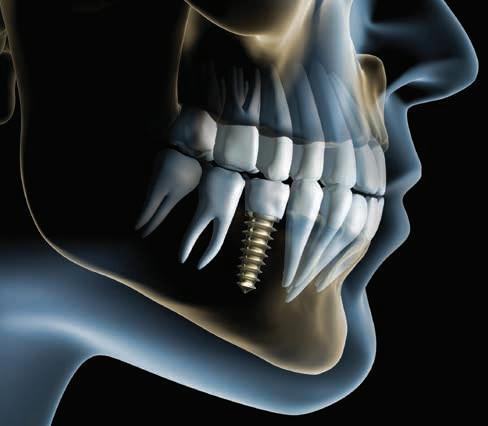


 By Phil Nieto, President, Best Card