14 THE WORLD’S SECOND FUNKY OPHTHALMOLOGY MAGAZINE Better Together When ophthalmologists and optometrists work together, patients win THE OPHTHALMOLOGY-OPTOMETRYCROSSOVERISSUE June/July cakemagazine.org2022 p20


If what I’m describing sounds a bit like clinical practice is being “optimized” to be the clinical correlate of Henry Ford's Model T production line, all in the name of efficiency, where people are assigned certain tasks, and that’s all they do, well, yeah. I think there is an element of that, particularly in practices that have a particular focus on one procedure, such as cataract surgery. Would I like to be so aggressively niched into such a role?
For people like cataract surgeons, volume will definitely increase — the demographics of a globally aging generation of baby boomers will see to that. So increasing efficiency is the economists’ — but not a particularly fun — answer. By its nature, finding efficiencies tends to be something with diminishing returns over time: The more you look for them, the less you're able to find them.
All
LETTER TO READERS
Goodness, no. I'd be bored to tears in a week. But I think you can see the pressures that are shaping everyone’s roles: As margins drop on procedures, volumes have to increase, hopefully without increasing costs too much.
I can see how a (small) ophthalmic practice could survive without an optometrist, but I can’t see how one can survive in the future without them.
| June/July 2022
Capitalism is getting you depressed, right? Well, let's lighten the mood. You have the team. If you’re doing it right, the team is tight, working well, and generally being awesome. And if you’re fortunate to be working with a great team, then you’re likely to be having a good time. And if I understand my American football idioms correctly, you’re the star quarterback. Or in terms of being a rockstar, you’re the band's Deryck Whibley. Even if you’re not Canadian, you rock! And you get to be your team’s Deryck right through to retirement.
Best,Hooray!
Mark Hillen, PhD Director of Communications ELZA Institute, Zurich, Switzerland Editor-At-Large | CAKE
I work in a successful, premium refractive surgery practice. We have the luxury of a high patient volume. We have a couple of big-name surgeons and several big-name consultants. Every patient wants to see these rockstars — I mean, that’s one of the main reasons they come to the clinic. But in a busy practice, every second counts. Actual rockstars tend to want to “spread the love” as widely as possible, but our ophthalmic rockstars need to make every moment they spend with their patients as effective as possible. To do that, they need to be as informed and as up-to-speed as can be on the patient. To do that, they need the team to do the prep work before the patient walks through the door. To do that, they need optometrists. I view it as being a bit like being Deryck Whibley, the lead singer of the Canadian rock band, Sum 41. He has a lot of autographs to sign, he wants every fan to walk away happy. But that queue isn't getting smaller and the people at the back of the queue are growing frustrated. Much like the albums, the interaction needs to be like the name of the band’s debut album: All Killer, No Filler. Achieving this is less rockstar-like — and more management-like. Hiring the right people. Training the staff. If you want to be the best, and want to beat the rest, delegation’s what you need. If you find one of our doctors performing a refraction, something has gone badly wrong somewhere. I get that there’s been quite a bit of resistance to this trend over the years. The general ophthalmologist that does everything — and this “everything” increasingly overlaps with what the optometrists do. Nobody likes competitors trying to steal your lunch money. But let’s look at how things are going to pan out in the future. In Sub-Saharan Africa, we’ve seen the advent of non-physician cataract surgeons. Why? Because there aren’t enough MDs in the region to meet the demand for the procedure. This is a pretty extreme example, but the principle is worth highlighting: What do you really need the doctor to do? (For me, the actual surgery is definitely one of those things.) Should a retinal physician be taking OCT scans on patients? Should the LASIK surgeon really be running the Pentacam on the patient? Do they need to be the person explaining the post-operative drop regimen? Fit and order scleral lenses? It takes a highly trained professional to do most of these, but it doesn't necessarily take a doctor to do it.
Mark Hillen Killer, Filler
I’m2 sorry, but this is a no-brainer.
No
How Ophthalmology and Optometry Has To Work Together


| June/July 2022 3 IN THIS ISSUE... We are looking for eye doctors who can contribute articles to CAKE magazine. Interested? Let's talk! Send us an email at editor@mediamice.com. To place an advertisement, advertorial, symposium highlight, video, email blast, or other promotion in CAKE magazine contact sales@mediamice.com. 25 343230 Texan Study Highlights Advance in SmartphoneCataractScreeningConferenceKudosCataract Highlights Cover Story Deliver withPatientsPresbyopicOutcomestheWantWELLFusion Ocular Tattoos Yes, this is a Topthing5 Pearls for Astigmatism…orManaging‘crumbs’ since we’re NewCAKEHarvard Research Studies Adenovirus & Cornea When Glaucoma Goes Rogue A case study of post-cataract surgery malignant glaucoma Using the Oculus Pentacam Preoperatively for Cataract Surgery WhenSuccessophthalmologists and optometrists work together, patients win Anterior Segment 181716121008 Media MICE Pte. Ltd. 6001 Beach Road, #19-06 Golden Mile Tower, Singapore 199589 Tel: +65 8186 7677 / +1 302 261 5379 Email:www.mediaMICE.comenquiry@mediamice.comPublishedby Matt Young CEO & Publisher Hannah Nguyen COO & CFO Robert Anderson Media Director Gloria D. Gamat Chief Editor Brooke Herron Editor Mark Hillen InternationalEditor-At-LargeBusinessDevelopment Ruchi Mahajan Ranga Brandon Winkeler Writers Andrew Sweeney April Ingram Ben MaricelHazlinCollinsHassanJoannaLeeMattHermanNickEusticeRogerShitakiTanSherLynnSalvador Graphic Designer The Great Cambodian Cataract Collective A troubled past, a challenging future Getting LGBTQ+ Inclusion ‘Right’ in Ophthalmology Assessing Johnson & Johnson Vision’s Next Generation IOLs Getting to the Core of Glaucoma Insults with Inflammatory Responses Improve Education and Access to BlindnessPreventableAlleviate Rayner’s Unique Trifocal Technology Offers Reversible Trifocality and More Highlights from Rayner’s trifocal user Practicalmeeting Tips for Achieving Greater Success in Your Solo Practice3836 Enlightenment Better Together 20 06 28


ADVISORY BOARD MEMBERS
Dr. George H.H. Beiko is a lecturer at University of Toronto and an assistant clinical professor at McMaster University in Canada. Dr. Beiko is a medical graduate of Oxford University and completed ophthalmology specialty training at Queens University in Canada. After his residency, he worked for one year at the St. John Ophthalmic Hospital in Jerusalem. He is currently a cataract, anterior segment and refractive surgeon practicing in St. Catharines, Ontario. His research interests include development of advanced cataract techniques and new intraocular implants. He has been an investigator in a number of Phase 1 FDA trials on intraocular lenses and he has done extensive work investigating multifocal, accommodating and aspheric IOLs. Dr. Beiko has published numerous peerreviewed articles in Ophthalmology, Journal of Cataract and Refractive Surgery and the Canadian Journal of Ophthalmology. He has also authored 10 book chapters. He has given over 500 scientific presentations at meetings throughout the world, including the annual meetings of the AAO, ASCRS, COS, CSCRS, ESCRS, WOC and george.beiko@sympatico.caISRS.
Dr. Chelvin Sng, BA, MBBChir, MA(Cambridge), MRCSEd, FRCSEd, MMed, FAMS, is a consultant at the National University Hospital (NUH) and assistant professor at National University of Singapore (NUS). She is also an honorary consultant at Moorfields Eye Hospital, London, and adjunct clinic investigator at the Singapore Eye Research Institute (SERI). A pioneer of minimally invasive glaucoma surgery (MIGS), Dr. Sng was the first surgeon in Asia to perform XEN, InnFocus Microshunt and iStent Inject implantation. A co-author of The Ophthalmology Examinations Review, Dr. Sng has also written several book chapters and publications in various international journals. Proficient in conventional glaucoma surgery and trained in complex cataract surgery, Dr. Sng co-invented a new glaucoma drainage device, which was patented in 2015. When not working, Dr. Sng can be found volunteering in medical missions in India and across Southeast Asia. chelvin@gmail.com
Dr. Boris Malyugin Dr. Harvey S. Uy Dr. Chelvin Sng Dr. George H.H. Beiko
Dr. Harvey S. Uy, MD, is a clinical associate professor of ophthalmology at the University of the Philippines, and medical director at the Peregrine Eye and Laser Institute in Makati, Philippines. He completed his fellowships at St. Luke’s Medical Center (Philippines) and the Massachusetts Eye and Ear Infirmary (USA). Dr. Uy is a pioneer in femtosecond cataract surgery, accommodation restoration by lens softening, modular intraocular lenses and intravitreal drugs. He has published over 30 peer-reviewed articles and is on the editorial board of the American Journal of Ophthalmology Case Reports. He is a former president of the Philippine Academy of Ophthalmology (PAO) and current council member of the APVRS. harveyuy@gmail.com
| June/July 20224
boris.malyugin@gmail.com
Dr. Boris Malyugin is a professor of ophthalmology and is the deputy director general (R&D, Edu) of the S. Fyodorov Eye Microsurgery Institution in Moscow, Russia. He is also the president of the Russian Ophthalmology Society (RSO). Dr. Malyugin is a world-renowned authority and expert in the field of anterior segment surgery. He has established himself at the forefront of advanced cataract surgery by pioneering numerous techniques and technologies. He is well known for his development of the Malyugin Ring, for use in small pupil cataract surgery. Dr. Malyugin has received multiple international awards and was invited to participate with named and keynote lectures and live surgery sessions during several national and international meetings. He is a member of the ESCRS Program Committee, Academia Ophthalmologica Internationalis (member since 2012), International Intraocular Implant Club (member since 2009), as well as the ICO and AAO Advisory Committees.


| June/July 2022 5
Prof. Jodhbir S. Mehta, MBBS, FRCOphth, FRCS(Ed), FAMS, PhD(UK), is the executive director and head of the Tissue Engineering and Cell Group at the Singapore Eye Research Institute (SERI), head of Cornea External Disease and senior consultant in the Refractive Service at Singapore National Eye Centre (SNEC), deputy executive director at Singapore Eye Research Institute (SERI), as well as a full tenured professor with Duke-NUS Medical School in Singapore. With a main interest in corneal transplantation, he completed a corneal external disease and refractive fellowship at Moorfields Eye Hospital in London and at SNEC. He has co-authored nearly 20 textbooks and 333 citations, and holds 16 patents, six of which have been licensed. Prof. Mehta has won several awards from the AAO and ARVO, among others, the latest of which was from the ASCRS in 2018. Prof. Mehta is also a favorite keynote speaker and presenter in several internationaljodmehta@gmail.comconferences.
Prof. Jodhbir S. Mehta Dr. William B. Trattler ASEAN Ophthalmology Society
Dr. William B. Trattler, MD, is a refractive, corneal and cataract eye surgeon at the Center For Excellence In Eye Care in Miami, Florida, USA. He performs a wide variety of cataract and refractive surgeries, including PRK; all laser LASIK; no injection sutureless cataract surgery; as well as laser cataract surgery. He has been an investigator for next generation technologies (like the Tetraflex accommodating intraocular lens) and procedures like corneal collagen crosslinking (CXL). His involvement in the FDA-approval study for CXL led to its approval in 2016. In addition to his private practice, Dr. Trattler is on the Volunteer Faculty at the Florida International University Wertheim College of Medicine, as well as the University of Miami’s Bascom Palmer Eye Institute. He is board certified by the American Board of Ophthalmology and has been an author of several articles and abstracts. In 2016, Dr. Trattler received the Catalyst Award in Advancing Diversity in Leadership from the Ophthalmic World Leaders (OWL), an association of interdisciplinary ophthalmic professionals dedicated to driving innovation and patient care by advancing diversity in leadership. wtrattler@gmail.com
Ophthalmology Innovation Summit Arunodaya Charitable Trust (ACT) Russian Ophthalmology Society (ROS) Asia-Pacific Academy of Ophthalmology He Eye Specialist Hospital Young Ophthalmologists Society of India ( YOSI ) SOCIETY FRIENDS Orbis Singapore World Ophthalmology Congress


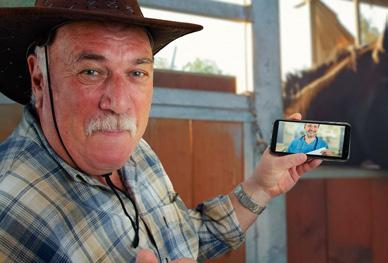
by Andrew Sweeney D o you remember something called COVID-19? It was this coronavirus that emerged at the end of 2019 and we’re pretty sure it didn’t have that much of an impact on our lives… well, perhaps, just slightly. Okay, fine, it was one of the most transformative experiences of our lives and we will all likely live in its shadow for the rest of our lives. Media MICE was no less affected in a number of ways, from disruption to our team spread across the world, to the themes of the content we produced during that time. One of the topics that dominated the medical headlines during much of the COVID-19 pandemic was telemedicine and how clinics across the healthcare spectrum, from cardiology and oncology to our own ophthalmology, were using it to continue treatments during stringent social distancing. Then the discussion moved from using the technology as a stop-gap measure to alleviate patient backlogs, toward becoming an accepted standardized option in ocular treatment. One of the conditions that has benefited the most from the increased options that telemedicine can offer is cataract, one of the most common conditions ophthalmologists face on a day-today basis. Cataract accounts for 51% of all eye diseases in the U.S. and as a progressive disease, early diagnosis, intervention and treatment are absolutely crucial in achieving the best possible patient outcomes.1 Thus, the first line of defense is imaging technology, the means by which the first cataract diagnosis can be made — so, what if we could find a way to make screening more accessible than ever before by using smartphones? That’s a mighty fine smartphone you’ve got there
| June/July 20226 ATARACT SCREENING TOOLS
Texan Study Highlights Advance in SmartphoneCataractScreening
That was the question a group of researchers based in the lone star state of Texas wanted to examine in their paper Detecting Cataract Using Smartphones. The Texans resolved to apply an efficient approach to identify cataract disease by adopting luminance features using a smartphone. The problem they said they had encountered in other studies was that results could vary from a wide variety of factors including camera quality and processing power (among others), and as a result, they needed to identify a methodology using luminance technology as it is not color-based, and thus dependent on camera sensor characteristics and environmental conditions.1
To justify their position our Texas rangers (of science) pointed to a study comparing standard hue, saturation, value (HSV) — also known as redblue-green (RBG) — with luminance technology. The study concluded that the luminance-based method had 86.67% accuracy, while the HSV colorbased method had only 33.4% accuracy in detecting cancer cells.2 Based on these findings, they moved on to outlining their cataract methodology. In the Texan tests, subjects would sit


August 20-21 in Da Nang,
References 1. Askarian B, Ho P, Woon Chong J. Detecting Cataract Using Smartphones. IEEE J Transl Eng Health Med. 2021; 9: 3800110.
To register or for more info: Visit www.mediamice.com/expo.
INDUSTRY UPDATE Hold onto your Banana Shirts for CAKE & PIE
2. Vaghela H, Modi H, Pandya M, Potdar MB. A Comparative Study of the HSV Color Model and YCbCr Color Model to Detect the Nucleus of White Cells. Int. J. Comput. 2016; 150(8):3842. Editor’s Note: A version of this article was first published on cakemagazine.org in a stable position and align their eye with a smartphone’s rear camera, which was located 10-50 cm from the eye with autofocus. After the images were captured, the smartphone processed them and presented the results. All in all, 100 eye model images were captured, 50 from healthy eye models, and 50 from diseased eye models.1
Why Da Nang?
Let’s round up them steers (I mean patients)
The CAKE & PIE Expo 2.0 will take place in Da Nang, Vietnam, from August 20-21, 2022. As Media MICE’s signature eye care event, C&PE 2.0 will feature educational sessions in ophthalmology and optometry, an exhibition hall, networking opportunities and more — all in the company's well known and unique Durstyle.ing the two-day event, C&PE 2.0 will include talks on both the anterior and posterior segments, as well as optometry — all led by renowned industry KOLs. The event is free for doctors to Forattend.those who are unable to travel to Vietnam, C&PE 2.0 will also host a live stream with highlights from sessions and around the event. The online component will be held over Zoom.
The researchers found that changing the camera angle, distance and smartphone had 2.2%, 3.3% and 3% impact on luminance values and 9.2%, 13.3% and 8.5% impact on RGB values, respectively. However, changing the ambient light had a 36% difference impact on the luminance values, which was similar to the 32% difference impact it had on the RGB values.1 So we can therefore see (baddum tish) that the luminance technique is arguably more reliable, but how about accurate? Our top Texan team reported that of the 100 eyes that were screened as part of their study they were able to achieve accurate results in 96.6% of cases, while also achieving 93.75% sensitivity and 93.4% specificity. While this is important, what also needs to be noted is that changing environmental factors had very limited impact (at an average of 2.8%) on the outcome results. This means that the luminance technique could be applied effectively in almost any setting, significantly improving the accessibility of screening. No doubt that will help improve access to treatment in the lone star state, which is absolutely huge by Expo 2.0 Vietnam
| June/July 2022 7 Y ou’ve been to eye care meetings before … but you’ve never attended one created by ophthalmology’s “funkiest” content agency and publishing company: Media MICE. That’s right, this year, the CAKE & PIE Expo (C&PE 2.0) will be coming to you LIVE — as an inperson meeting in tropical Da Nang, Vietnam, complemented by an online stream for overseas attendees. What is C&PE 2.0?
“We recently opened the Media MICE office in Da Nang,” said CEO Matt Young. “Now that COVID-19 restrictions are easing, we decided to hold our expo [C&PE 2.0] from our home — and our newest company location — in Vietnam.” C&PE 2.0 will be held at the Four Points by Sheraton Da Nang. Situated across the street from the city’s gorgeous beaches, and near the base of Son Tra Peninsula, attendees will find plenty of diversions — whether sight-seeing, relaxing, shopping or eating — once the expo finishes for thetheday.way, and also around the world as smartphone screening can get almost anywhere. Kudos!

In their case study Malignant glaucoma presenting with uncontrolled intraocular pressure and myopic refractive surprise after cataract surgery,* Drs. Albert Xiong and Doohoo Kim deal with one particularly dastardly head of the hydra in malignant glaucoma. Also known as aqueous misdirection syndrome, ciliary block glaucoma, lens block angle closure, or simply by the symptoms it presents with, this fiendish foe surfaced in the doctors’ care after cataract surgery in a 74-year-old Caucasian male. The humble beginnings of a highly complex issue
| June/July 20228 ATARACT SURGICAL CASE
When Glaucoma Goes Rogue
A Case Study of Post-Cataract Surgery
Malignant Glaucoma by Matt Herman D rs. Albert Xiong and Doohoo Kim dive into the successful management of a deceptively thorny case of the poorly understood malignant glaucoma.
Glaucoma is a devilish disease recognized worldwide as a leading cause of blindness; it strikes the old, the young, the wealthy, the healthy, and the poor in nearly equal measure. Stacks of research stretching back decades have been compiled on the topic of elevated intraocular pressure (IOP) and its ravages, but glaucoma is a crafty adversary. Like Hercules’ hydra, every time researchers classify and develop treatment for a new subvariant of the disease, a new one inevitably appears in its place.
It all kicked off with a routine examination that revealed something

Malignant glaucoma is stubbornly resistant to traditional glaucoma therapies, but once it is recognized, selective treatment centering on lessening anterior displacement of the lens-iris diaphragm and vitreous volume reduction is key. In other words, this is one head of the glaucoma hydra that needs no Herculean effort to tackle.
Time for some action and the aftermath Time was working against the doctors, and the moment had arrived for decisive and drastic action in the form of iridozonulo-hyaloido-vitrectomy through the previous PI site. The surgery was uneventful, and just like that, the scramble was over. Post-op day one saw uncorrected visual acuity improve to 20/40, manifest refraction to -0.25 + .50 x 166, an IOP level improvement to 17 mmHg, and more anatomically typical IOL positioning in the capsular bag, with further improvement to 20/20-2 with manifest refraction of −0.50 spherical after another week. The authors concluded the case study by remarking on the difficulty of recognizing malignant glaucoma. It is a disease diagnosed mainly in the absence of pathologies, and the doctors recommended the use of ultrasound biomicroscopy to exclude other potential conditions like suprachoroidal hemorrhage, choroidal effusion or ciliary body Thoughtumor.itmay seem like bilateral LPI would have been the right choice at the get-go, the authors defended the choice to proceed with cataract surgery due to the added cost of LPI. Besides, the LPI performed eventually on the left eye failed, making it doubly unnecessary in Malignanthindsight. glaucoma is anything but just another kind of glaucoma, and doctors should be aware of its signs.
| June/July 2022 9 more. The patient presented at the clinic complaining of poor vision, and after a battery of tests, IOP by Goldmann applanation was 40 mmHg in the right eye and 30 mmHg in the left. On closer examination, bilateral angle closure was diagnosed and sameday YAG laser peripheral iridotomy (PI) decided on for the right eye. Surgery was successful but made more difficult by peripheral iris-corneal touch. Intraocular pressure in the right eye dropped postoperatively to 15 mmHg, while the left eye settled at 28 mmHg after bilateral timolol maleate/ brimonidine tartrate and travoprost eye Thedrops.patient’s poor vision was determined to be from a cataract in the left eye, so the doctors decided to forgo PI and went full steam ahead with cataract surgery. Another successful surgery resulted — or so it seemed. When it all went south As uneventful as both surgeries were, things were unfortunately not so rosy in reality. Examination of the anterior chamber showed a visibly shallow chamber both peripherally and centrally, and a particularly puzzling anterior displacement of the IOL. Furthermore, the left eye showed a significant myopic shift (-3.50 spherical refraction with 20/400 visual acuity) and elevated IOP, which spiked after temporary cessation of prescribed atropine eye drops. At this point with this response to the eye drops, the closed angle anatomy, axial shallowing of the anterior chamber, IOL displacement and myopic shift, malignant glaucoma was on the menu. The race was on to save the patient’s sight and bring his IOP down. The doctors proceeded with a laser iridotomy with hyaloidotomy on the ailing left eye, to no avail. The myopic shift remained with visual acuity of 20/400, and IOP increased to 35 mmHg despite timolol/dorzolamide BID and latanoprost eye drops. The symptoms and reaction to treatment were now a glaring neon sign in the depth of night pointing to malignant glaucoma.
* Xiong AS, Kim DB. Malignant glaucoma presenting with uncontrolled intraocular pressure and myopic refractive surprise after cataract surgery. Clin Case Rep. 2022;10(6):e05810.
The authors note an overall incidence of the disease as 1-3% after surgeries like trabeculectomy, cataract, pars plana vitrectomy, laser capsulotomy and laser iridotomy. It rarely rears its ugly head after miotic agent use, or even spontaneously. The most important thing, though, is to hone in on the diagnosis as quickly as possible.

Using the forPentacamOCULUSPreoperativelyCataractSurgerySuccess
| June/July 202210 OCULUS CORNER
The many biometrics provided by the OCULUS Pentacam can equip patients with the knowledge needed for confidence in the procedure. “The Pentacam can perform AXL measurements and posterior cornea measurements,” explained Dr. Hutauruk. “This makes it easy to see if there is any irregular astigmatism so I can manage the patient’s expectations.” But in the end, it is doctors who rely most on a successful preoperative routine. And for maximum confidence before setting foot in the operating theater, Dr. Hutauruk knows he can rely on the Pentacam. “There’s a lot of critical information I get from the Pentacam,” he said. “With corneal tomography it is easy to see abnormalities, irregular astigmatism, true net power and posterior cornea Themeasurements.”listgoeson with the range and depth of measurements that the Pentacam places at doctors’ fingertips. But the Pentacam’s long-standing reputation for efficiency and accuracy are what makes it the go-to biometry suite for Dr. Hutauruk. “The other main advantage is that the measurement is very quick, which makes it very convenient for patients. And of course, it provides reliable results.”
Contributing Doctor Johan A. Hutauruk , MD, is a senior consultant ophthalmologist in cornea, cataract and refractive surgery and currently the president director of JEC Eye Hospitals and Clinics. Dr. Hutauruk is the president of Indonesian Cornea Society (INACORS), vice president of the Indonesian Ophthalmologists Association (PERDAMI), vice president of Indonesian Eye Hospitals Association (ARSAMI) and the past president of the Indonesian Society of Cataract and Refractive Surgery (INASCRS). He is actively involved as a scientific committee member for the Indonesian Ophthalmologist Annual johan.hutauruk@jec.co.idMeeting.
by Matt Herman D r. Johan Hutauruk of Indonesia talks tips for optimizing cataract surgery outcomes, and the Pentacam from OCULUS plays a starring role. Cataracts — once a fearsome harbinger of blindness for sufferers worldwide — have largely become a common medical problem with a routine solution. Today, the modern cataract patient expects a smooth outpatient procedure with little disruption to their daily life … and excellent visual Ophthalmologistsoutcomes.knowthat to get excellent visual outcomes there is a laundry list of highly complex measurements that must be taken with incredible precision. And those in the know understand that a positive cataract surgery experience starts with a well-executed preoperative process — optimized by the proven accuracy and efficiency of the Pentacam (OCULUS Optikgeräte GmbH, Wetzlar, Germany). To discover some pearls for great cataract surgery outcomes, we sat down with Dr. Johan Hutauruk of Indonesia to talk about how the Pentacam has enhanced his preoperative routine. Meeting evolving patient expectations in the age of premium IOLs Challenges of all shapes and sizes abound for surgical teams preparing for cataract surgery, but for Dr. Hutauruk, the most difficult of all comes from a surprising source. “The greatest challenge [in preparing for cataract surgery] is to meet patient expectations,” he said. But with the advent of breakthroughs like multifocal and toric intraocular lenses (IOLs), this expectation has evolved. This was according to a talk by Dr. Hutauruk at the recently held cyber course of the Indonesian Society of Cataract and Refractive Surgery (INASCRS INDEPTH 2022): “Cataract surgery has now become cataract refractive surgery,” he explained. “The target is not only visual rehabilitation by removing the cloudy lens, but also to optimize visual acuity postoperatively so our patients can expect to be free of Withglasses.”itsrobust suite of tools and features, the Pentacam range is uniquely suited to embrace this new wave of IOLs and the accompanying increase in preoperative demands. Dr. Hutauruk sees the Pentacam as indispensable. “The Pentacam is particularly important for the implantation of premium IOLs, especially toric and multifocal lenses,” he Forshared.preoperative IOL calculations, the Pentacam comes with the whole suite of tools to make the fine measurements and calculations demanded by nextgen IOLs. “Especially for multifocal IOLs, I need to consider the corneal topography, posterior cornea and angle kappa,” Dr. Hutauruk shared. The Pentacam measures these key parameters, among others. Another key to success is in choosing the right IOL formula. Though there are many out there, Dr. Hutauruk says the Barrett Universal II formula is the right choice for all axial lengths. But no matter the physician’s personal preference, getting it right from measurement to formula input is a breeze with Pentacam, saving doctors time and reducing transcription errors.
Building doctor AND patient confidence with Pentacam
The patient and what they hope to get out of their procedure are the most mercurial of all variables leading up to cataract surgery. The biometrics of the eye are a fixed quantity compared to the whims and fancies of the human brain, and keeping the latter aligned with surgical outcomes is a minefield.


2.0 AN ASIAN-BASED, GLOBALLY-MINDED FUNKY OPHTHALMOLOGY AND OPTOMETRY EYE CONFERENCE A unique hybrid show with hundreds of in person guests, international speakers and exhibitors as well as engaging and entertaining coverage available online. FOUR POINTS BY SHERATON DA NANG, VIETNAM 20TH & 21ST 2022 AUGUST Scan here or forwww.mediamice.com/expovisitmoreinformation. FREE DOCTORSFORTOATTEND Email expo@mediamice.com to register.


Lack of vision at all distances coupled with photic phenomena has created a clear unmet need in presbyopia correction — and one that SIFI, an Italian group of companies focusing on R&D, manufacturing, and commercialization of ophthalmic products, is addressing through its one-of-a-kind WELL Fusion optical system. This system utilizes two bilaterally implanted EDOF IOLs, the Mini WELL and Mini WELL PROXA, to provide clear, spectacle-free vision at all distances with negligible photic phenomena and minimal to no loss of contrast sensitivity. Using the same non-diffractive extended depth of focus (EDOF) said that all of the studied IOLs show similar, extremely low halo and glare with logCS (contrast sensitivity) ranging from 1.67 to 1.82, which is closer to monofocal contrast sensitivity than a diffractive multifocal. Similar results for the EDOF Mini WELL were reported by Auffarth et al., in 2020: Mean UDVA was -0.01 ± 0.15; UIVA was 0.03 ± 0.10, and UNVA was 0.10 ± 0.11 logMAR. Further, he reported a mean halo size of 33.06 ± 14.25, mean halo intensity of 38.00 ± 18.51, mean glare size of 23.85 ± 10.43, and mean glare intensity of 42.23 ± 13.22.3 They concluded that the Mini WELL “provides good visual acuity across various distances and functional reading ability provided at a near range, and delivers an enhanced contrast sensitivity while causing a low incidence of photic phenomena.” In October 2020, Dr. Caparas began his study of WELL Fusion with 40 eyes of 20 patients. They were followed for 90105 days; 19 patients completed their platform and with wavefrontengineered complementary IOL design, both quantity and quality of vision are improved with WELL Fusion. In this system, extended depth of focus is created by inducing targeted amounts of spherical aberration in the concentric optical zones in the central part of the IOLs. This creates one continuous focus — without dividing the light beam — resulting in a lower risk of photic phenomena compared to other multifocal IOLs. That means with WELL Fusion, patients can achieve uninterrupted vision across all distances without compromise, including excellent near vision and stable and consistent visual performance up to -3.5 D defocus.2 Clinical experience and results Recognizing the capabilities of this new system is Dr. Victor Caparas, an ophthalmologist in Manila, Philippines. He has been studying and implanting the SIFI family of IOLs for the past six years, including the Mini WELL (from 2017), the Mini WELL Toric (from mid-2018), and the Mini WELL PROXA (from late 2020). As a whole, Dr. Caparas reported similar and good visual results, with patients reporting high satisfaction with these three IOLs. For quantity of vision, binocular distance (4 m) was better than -0.1 logMAR; intermediate (63 cm) was 0.1 to 0.2 logMAR; and near (40 cm) ranged from 0.0 to 0.2 logMAR. Further, with WELL Fusion (and the Mini WELL PROXA) near vision at 33 cm was better than -0.1. For quality of binocular vision, Dr. Caparas
NTERIOR SEGMENT EDOF IOL s
| June/July 202212
Deliver the Outcomes Presbyopic Patients Want with WELL Fusion by Brooke Herron M ost often, the topic of intraocular lenses (or IOLs) arises when discussing cataract surgery. After all, it’s during this procedure that the clouded lens is replaced by a new, artificial one. However today, surgeons also have a new batch of patients looking for clear vision: those with presbyopia. When IOLs were first introduced, the monofocal was king. Now considered a basic or standard IOL, this lens gives good distance vision but lacks in intermediate and near — and as a result, leaves patients in spectacles, postoperatively. As patients demanded better outcomes — and clearer vision at all distances — premium IOLs, like trifocals and multifocals, entered the market. But unfortunately, these IOLs still struggle to deliver spectacle-free vision at all distances; they can also be plagued by photic phenomena like glare and halos.1
The unmet need in presbyopia correction


postoperative visits. In this clinical study, Dr. Caparas evaluated the visual function, quality of vision, subjective outcomes, and safety after implantation with Mini WELL and Mini WELL PROXA. This was a part of a multicenter, observational, prospective, single-arm, and (40wasintermediate(4vision,RegardingofDr.competingFusionTosecond3andstudyinvestigator-drivenwithvisualfunctionqualityevaluatedatmonthsfollowingtheeyeimplantation.illustratehowWELLgoesastepaboveIOLplatforms,Caparassharedsomehispostoperativedata:quantityofdistanceVAm)was-0.22logMAR;VA(63cm)0.1logMAR;nearVAcm)was-0.05logMAR; and near VA (33 cm) was -0.12 logMAR. Snellen results showed the following: distance VA of 20/12; intermediate VA of 20/25; near (40 cm) VA of 20/18; and near (33 cm) VA of 20/15. “[With WELL Fusion,] the results have been great. Now, we have patients that can read up to 30-35 cm as opposed to the first Mini WELL studies,” he shared. Further, thanks to its wider depth of focus, gaps in near vision are filled. “The Mini WELL gives us good far and intermediate vision,” said Dr. Caparas. “On the other hand, the Mini WELL PROXA gives us excellent near vision. Together, these [IOLs] give us an uninterrupted range of vision from infinity to all the way to about 30-35 Further,cm.” when assessing quality of vision, using the Halo and Glare Simulator software, Dr. Caparas found extremely low glare and haloes: halo size (20); halo intensity (28); glare size (15) and glare intensity (17). Meanwhile, he reported contrast sensitivity at 40 cm was 1.82 (logCS). With such impressive data, it’s clear that SIFI’s focus on improving both quantity and quality of vision is resulting in improved outcomes. “WELL less light dependence,” he continued. “For doctors, this means less complicated options can be presented to patients — for example, no mix-and-match or micromonovision options — and it provides reliability in both visual acuity and near absence of photic phenomena,” he said. “All of this equates to happier, more satisfied patients.” The ‘right’ IOL in the ‘right’ patient Before implanting a premium IOL, surgeons must consider numerous elements — from anatomic to lifestyle — to achieve the best possible visual outcome. Surgeons must obtain exact measurements, understand the IOL’s characteristics, and discuss the patients’ demands and Forlifestyle.example, the patient’s age and activity level should be considered, as well as their occupation and lifestyle needs. “It’s important to understand the needs and preferences of the patient: Does the patient have reasonable demands? Do they drive at night? Do they read or use laptops or smartphones
Postoperative data from Dr. Caparas illustrating WELL Fusion's performance in terms of quantitative outcomes: distance VA (4 m) was -0.22 logMAR; intermediate VA (63 cm) was 0.1 logMAR; near VA (40 cm) was -0.05 logMAR; and near VA (33 cm) was -0.12 logMAR. Mini WELL and Mini WELL PROXA IOLs share the same EDOF nondiffractive platform which creates one continuous, extended focus by inducing targeted amounts of spherical aberration in concentric optical zones in the central part of the optics without dividing the light beam. Mini WELL PROXA has a number of zones higher than Mini WELL, allowing the range of vision to extend up to 3.0 D (33 cm). Both lenses share the same aspheric monofocal design in the periphery. Fusion fulfills the need for excellent visual acuity at all distances: far, intermediate, near, and very near. Just as important, it affords this without the disabling photic phenomena that very often, for the patient, negates the good results in visual acuity,” said Dr. “ThisCaparas.offers patients more functionality, more spectacle independence, and
| June/July 2022 13


Postoperative data from Dr. Caparas illustrating WELL Fusion's performance in terms of qualitative outcomes. Using the Halo and Glare Simulator software, WELL Fusion showed negligible photic phenomena compared to diffractive IOL platform.
• Spectacle tolerance: Does the patient understand the possibility of spectacle use for prolonged, visuallydemanding tasks?
| June/July 2022
• Eye status: Do they have dry eye, refractive error, cataract, or any posterior segment pathology? And although these are all key considerations when choosing an IOL, Dr. Caparas shared that some of these factors are not as critical with WELL Fusion — and that’s because the optical system allows the patient profile to be extended, thanks to a full range of focus from far through near along with a very high quality of vision.
Dr. Caparas shared that he follows SIFI recommendations by implanting the Mini WELL in the dominant eye and the Mini WELL PROXA in the non-dominant eye. “I don’t have any experience doing so otherwise [implanting the Mini WELL in the non-dominant eye and vice versa], but I do have extensive experience with monovision using monofocal IOLs, and I’ve hardly seen any difference in using the dominant eye for distance or for near vision,” he said. Through his experience with WELL Fusion, Dr. Caparas has identified further pearls that allow him to achieve the best results for patients. “We discovered early in our studies that aiming for a slightly hyperopic
NTERIOR SEGMENT EDOF IOL s
• Personal motivation: Do they understand the need for neuroadaptation and can they wait for visual outcomes to improve?
• Psychological health: Are there any mental health issues?
Pre-op: Measure and counsel Preoperatively counseling patients can help improve postoperative outcomes: Patients who know what to expect will generally be happier. Dr. Caparas shared that before proceeding with WELL Fusion, he counsels patients on the effects dry eye could have, as well as the time required for neuroadaptation (albeit it rapid). Dr. Caparas then described some of his preoperative processes with WELL Fusion. For example, he checks for eye dominance, dry eye status, and pupil status. He reviews the biometry to ensure a good quality scan and that the patient has normal K values (40 to 47 D) and astigmatism of less than 0.75 D. Using the Pentacam AXL-Wave (Oculus Optikgeräte GmbH, Wetzlar, Germany), he shared patients should be within the following ranges: chord mu of <0.4 mm (surrogate for angle kappa); spherical aberration/Z40 (6.0 mm) of <0.3-0.5 µm; HOA (4.0 mm) of <0.3-0.5 µm; and Q value/asphericity should be negative (prolate cornea). Meanwhile for aberrometry, Dr. Caparas uses the iTrace (Tracey Technologies, Houston, Texas, USA). He noted that patients should be within angle alpha (limbal center and visual axis) of <0.5 mm; and angle kappa (pupil center and visual axis) of <0.5 mm. He also measures the corneal, internal, and total eye aberrations using Chang analysis. Intra-op: Techniques and considerations While WELL Fusion may provide revolutionary outcomes, the surgical procedure to implant the IOLs is more traditional. “I use the same technique as with all other of my phacoemulsification surgeries: I make a 2.2 mm temporal incision and a 5.5 mm capsulorhexis, I fill the capsular bag with sodium hyaluronate/HPMC viscoelastic for implantation of the IOL, and then I meticulously polish the capsule and remove the OVD (ophthalmic viscosurgical device).”
“Any patient who desires both good distance and near vision, and understands the need for adaptation will be an ideal candidate for WELL Fusion,” continued Dr. Caparas. “This is based on the visual results, absence of photic phenomena, good contrast sensitivity, — and because WELL Fusion provides the whole range of very good to excellent vision, with excellent quality.”
14often? Do they prefer distance vision over near, or vice versa? These all must be considered,” said Dr. Caparas. Other factors include:

He continued: “Also, I avoid very small pupils (<2.0 mm, photopic) as we have observed distance vision to suffer under bright conditions, like driving in very bright daylight.”
Dr. Caparas shared that the quality of vision questionnaire reported the mean score assessing photic phenomena was 83% (the higher, the better). The mean overall spectacle independence scores (1 is best and 5 is worst) were 1.12 for distance vision; 1.18 for intermediate vision; and 1.18 for near vision.
In addition, Dr. Caparas shared that dry eye can significantly affect performance, so he recommends managing dry eye and using lubricants early, even before surgery.
Further, it’s so uncommon for patients to report haloes or glare with WELL Fusion, that Dr. Caparas said he’s had to describe them to patients. “Often when performing the simulator test, halo and glare have to be explained, as patients report that they have not experienced any — even when driving at night,” he shared. “This is in contrast to other IOLs, especially diffractive IOLs, where haloes and glare are significant — even after months and years of neuroadaptation and learning to tolerate the dysphotopsia,” continued Dr. FollowingCaparas.
Another added benefit? “WELL Fusion retains the same excellent visual qualities of the Mini WELL, while achieving even better near vision at 33 cm,” said Dr. Caparas. It’s clear that WELL Fusion offers numerous benefits to doctors and patients. But perhaps the most poignant takeaway is this: The results and satisfaction with WELL Fusion is changing the face — and expectations — of presbyopia correction.
| June/July 2022 15 postoperative refraction resulted in better distance vision and happier patients,” he explained. “So we have adjusted our target, or alternatively, adjusted the recommended A constant.”
Post-op: Show me the outcomes Through his study of WELL Fusion, Dr. Caparas reports that patient satisfaction is high following surgery. “All subjects in our study had excellent vision — good enough for them to rate their quality of vision and spectacle independence very highly at three months post-op,” he said. In general, it takes most patients between one and three months to adapt, although some patients may have significant adaptation by one month. This shows that patients are adapting rapidly — and what’s more, most of them are spectacle-free: “Sixteen of 18 subjects from our WELL Fusion studies are 100 percent spectacle independent (score 1.0 out of 5, where 1 is “never” and 5 is “all the time”). Of the remaining subjects, one scored 1.67, while the other scored 3.0.”
But what are the patients actually saying? Survey says they’re happy — with a mean score of 1.06 in the study’s spectacle and light dependence questionnaire (where 1 is best and 5 is the worst). Patients also reported that they would have the same lens implanted again and would recommend the IOLs to a friend (both scores of 1.0). In total, only one patient scored less than a 1.0 in regard to satisfaction at 1.25.
Contributing Doctor Dr. Victor Caparas obtained his medical degree and ophthalmology training from the University of Philippines–Philippine General Hospital and completed fellowships in cornea and anterior segment at the University of the Philippines Institute of Ophthalmology and the Schepens Eye Research Institute of the Harvard Medical School. He also holds a Masters degree in Public Health from the Harvard School of Public Health. Dr. Caparas is among the top cornea specialists in the Philippines. He is a founding member of the Cornea Society of the Philippines, the Philippine Society of Cataract and Refractive Surgery, and the International Ocular Surface Society. Currently, Dr. Caparas serves as director of the Eye and Vision Institute of The Medical City (TMC, Ortigas, Manila, Philippines), as well as head of the section of Cornea and External Diseases, and founder and driving force behind the TMC Dry Eye Clinic.victor.caparas@gmail.com
Editor’s Note: A version of this article was first published on cakemagazine.org
Deliver results with WELL Fusion With so many “answers” to presbyopic correction, it’s refreshing to come across a real solution in terms of quantity and quality of vision. Indeed, when compared to other IOLs, WELL Fusion provides excellent VA, range of vision, and visual quality with close to zero photic phenomena (Figure 4).
surgery, very few side effects have been reported, which Dr. Caparas attributes to small pupils and dry eye. “A small number of patients have mentioned decreased distance vision in very bright conditions — we attribute this to very small (<2.0 mm) pupils in photopic conditions,” he explained. “Also, the effects of dry eye are pronounced with this type of IOL, as with other similar lenses.”
“My attitude to presbyopia-correcting lenses has changed,” said Dr. Caparas. “I have more confidence in results and their predictability, I have more happy patients, with hardly any complaints. And finally, implantation of premium IOLs has also increased significantly.”
References 1. Sieburth R, Chen M. Intraocular lens correction of presbyopia. Taiwan J Ophthalmol. 2019; 9(1): 4–17. 2. SIFI. (2021). Clinical Experiences with WELL Fusion [White paper]. 3. Auffarth GU, Moraru O, Munteanu M, et al. European, Multicenter, Prospective, Noncomparative Clinical Evaluation of an Extended Depth of Focus Intraocular Lens. J Refract Surg. 2020;36(7):426-434.

Dermatological and Ophthalmological Inflammatory, Infectious, and Tumoral TattooRelated Reactions:Review*SystematicA is as the effectsstudysuggests,nameaintotheoftattoos on the body — and it also offers particular insight into ocular tattoos. Authored by researchers based at a number of institutions in Bogota, Colombia, the review examined 104 studies primarily from Europe and North America, but also from other continents. It looked at 52 case reports, 21 cross-sectional studies, 20 case series, 10 case-control studies, and one cohort study. Eighty-six studies described skin tattoos, of which seven were publications about eyebrow tattoos and six of eyelid tattoos, and five articles included cases of subconjunctival tissue. Naturally, we’re going to focus on the ocular tattoos for this article — and it should come as no surprise that the Colombian researchers found that subconjunctival tissue tattoos are associated with significant comorbidities. This is because the eye is an “immunologically privileged organ that reacts with different inflammatory processes in the face of an immunological affront.” Despite this fact, the popularity of such tattoos has grown, and they are delivered via the application of some type of pigment directly under the bulbar conjunctiva of the eye with a needle.
Yes, This is a Thing by Andrew Sweeney F or the older generation of Media MICE readers (though we’re sure you’re young at heart), the modern popularity of tattoos amongst the younger generation must have come as something of a thePreviously,shock.preserve of
onIndeed,partaresailors,criminalssubcultures,undergroundandtattoosnowverymuchofthemainstream.severalMICErsthewritingstaffhave tattoos — including the author of this article, who has several of them. If you think, however, that tattoos are purely a product of modernity, then think again. In fact, they are as old as time and frequently appear in ancient artwork and on the bodies of people from eons ago. Cultures as far afield as the Americas, the Eurasian Steppe and the islands of Polynesia have used tattoos to mark social status, tribal identity or a number of other social factors. Arms, torsos, legs, and even faces with tattoos are not unusual according to the historical record. What is notably absent, however, is the presence of eye tattoos. (And perhaps for good reason?!) Imagine getting your eye tattooed… Yes, eye tattoos. We hope that didn’t make you spit out your tea — but eye tattoos have become more common amongst the practitioners of extreme body modification (this group of people can often be spotted sporting alterations like split tongues, pointed ears and more). For sure, getting one’s eyes tattooed remains rare amongst the general public — and although you may be unlikely to encounter someone who has undergone this modification — but the trend is out there. So, naturally, the Media MICE team became interested in this subject and we came across an interesting study into the phenomenon.
What color would you go for?
Editor’s Note: A version of this article was first published on cakemagazine.org
TattoosOcular
| June/July 202216 NTERIOR SEGMENT OCULAR TATTOOS
Intraocular pigments (also called episcleral tattoos) have been associated with considerable short and longterm complications secondary to this practice, possibly due to the lack of standardization on the type of pigments, the technique, and the appropriate personnel to perform it. The most common include proptosis; deposits of pigment in the conjunctiva, corneal endothelium, iridocorneal angle, iris and anterior capsule of the lens and vitreous; inflammatory processes such as nodular episcleritis, chemosis, anterior nongranulomatous uveitis and hypopyon; as well as cataract, secondary glaucoma, vitritis, serous retinal detachment associated with vitreoretinal proliferation, and choroidal detachment. The Colombian researchers reported that these issues can trigger endophthalmitis and blindness — and could even require the enucleation of the organ — given the severity of the immune response that occurs in these Thetissues.study concludes, rather wisely, that ophthalmologists should be aware of the severe damage caused by even small amounts of tattoo ink on the skin and in the eyes, thus requiring strict regulations for its use. Indeed, if ocular tattoos have to happen (as they’re not recommended) the researchers stated that such procedures should be performed using a surgical microscope and in sterile conditions with trained medical However,personnel.theyrightly pointed out that more evidence and research are required, so please, in the meantime, don’t get your eyeballs tattooed.
* Muñoz-Ortiz J, Gómez-López MT, EcheverryHernánde P, et al. Dermatological and Ophthalmological Inflammatory, Infectious, and Tumoral Tattoo-Related Reactions: A Systematic Review. Perm J. 2021; 25: 20.225.

Crumb ofAccurate#2:pre-opassessmentastigmatism If we can’t accurately assess astigmatism, then we can’t properly treat it. Dr. Rubenstein suggests the use of topography,assessmentmultipletools:“Themoreinformation,thebetter,”hesaid.Whilebiometry,cornealmanual
It’s imperative that PCRIs are performed with the utmost precision. It’s important to make sure the epithelium is smooth, and the length and depth of incisions are exact. “I think a lot of people give manual PCRIs a bad reputation because they are not performed with proper technique,” said Dr. Rubenstein. Toric IOL’s also require proper technique. Thus, a clean surgery, controlled corneal incisions, ensuring anterior and posterior capsule and zonules are intact, as well as precise lens alignment are crucial for a positive outcome.
Top 5 Pearls for AstigmatismManaging ‘crumbs’ since we’re CAKE by Ben Collins A stigmatism results from at night. On Day 2 of the annual meeting of the American Society of Cataract and Refractive Surgery (ASCRS 2022), Dr. B. Rubenstein, MD, Deutsch Family Professor and Chairman of the Department of Ophthalmology at Rush University Medical Center, Chicago, Illinois, dropped his top five tidbits of ophthalmological genius for dealing with the condition … and CAKE magazine was on hand to greedily gobble up every last crumb!
…or
Editor’s Note: ASCRS 2022 was held on April 22-26, as a physical show in Washington DC, USA. Reporting for this story took place during the event. A version of this article was first published on cakemagazine.org
Crumb #1: astigmatism!Think It sounds simple, but Dr. Rubenstein says this is a common diagnostic error that can lead to the mismanagement of astigmatism during cataract surgery. “You gotta think about it! Obviously, the first step is to decide if the patient needs cataract surgery. Is it visually significant? Does it interfere with their daily activities? Could there be a significant refractive advantage to cataract surgery? Once you’ve decided ‘yes, this patient needs cataract surgery,’ your first consideration should then be does this patient have treatable astigmatism? I consider this before doing any IOL calculations, is there astigmatism and do I need to correct it? Our goal is really to eliminate astigmatism (if possible), therefore less than a half a diopter is ideal to minimize a patient's need for spectacles post surgery.”
| June/July 2022 17 NTERIOR SEGMENTASTIGMATISM
There are multiple techniques for correcting astigmatism, including manual PCRIs (peripheral corneal relaxing incisions), femto PCRIs, toric IOLs, or a combination of these. “We don’t need to be married to one particular technique,” stressed Dr. Rubenstein. Factors that could determine which treatment is best for a particular patient may include cost (some procedures are more expensive than others), as well as axis and magnitude of the astigmatism. For most of us, a toric IOL is the first choice for treatment. These work best with regular, predictable astigmatisms, and can correct up to 4.75 D. Manual PCRIs have a lower cost and are good for correcting less severe astigmatism. They are also useful for treating mild non-orthogonal astigmatism. Femto PCRIs are also useful for correcting small amounts of astigmatism, and can be used as an adjunct to toric IOL’s.
keratometry and elevation mapping all have their merits, Dr. Rubenstein prefers biometry (and sometimes manual keratometry) for assessing axis and magnitude, and uses corneal topography as a qualitative guide. “Preoperative corneal topography is essential for all cataract surgeries,” he continued. “It’s good for comparing axis and magnitude, assessing regular versus irregular astigmatism, and screening out patients with irregular mires. Don’t just look at the values on the biometry, because if their topography is showing something different, you know you can’t really trust those values.”
Crumb #3: Plan the appropriate surgical technique
Crumb #4: Accurate Alignment
alsoheadaches.eyeorsufferastigmatismcornea.misshapenaPeoplewithtypicallyfromblurrydistortedvision,strainandTheycanhavedifficultyseeing
This can be achieved multiple ways: Manual, automated, or intraoperative. The simplest way to do this is to anesthetize the eye and mark either the 6 o’clock, or 3 and 9 o’clock positions with the patient looking straight ahead and both eyes open. Fancier techniques include Callisto Toric IOL Alignment, which overlays real time imagery with data from preoperative measurements.
Crumb #5: Precise surgical technique

New &AdenovirusResearchHarvardStudiesCornea
A t CAKE magazine — and across the Media MICE empire — we always strive to thatawesomeprovidearticlesareamazing and never anodyne, acing the articulation of the all-and-all about our industry. We also apparently have a penchant for alliteration in our content, or at least, some of our article-crafters do, especially when they come upon a subject that they haven’t covered before. So when the term “adenovirus” (AdV) came across the editorial desk it became one writer's ambition to perform a research deep-dive into the condition and to think of as many adjectives beginning with the letter “A” as possible.
| June/July 202218 NTERIOR SEGMENT VISION RESEARCH
To the (somewhat) casual observer it may seem that AdV might have little to do with an publication,ophthalmologybutit has a number of impactful and fascinating ocular associations. For starters, it was only officially discovered in 1953 and since then, more than 120 speciesspecific adenoviral serotypes have been identified in humans, mammals, birds, fish and reptiles. The established wisdom has been that human adenoviruses are not generally associated with causing severe disease in immunocompetent humans, but they may cause severe infections in immunocompromised people.1 But is that the case? Firstly by Andrew Sweeney

‘Kounting’ your K’s
adenoviruses can attack several areas of the body with mild infections, usually involving the upper or lower respiratory tract, gastrointestinal tract, or conjunctiva. Rare and typically more serious manifestations of AdV include hemorrhagic cystitis, hepatitis, hemorrhagic colitis, pancreatitis, nephritis and meningoencephalitis,2 most of which are rather nasty and can cause severe disease in even the healthiest of patients. So much for perceived wisdom, and when it comes to the more ocular of complications it is becoming apparent that AdV can involve some severe ocular consequences beyond a nasty case of pink eye. Red eye: Awful in the afternoon One of the most prominent things we should be aware of is AdV’s propensity toward causing corneal inflammation, also known as keratitis, thanks to the virus’s ability to persist or recur for months to years after infection. Keratitis is a common condition familiar to most ophthalmologists
One of the lead researchers of the study, Dr. Jaya Rajaiya, had previously worked on a 3D in vitro model of the human cornea, the “human corneal facsimile,” and he put his research to good use in this AdV study. For the facsimile, primary cultured human keratocytes were mixed with type I collagen overlayed with Matrigel (an epithelial basement membrane-like layer), to simulate a human corneal stroma and epithelial basement membrane. AdV was then introduced via the section of the eye overlayed with Matrigel via keratocytes, co-localized with heparan sulfate in a multifocal pattern.4
3. Koganti R, Yadavalli T, Naqvi RA, Shukla D, Naqvi AR. Pathobiology and Treatment of Viral Keratitis. Exp Eye Res. 2021 Apr;205:108483.
Keratoconjunctivitis is a condition where the patient suffers from both keratitis and conjunctivitis at the same time, and it is certainly not a great afternoon either. Our big brain guys from Harvard also found that there was “an abundance of evidence supporting corneal epithelial cell infection by AdVs,” in fact, typical adenovirus inclusions were observed at a rate of 85% among corneal epithelial scrapings of studied patients. Another of their findings — one that is perhaps even more fascinating — is that HAdV-D8, the most common cause of keratoconjunctivitis worldwide, can replicate in primary human corneal epithelial cells cultured in vitro.4
4. Rajaiya J, Saha A, Ismail AM, Zhou X, Su T, Chodosh J. Adenovirus and the Cornea: More Than Meets the Eye. Viruses. 2021 Feb; 13(2): 293. infection by adenoviruses, including corneal epithelial cell receptors and determinants of corneal tropism. Their paper is titled, Adenovirus and the Cornea: More Than Meets the Eye.4
Adenovirus and the Cornea: More Than Meets the Eye is a complex, unique and fascinating study that is absolutely worth taking time to read in full — so make sure you check it out. In their concluding remarks, the Harvard researchers, while recognizing that there remains a long way to go in AdV research, stated that their findings suggested hope for the development and the non-infectious variant is usually fairly minor in nature. However, the infectious type can lead to reduced vision, discomfort and light sensitivity. In more serious and rare cases, keratitis can end up permanently damaging vision, which is nobody’s idea of a good Treatmentafternoon.3ofsevere
References 1. Kulanayake S, Tikoo SK. Adenovirus Core Proteins: Structure and Function. Viruses. 2021 Feb 28;13(3):388.
keratitis caused by AdV often relies on topical corticosteroids, which are noted to work effectively, but they can cause major side effects including some that are vision-threatening. Obviously, that’s not an ideal outcome for anyone involved — and the search for alternative treatments is already underway. Some of the trailblazers in this field include a group of researchers based at Harvard Medical School (Cambridge, Massachusetts), who drew up a review into AdV and keratitis, which covers current knowledge on corneal After that, when leukocytes derived from human peripheral blood were inserted, the neutrophils migrated upward. According to Dr. Rajaiya et al., this “supports a mechanism for stromal keratitis in which infected keratocytes express chemokines that deposit at negatively charged moieties in the corneal epithelial basement membrane.” However, they did add that evidence to support infection of corneal stromal cells in the intact human cornea by AdV is still lacking, and requires further research.4 of effective therapy to mitigate immunopathology in keratitis and AdV
summerfindings,deliberatelybutdeepkeratoconjunctivitis.originatingIt’sadive,denseattimes,damnedinterestingandupliftinginitssoaddthisresearchtoyourreadingmaterial.
2. Lynch III JP, Kajon AE. Adenovirus: Epidemiology, Global Spread of Novel Serotypes, and Advances in Treatment and Prevention. Semin Respir Crit Care Med. 2016 Aug;37(4):586-602.
| June/July 2022 19

| June/July 202220
And it is teamwork where we see this old adage applied most often. When people are passionate about what they are doing, and focus on doing their part in a cooperative effort, their efforts really start to shine. No one person can do everything at once, but working as a team and striving to do their best at their specialty leads to a better result on the Nowherewhole.isthis truer than in patient care, where teams are vital to supporting the public’s health and wellbeing. We see this in the operating room with nurses supporting surgeons, and in oral care, where hygienists support dentists who refer out their orthodontic and endodontic procedures.
COVER
When ophthalmologists and optometrists work together, patients win STORY by Nick Eustice T here’s an old saying, often attributed to Aristotle: “The whole is greater than the sum of its parts.” Whether Aristotle said it or not, there’s no doubt that the quote has been around a pretty long time. And it’s stuck around for a good reason, as it rings true in a lot of different settings.
A pile of car parts and a can of gas won’t get you to work, unless they happen to be put together in just the right configuration. A list of ingredients is one thing, but a great soup is a whole other story. Five basketball players all in it for themselves may each look good sometimes, but it’s almost always the ones who play together as a team that end up winning.
And of course, this is also very true in eye care. We have opticians and optometrists, ortho-keratologists and ophthalmologists. Specialists like neuro-optometrists and retinal surgeons — there is a long list of hard-working caregivers in the eye care industry.
While there is a lot of overlap between the various specializations, just about every area of the field has its own special place. And that place is best expressed as part of a team, building a holistic and patient-centered approach to care. This kind of approach lends itself to a wider variety of skills and innovations, and has the benefit of improving patient education as well.
Better Together

In this issue, we’re going to be focusing on that whole that’s better than the sum of its parts — how optometrists, ophthalmologists, and all the rest of the eye care field are indeed better together — and how working together leads to healthier, happier patients with better visual outcomes. As CAKE focuses on the anterior segment, we’re going to be giving special treatment to that aspect of the eye, while our sister publications PIE and COOKIE will take a look at other aspects of eye care.
On the local level, Dr. Trattler spoke to the many different areas in which he works closely with optometrists. He said that the relationship he has with these optometrists is excellent, and that much of the time, each doctor understands their role as part of a team to improve the overall health of a “Locally,patient. for patient care, we work together for managing patients. Whether it’s for dry eye, or pre- and postoperative care for surgeries, or with many of the experts in South Florida who are specialists in fitting scleral lenses or treating keratoconus, there’s really a lot of opportunity to work collaboratively with optometry in our Wecommunity.”askedDr. Trattler about referrals, and what avenues he receives most of his patients from. He replied that this is a complete mix, as it always has been. Some patients are referred by optometrists, while still others come from general practice physicians. But, citing that “it is now 2022,” Dr. Trattler pointed out another first point of contact for many of his patients: AsGoogle.aspecialist with a rather high profile in the industry, Dr. Trattler has found that many of his patients find him online, and seek out his help directly. Often enough, this results in him referring as many patients out to optometrists as they refer to him. he, as a sub-specialist, refers out to optometry for care. Often enough there are treatments which these colleagues may specialize in, and can help with more readily than he can. For example, a patient with keratoconus may seek him out for corneal cross-linking treatment. Following that procedure, Dr. Trattler would in turn refer that patient to an optometrist who fits scleral lenses in their local area.
An ophthalmologist’s take on a vibrant medical community
This led Dr. Trattler to talk about a remarkable networking platform called The Chiasm, which was developed by a colleague of his in South Florida. The platform allows doctors to expand their collegial network and provide their patients with the best specialist referrals they can.
To find out more about the relationships of which ophthalmologists and optometrists build to provide better patient care, we spoke with Dr. William Trattler, an ophthalmologist specializing in the anterior segment at the at the Center For Excellence In Eye Care, in Miami, Florida. Dr. Trattler was eager to talk about what he called a “really great and vibrant optometric and ophthalmic community” in South Florida. Dr. Trattler described a truly positive working network with optometrists, both locally and nationally. On the local level, he has many optometrist contacts with whom he has a close working relationship, while nationally, he has an even broader network of connections as a key opinion leader (KOL). Though far away geographically, he says that these colleagues are a vital part of his work, in attending and speaking at conferences together, coordinating webinars, and numerous other capacities. Working together with these nationally recognized experts he sees as a tremendous asset for improving education, both among patients and among the general public.
The Chiasm was developed by Dr. Giannie Castellanos, an optometrist at Infinite Vision Eye Care in Miami Lakes, Florida, and a frequent collaborator with Dr. Trattler. Its various networking functions are neatly tailored to the needs of health care professionals, and are especially conducive to building and maintaining relationships with Speaking in more detail on this referral process, Dr. Trattler described a number of patients who find him on Google, and
Building up a robust referral network While doctors use different social networks and online platforms to communicate in a lot of different capacities, The Chiasm is somewhat unique in that its focus is to provide a space for doctors to focus on referrals. To this end, it uses a high level of security to allow doctors to communicate about patients’ specific needs without any concern about compromising the patients’ privacy in any Dr.way.Trattler described the simple process by which a doctor can use this platform to network and find a doctor who can provide specialist care for a patient. When a doctor wants to find a referral, they can use the program to filter through lists and select the appropriate physician, corresponding using the online platform and securely sharing patientspecific information with only the other doctor.
| June/July 2022 21

Just as a different perspective alters what the eye can see, it can also bring us a fuller picture of how a team works. To get a broader perspective on the many ways that cooperative care brings better results to the anterior segment, we spoke with one of the many optometrists in Dr. Trattler’s network of colleagues in South Florida.
Otherophthalmologist.conditions
In addition, referrals are often made when patients experience complications from procedures requiring further Wheninterventions.askedabout how networking with ophthalmologists and other optometrists help her to provide better patient care, Dr. Kramer began by speaking about the importance of mutual education within the eye care industry. It is especially important, she said, to inform fellow optometrists and ophthalmologists about the work that she does, because more often than not they have a patient who needs specialized services. This collegial education helps to provide better care, and among colleagues allows for greater focus on their area or areas of expertise. By keeping in regular communication about what her practice has to offer, Dr. Kramer says that she enables colleagues to provide more robust care options in a way that requires no research, time, or even desire to expand into other practice
COVER STORY
| June/July 2022
Thisareas.communication, she pointed out, is often necessary for a holistic approach to patient care. And when it comes to referrals, educating patients is just as important — if not even more important — than raising awareness among “Lettingcolleagues.apatient know that you’re sending them somewhere else definitely requires education,” Dr. Kramer said. It’s important to let them know that you’re not just getting rid of them, but you’re comanaging with partnership. Education is really important to Wepatients.”asked Dr. Kramer about how the relationships within the eye care community have changed over the years. She answered that there has been a big change, and that change has definitely been one for the better. In the past there was sometimes animosity between the two specialties of optometry and ophthalmology. This often came about because “We regularly discuss options for the most effective treatments, as well as give and receive referrals. We also take measurements, and work with lens manufacturers as part of that communication loop to make sure patients are receiving the best care. Because of the complexities of ocular surface disease, I and other doctors in my network will often see patients multiple times, sharing information back and forth.” — Dr. Elise Kramer
While face-to-face and telephone interactions remain the backbone of eye care collaboration, new forms of online communication which help to safeguard patients’ confidential information can do a lot to facilitate a cooperative approach to eye care.
are quite common reasons for an ophthalmologist to refer a patient to Dr. Kramer’s specialized practice as well. Keratoconus is one condition which she specializes in treating, and for which she receives frequent referrals for cross-linking procedures. Corneal transplants, corneal scarring and Stevens-Johnson syndrome are also conditions for which she sees a lot of referrals from ophthalmologists, as well as fellow optometrists who do not specialize in treating these particular conditions. On the other hand, Dr. Kramer said that there are a number of conditions for which she refers patients to ophthalmologists. Specifically, she said that she refers patients to ophthalmologists with whom she has a good working relationship when they need surgical treatments. These can be for anterior segment procedures such as cataract and glaucoma surgery.
Evolving into a holistic eye care community
One of the features The Chiasm has implemented in order to foster the development of cooperative patient care is a referral tracking system. This helps doctors and their staff keep track of referrals on both ends, and allows for back-and-forth updating on patients’ progress and recovery.
22other doctors and providing better patient care.
Through the use of online platforms like this one, doctors can evolve their existing networks in order to provide better care for the patients they serve.
Dr. Elise Kramer is an optometrist who specializes in treating ocular surface diseases and fitting scleral lenses for long-term treatment of anterior eye conditions. As a specialist in these areas, Dr. Kramer said that cooperation and referrals are an intrinsic part of her practice. When asked how her specialty lends itself to close cooperation with ophthalmologists and other optometrists, Dr. Kramer did not mince “Itwords.requires it,” she said. “We regularly discuss options for the most effective treatments, as well as give and receive referrals. We also take measurements, and work with lens manufacturers as part of that communication loop to make sure patients are receiving the best care. Because of the complexities of ocular surface disease, I and other doctors in my network will often see patients multiple times, sharing information back and forth.”
When asked about referrals, Dr. Kramer said that there are numerous times when patients are referred to her practice from ophthalmologists, and when she in turn refers them as well. When a patient requires a special lens, they will most often come in because of a recommendation from their
Contributing Doctors
| June/July 2022 23
ophthalmologists felt the need to be primary caregivers who had to take care of all of a patient’s problems themselves, and felt they had to do everything rather than focusing on their surgical Sincespecialties.thattime, Dr. Kramer notes that there has been a significant shift, where ophthalmologists have let optometrists take their spot as primary caregivers. “The field has developed to become much more cooperative,” she said. Each specialty within the field has become more aware of the others, and of how they can focus on better care by working together. We asked Dr. Kramer what changes could help to make patient care more holistic and cooperative. She answered that the field could benefit from more courses on approaches to delegation and cooperative care. While the industry has made great strides, she pointed out that most everyone is afraid to delegate due to losing patients at some “Whatpoint.people need to realize is that the best interest of the patients is always foremost,” Dr. Kramer said. “If there were more articles and thought leaders explaining how important cooperation is to eye care, I think we could see doctors being more comfortable and confident in working together to achieve the best result.”
In addition, whether a doctor is receiving a new referral or sending one out, staying in regular contact can make a world of difference for the patient’s sense of well-being. Education of patients and colleagues alike helps to strengthen this all the more. By working together, the different specialties in the eye care profession can achieve a whole that is in many different ways far better than merely the sum of its parts.
Dr. William Trattler is a board certified ophthalmologist at the Center For Excellence In Eye Care, in Miami, Florida, USA. He completed his ophthalmology residency at University of Pennsylvania, Scheie Eye Institute, and spent a year in training in cornea and refractive surgery at the University of Texas Southwestern Medical Center in Dallas. Dr. Trattler is chairman of the board for the American-European Congress of Ophthalmic Surgery (AECOS) through December 2022. Dr. Trattler has also served on the executive board of the International Society of Refractive Surgery (ISRS) from 2011 through 2019, as well as the Refractive Surgery Alliance (RSA) from 2015 to present. Dr. Trattler is the co-program director for AECOS Deer Valley, and is on the program committee for Hawaiian Eye, OSN NY, Modern Optometry Live, and CedarsAspens Annual Conference. Dr. Trattler has written hundreds of articles focused on cross-linking for keratoconus, cataract surgery, multifocal IOLs, LASIK, laser vision correction, dry eye and MGD.
Dr. Trattler has also given more than 400 presentations at regional, national and internationalwtrattler@gmail.comconferences. Dr. Elise Kramer is a residency-trained optometrist in Miami, Florida, USA, who specializes in ocular surface disease and specialty contact lens design and fitting. Her doctorate degree was awarded in optometry from the Université de Montréal in 2012. During her fourth year, she completed her internship in ocular disease at the Eye Centers of South Florida and went on to complete her residency at the Miami VA Medical Center. Her time there included training at the Bascom Palmer Eye Institute, the nation’s top eye hospital. After her residency, Dr. Kramer became a fellow of the Scleral Lens Education Society (SLS) and now serves as the treasurer for the SLS. Dr. Kramer is a member of the American Optometric Association (AOA), the International Association of Contact Lens Educators (IACLE), a Fellow of the American Academy of Optometry (AAO) and of the British Contact Lens Association (BCLA). She is also the Delegate of International Relations for the Italian Association of Scleral Lenses (AILeS). Dr. Kramer has published several articles and reviews and participates in clinical research trials. She enjoys lecturing all around the world in several different languages about ocular surface disease and specialty elise@miamicontactlens.comlenses.
A wholestrongerthanany part can achieve alone As Dr. Kramer pointed out, the anterior segment may be the area of eye care most conducive to teamwork. While optometrists have limited abilities to work directly with the retina and other parts of the eye’s posterior segment, there are a great many treatments and procedures in the anterior where optometrists are able to provide specialized care. As a patient’s usual first point of contact for eye care, optometrists have a wide variety of treatments which they can provide in the anterior segment. It is in this capacity that they are also of vital importance in recognizing how referring a patient out, whether to ophthalmologists or to specialist optometrists, can provide the best care. The same is entirely true for when ophthalmologists are that initial “port of call.”


Reference Cunningham DN, Whitley W. What is ‘Integrated Eye Care?’ Review of Optometry.
Published
Accessed
Available at: www.reviewofoptometry.com/article/what-is-integrated-eye-care. on March 15, 2012. on July 13, 2022.

| June/July 2022 25 UDOSOUTREACH PROGRAM
A Troubled Past, a Challenging Future
But dig a little deeper into this wonderful land's troubled past, and you might begin to understand some of the problems which linger and hinder the Kingdom's development socially and Aftereconomically.theKhmer Rouge (lead by the infamous Pol Pot) seized power in 1975 they began the systematic slaughter of Cambodian citizens. By the end of the regime, some 1.5 to 2 million people (a quarter of the country’s population at the time) had been senselessly Themurdered.genocide was carried out under political, ethnic, religious and racial grounds. Government officials, professionals and intellectuals in particular were targeted. Cambodia's previous military and political leadership, along with an entire generation of business leaders, journalists, students, doctors and lawyers were effectively wiped out. To this day, the country is still struggling to recover from this massive loss of knowledge and expertise.
A refugee returns with a remedy… Dr. Sean Ngu (co-founder of the Khmer Sight Foundation) escaped to Australia as a refugee in the 1970s. He returned in the 1990s with the lofty goal of tackling general health care for communities living in remote parts of the Kingdom, often where there are no medical services available. During his outbound medical missions, he witnessed unnecessary blindness in the region and set up a mobile eye clinic, which is still performing surgeries today. From these humble beginnings, together with his great friend and colleague, the late Dr. Kim Frumar, the Khmer Sight Foundation (KSF) was eventually formed in 2015. Further, it was Dr. Sunil Shah who saved the foundation from closing down when he spent a year with KSF to rebuild it to what it is today, shared Dr. Ngu. Established as a means for unifying and developing eye care infrastructure in Cambodia, the KSF aims to equip the country with the skills and resources to tackle avoidable blindness independently in the future. International connections within the ophthalmological community enable volunteer specialists to travel to the area, providing training as well as life changing sight-restoring surgery and equipment.
W hen this writer thinks of Cambodia, he imagines white sandy beaches, incredible cuisine and ancient ruins. Long a popular tourist destination, the country oozes culture, history, warmth and vibrancy for the intrepid traveler.
The Great Cambodian Cataract Collective by Ben Collins


A surgery drive in Cambodia however, is never a straightforward undertaking. Logistical and infrastructural challenges abound. Unfortunately, the population in rural areas is often cut off from medical care and has no opportunity to have an ophthalmological examination. Basically, there is a lack of well-trained doctors, nursing staff and medical facilities throughout the country. I was lucky enough to have a chat with Dr. Sean Ngu, who described firsthand some of the challenges and learning experiences KSF has faced bringing modern eye care to the people of Cambodia. He talked about the difficulties involved in simply transporting patients from rural areas to the operating rooms in Phnom Penh: Often located in rugged mountainous terrain, patients with limited or no sight are loaded firstly onto motorbikes, and then into air conditioned buses for the long journey to Phnom Penh. Early iterations of this system however, failed to take motion sickness into account — it turned out many of these Cambodians were traveling for the first time in an enclosed motor vehicle, and became immediately ill. Needless to say, fully functioning windows and regular bathroom stops became a prerequisite for future bus journeys. He went on to explain how many patients don't really seem to realize what is actually happening until the surgical gown is put over them. Then it finally dawns on them: This is really happening! The gratitude and kindness of the Cambodian people knows no bounds Despite these challenges, the rewards of providing sight restoring/life changing surgery far outweigh any difficulties. Dr. Ngu fondly remembers a rural patient who ran a buffalo farm, thanking him profusely, tears in his eyes — overwhelmed with joy that he could see his herd again! Dr. Kretz offers similar feedback on their work in the region. “Giving them the gift of sight, seeing them smile is the greatest payback ever,” he said.
| June/July 202226
These surgery missions are essential for making inroads on the huge backlog of patients awaiting treatment in Cambodia. There is a major lack of qualified ophthalmic surgeons and infrastructure, so international ophthalmologists with specialist skills are wholeheartedly welcomed. One such specialist ophthalmic surgeon, who has regularly volunteered his time and expertise with the Khmer Sight Foundation, is Dr. Florian Kretz of PVK Precise Vision in Germany.
Already involved with similar projects in Uganda and Nepal, and looking for a long-term relationship with an NGO, Dr. Kretz partnered with the Khmer Sight Foundation in 2016. Unable to travel to the region in recent years due to COVID-19 restrictions, the team at PVK Precise Vision gave online lectures and planned studies to perform in Cambodia that would support the Khmer Sight Foundation. Working together with their local Rotary Club, they were also able to send equipment worth around 100,000€.
UDOS OUTREACH PROGRAM
Photo credit: Dr. Florian Kretz and colleagues
Itchy feet ophthalmologists But they were itching to get back on the ground in Cambodia, and as travel restrictions began to ease in May of this year, that dream once again became a Thereality.surgical team (consisting of Drs. Florian Kretz, Karen Glandorf and Stephanie Henke, and OR managers Michaela Fischer and Kristin Pinke), traveled to Phnom Penh for a week to support Prof. Sunil Shah and the Khmer Sight Foundation Prof. Shah, who is the international medical director for the Khmer Sight Foundation, was integral in connecting Dr. Kretz and his team with KSF. Not only are he and Dr. Kretz esteemed colleagues, they are great friends as well. Prof. Shah dedicates untold hours to the Foundation, and travels to Cambodia several times a year to lead surgery missions
Ocular obstacles to eye health in a developing nation
Enter international expertise

Dr. Florian Kretz (FEBO), MD, is an internationally established ophthalmologist, surgeon, speaker and researcher, with research around cataract, refractive surgery, glaucoma and macular degeneration. As founder and CEO of PVK Precise Vision GmbH, he works at the locations: Rheine, Greven and Erlangen, Germany. Dr. Kretz has published more than 100 scientific articles in specialized ophthalmological press and is a frequent consultant for ophthalmic news magazines.f.kretz@precisevision.de
with the Khmer Sight Foundation is really not just to treat the poor. We want to establish a proper residency program, facilitate international exchanges, and give the opportunity for better eye health care. We did the first wet lab during our stay with freshly graduated medical students and experienced optometrists. We trained staff for conservative care and how to use modern diagnostics,” shared Dr. Kretz So, the success of this and previous surgery missions is multiplied through an educative and enabling approach. Internationally renowned surgeons pass on their expertise to local ophthalmologists, who can then train more staff, and so on and so forth. Dr. Ngu is at pains to explain how restoring the sight of an individual within a community also frees that patient's carer of their responsibilities. Thus restoring the sight of one person, leads to the return of two able-bodied citizens to the community. Warm fuzzies abound
| June/July 2022 27
The team at PVK Precise Vision, working with the Khmer Sight Foundation, continues to take away many positives from their time in Cambodia. Dr. Kretz admires the gratitude and kindness of the local people: “Cambodians are grateful. They are supportive and giving them the gift of sight, seeing them smile is the greatest payback ever. A Cambodian would give you his last bowl of rice.”
The benefits are exponential But “number of surgeries performed” is not the only measurement of success for the fine work the Khmer Sight Foundation undertakes during these surgery missions. Training local ophthalmologists and OR staff in the latest techniques and use of technologies is equally important. Appropriately dubbed “The Multiplier Strategy,” the idea behind this forwardthinking approach is ultimately to develop a robust and capable localized eye care industry for the future — one equipped to deal with the unique challenges facing developing countries like “OurCambodia.cooperation
Contributing Doctors
Perhaps a good indication of the appreciation Cambodians have for the amazing results PVK Precise Vision and the Khmer Sight Foundation have achieved on these missions is exemplified by the fact Dr. Kretz and Professor Shah were welcomed and honored for their ongoing commitment to tackling eye health in the region by Cambodia's High Prince Sisowath Tesso. Kudos to their success, recognition and dedication to saving sight in communities where it is most needed!
Photo credit: Dr. Florian Kretz and colleagues
This particular mission, Dr. Kretz and his team earned the everlasting gratitude of at least another 120 Cambodians — that being the number of sight-restoring cataract surgeries performed in the week they were there.
Dr. Sean Ngu co-founded the Khmer Sight Foundation along with Dr. Kim Frumar. Dr. Ngu escaped to Australia as a refugee in the 1970s during the Khmer Rouge regime. He returned in the 1990s with the lofty goal of tackling general health care for communities living in remote parts of the Kingdom, often where there are no medical services available. During his outbound medical missions, he witnessed unnecessary blindness in the region and set up a mobile eye clinic, which is still performing surgeries today. He has also served as the Cambodian Secretary of State.seanngu@me.com


By Andrew Sweeney
| June/July 202228
The sexual revolution of the 1960-70s brought about remarkable and longlasting changes in Western societies. And since then, in many countries (and the U.S. in particular), attitudes toward sexuality, including homosexuality, have witnessed positive change and higher rates of acceptance [Editor’s Note: Although more work still needs to be done.] For the positive shifts that have occurred, this change is not just limited to society at large, it is evident in the medical community, and ophthalmology is no exception. In June, to recognize and celebrate PRIDE Month, the CAKE team decided to do something different. You will be familiar with our “Women in Ophthalmology” series, where we focus on the achievements of women in the field. So, in the spirit of diversity and inclusion, this month we’ll show our support to those in ophthalmology’s LGBTQ+ community.
D o you remember when it was a crime to be gay in your country? (Fun fact: It was probably more recent than you think…) Or, do you live in a country that still criminalizes same-sex relations?
Getting inInclusionLGBTQ+‘Right’Ophthalmology
The Media MICE team lives across the continents: Some of us live in countries where those in the LGBTQ+* community have rightfully attained civil liberties; while in other countries, identifying as anything other than straight might be illegal or nearly so, and generally is not tolerated to any degree by wider society.
*LGBTQ+ is an acronym for “lesbian, gay, bisexual, transgender and queer” with a “+” sign to recognize the limitless sexual orientations and gender identities.
Challenges remain for LGBTQ+ in ophthalmology
Dr. César A. Briceño is an associate professor of ophthalmology and advisory dean at the Perelman School of Medicine at the University of Pennsylvania (Philadelphia, USA). He has special expertise in thyroid eye disease, and reconstructive surgery of the eyelids and upper face. Not only is he an expert in his field, he speaks four languages as well. He is also an open member of the ophthalmology LGBTQ+
Wecommunity.spokewith him to understand what the LGBTQ+ experience is like for both doctors and patients in ophthalmology
UDOS DIVERSITY IN OPHTHALMOLOGY

Contributing Doctor Dr. César A. Briceño is a specialist in ophthalmic plastic and reconstructive surgery, a field that involves aesthetic and reconstructive surgery of the eyelids, eye socket (orbit), and tear duct system. He has special expertise in thyroid eye disease, and reconstructive surgery of the eyelids and upper face. He is board certified by the American Academy of Ophthalmology (AAO) and the American Society of Ophthalmic Plastic and Reconstructive Surgery (ASOPRS). He is particularly interested in finding ways to enhance medical student and resident education, especially in surgical settings. He is also interested in finding novel ways to enhance provider diversity within the surgicalcesar.briceno@pennmedicine.upenn.edusubspecialties.
— as the result was mixed. Dr. Briceño pointed out that progress has been made in attitudes over the years, but challenges remain. In particular, discrimination still exists — and even in Western countries, LGBTQ+ people can face barriers to treatment.
“Getting LGBTQ+ inclusion right is mostly a matter of common courtesy and respect. Clinicians who make the effort to approach their patients in a non-judgmental manner are doing it right. Institutions that provide singleoccupancy, gender-neutral restrooms are doing it right. Staff who make an effort to use a patient's preferred pronouns or names are doing it right,” he said. “It is the intention and effort that matters and not necessarily perfection every time. It is easy to get these things right when leading with courtesy and kindness,” he added.
| June/July 2022 29
“The biggest problem that I have personally witnessed is discomfort on the part of providers when engaging in these topics with patients who present in a gender or sexually diverse way. We should normalize taking a complete social history from every person, and not shy away from questions that seem difficult or uncomfortable. It is our duty to treat all patients equally, and asking all of the difficult questions is a part of that,” said Dr. Briceño.
“LGBTQ+ ophthalmologists face many of the same challenges that professionals face in other industries. These include discriminatory practices in hiring (still legal in some U.S. states, overt social discrimination, e.g., slurs), or micro-aggressions in the workplace that make us feel unwelcome and/or unsafe,” shared Dr. Briceño. “While there have been several important steps forward that have happened in the past decade — such as the allowance for diverse gender identities to be recorded in the electronic health record, and an overall positive shift in cultural attitudes toward LGBTQ+ persons — there has been a loss of momentum,” he said.
Transgender patients face higher discrimination Dr. Briceño pointed out that societal attitudes toward and the treatment of transgender individuals is proving to be particularly challenging. This is particularly the case in the American context where, due to the country’s lack of a national healthcare system, many individuals oft-maligned by society like the trans community, face barriers to treatment. This is not predicated on attitudes, but is more often caused by financial constraints.
“Poverty rates among LGBTQ+ persons (especially among trans women) are much higher than average. Trans patients, in general, face higher levels of discrimination socially, as well as in healthcare. The Fenway Institute reported in 2011 that trans women are four-times more likely than the average American to live on less than $10,000 per year, and twice as likely to be unemployed,” said Dr. Briceño.
“These social and economic determinants have a direct impact on health and access to care. Clinicians can make an impact by training themselves and their staff on cultural competence and compassion when it comes to gender and sexual minority persons,” he said. Kindness: Make the effort If there's a key takeaway from our conversation with Dr. Briceño, it’s this: The foremost — and entirely achievable — factor that can help improve patient care for LGBTQ+ people (and especially those in the trans community) is compassion and communication. This is not just a matter of finding a way to be empathetic with patients. Rather it is a recognition of the specific medical needs of individuals in ways one may not immediately think of. Dr. Briceño shared that members of the LGBTQ+ community (and again, especially trans people) are more likely to experience partner abuse, problems with illicit drugs, and a number of other medically important factors. All of these can significantly impact ophthalmic treatment — and especially so if transgender people are undergoing hormonal treatment. It is this latter point that proves particularly concerning for Dr. Briceño.


NextJohnsonAssessing&JohnsonVision’sGenerationIOLs
by Matt Herman T he latest research on Johnson & Johnson Vision’s TECNIS Eyhance and TECNIS Synergy IOLs shows outcomes are more than promising. A steady wave of intraocular lenses (IOLs) has hit the market over the past few years, and the arrival of new, cutting-edge options gives both doctors and patients a panoply of choice. With these IOLs, previously unmet needs in conditions like presbyopia and astigmatism are being swept away by a rising tide of new tech. But with such a dizzying array of options, sorting the pretenders from the contenders has become a challenge unto itself. Fortunately, exciting research on this next wave of IOLs is finally catching up — and Johnson & Johnson Vision’s futuristic TECNIS family of IOL offerings are distinguishing themselves as the crème de la crème.
TECNIS Eyhance a wildly successful twist on an old classic Monofocal IOLs represent the triedand-true old guard of lens design. Focused for one distance (usually far), patients then use vision correction like eyeglasses to make up for the distances lacking in power. This has been the status quo for many years in the IOL space, but one with glaring downsides in visual range. Innovation in monofocal technology is past due, and the team at Johnson & Johnson Vision have answered the call with their TECNIS Eyhance IOL. This lens is designated as a monofocal plus IOL, meaning it pushes its focal range beyond traditional monofocal boundaries. But does this design concept match real world performance?
ESCRS President and IOL guru Dr. Oliver Findl answered that question with a resounding “yes” in an interview during APACRS 2022 in Seoul, South Korea. “I use the Eyhance as my standard monofocal lens,” he stated Oneemphatically.reasonfor his preference is the coveted extended range of vision that TECNIS Eyhance offers, especially into the intermediate range. “[The Eyhance] increases what I call the ‘landing zone.’
The defocus curve is a little broader. So even if you're a quarter or half a diopter off, the patient still has very good unaided visual acuity,” explained Dr. Findl. “If you really hit emmetropia, or if you go a little monovision on the other eye … these patients actually have pretty good intermediate vision as Highwell.”praise from one of the field’s preeminent doctors, but why take it from just one doctor? In a presentation from APACRS 2022 entitled Laboratory and Clinical Evaluation of Multifocal IOLs, Dr. Chul Young Choi presented optical bench test MTF curve data on the Eyhance versus an older monofocal “Eyhancelens. showed really good outcomes, especially with a constricted pupil,” he concluded, pointing to an MTF curve showing an almost 0.7D power improvement in the intermediate range. “This is the reason it can cover So,intermediate.”theTECNIS Eyhance is a monofocal plus lens with excellent results in the intermediate range. But extended visual range does not make an all-purpose monofocal IOL. A go-to IOL must also be appropriate for a wide range of patients, and this is what puts the TECNIS Eyhance over the edge for Dr. Findl. “We have glaucoma patients, vitreo-retina patients, macular patients, diabetics … I don’t really have any inclusion or exclusion criteria. I really use this as my monofocal lens, and I’m not selective,” he said. “We have a lot of patients who have comorbidities and we use the lens, and we have not seen any drawbacks.”
| June/July 202230
TECNIS Synergy at the vanguard of multifocal IOL technology Presbyopia, an aging eye condition — that age-old bane of optics and IOL technology — has traditionally been corrected with some form of monofocal IOL and reading glasses. But after the advent of multifocal technology, the landscape shifted, and the race was on to give presbyopic patients a premium option to functionally cover two, if not all three, ranges of vision.
NLIGHTENMENT IOL s


When comparing binocular TECNIS Synergy with top multifocal competitor AcrySof IQ PanOptix, the TECNIS Synergy proved far superior. For range of vision, it was no contest. “Across every distance, the Synergy performed better,” he said. TECNIS Synergy especially distinguished itself entering the near 40-25 cm range. Dr. Fam reported a 0.8 line of visual acuity improvement over PanOptix in the 4033 cm range, increasing to 1.1 lines of visual acuity at 25 cm. Dr. Choi’s optical bench study comparing TECNIS Synergy and two of its multifocal competitors led to the same sparkling results. Average MTF scores were relatively similar at far distances to the ZEISS AT LISA tri and Alcon PanOptix, but as the curves approached intermediate ranges and the critical 33-27 cm reading distance at 3 mm aperture, TECNIS Synergy pulled away from the pack. Where the AT LISA tri and PanOptix peaked at 33 cm with an average MTF of 0.2, the Synergy increased to a peak of 0.3 in the 33-27 cm reading range.
The TECNIS Toric II’s answer to this are its frosted haptics, and research presented by Dr. Tetsuro Oshika during a talk at APACRS 2022 entitled Maximizing Surgical Success with Phaco and Toric IOLs Using Latest Innovations, indicated a huge leap forward with Johnson and Johnson Vision’s newest offering for patients with astigmatism. “We found that the largest rotation occurs within one hour after surgery … Thereafter, the axis orientation was very, very stable,” said Dr. Oshika. An immediate firm grip on the eye is what the frosted haptics are designed to do. As the coup de grâce, Dr. Oshika displayed data showcasing the significant advantages of the TECNIS Toric II over others in misalignment percentage. At three months postop, the TECNIS Toric II more than halved the misalignment percentage of its predecessor from around 5.5% to an impressive 2.5%. Another tip Dr. Oshika gave to prevent misalignment is to be patient as the IOL unfolds, as many lenses can be very slow to open. But the TECNIS Toric II is not like any other lens, and just as it proved superior in preventing misalignment, it also showed significant advantages over previous generations in unfolding speed, registering an almost 15 second improvement in the time it took the lens to unfold to 11 mm. All in all, the TECNIS Eyhance and TECNIS Synergy together with TECNIS Toric II platform represent some of the most advanced technology in the IOL world. And with a new round of research showing unprecedented results in visual outcomes and quality, one thing is clear: This is just the beginning with next generation IOLs, and Johnson & Johnson Vision’s TECNIS Synergy and TECNIS Eyhance are blazing the trail forward. Editor’s Note: The annual meeting of the Asia-Pacific Association of Cataract and Refractive Surgeons (APACRS 2022) was held from June 11-12, 2002 in Seoul, South Korea. Reporting for this story took place during the event. A version of this article was first published on cakemagazine.org © Johnson & Johnson Surgical Vision, Inc. 2022. PP2022MLT5759
All this adds up to a significant need for an IOL with near performance, and the TECNIS Synergy is Johnson and Johnson Vision’s next generation IOL to meet this need. A synthesis of diffractive multifocal technology and extended depth of focus (EDoF) principles, TECNIS Synergy purports to provide a continuous range of vision clear through distance to near Butranges.are these claims just impressivesounding jargon, or do they translate to real world results? According to a clinical study presented by Dr. Fam during his APACRS 2022 talk, Redefining Near Vision Needs with Presbyopia-Correcting IOLs, the answer to this question is a resounding “yes ... and more importantly, how [do they translate to real world results]?”
Nowhere else in the world is this more important than Asia, where near vision is particularly critical. And not many in the game know this better than Dr. Fam Han Bor, head of Cataract, Implant, & Anterior Segment Service at The Eye Institute of Tan Tock Seng Hospital in Singapore. “I think most Asians, and East Asians especially — we are quite short-sighted. Habitually we like to hold things a little bit closer, at 33 cm instead of 40 cm,” he said. “If you look at [Asian writing] characters, and especially Chinese — they’re blockish with a lot of strokes.”
| June/July 2022 31
Constricted pupils, represented by an aperture of 2 mm on the optical bench, further crowned TECNIS Synergy king. At the intermediate peak, average MTF for TECNIS Synergy was 0.5, compared to 0.3 for the PanOptix and 0.2 for the AT LISA tri. For the 33-27 cm reading range, TECNIS Synergy’s average MTF near peak was 0.4, compared to under 0.2 for the PanOptix and AT LISA tri. “The superiority of the TECNIS Synergy over the other two multifocal IOLs was immediately clear,” said Dr. Choi. “TECNIS Synergy lenses can cover a much higher and wider peak reaching up to the reading distance. We could easily see which one [of the three tested] provides better visual performance especially at intermediate and near.”
The results for the TECNIS Eyhance and TECNIS Synergy IOLs — from studies to the real world — speak for themselves. The future of IOLs is here, and patients with astigmatism are invited to the party with the TECNIS Toric II platform. Both the TECNIS Synergy and TECNIS Eyhance are available on the TECNIS Toric II platform, and both doctor and patient can rest happy knowing that these IOLs stay put. Axis rotation and misalignment are two of the greatest sources of IOL failure, so any IOL design needs a solution to hold the lens fast.
Results you can count on with TECNIS Toric II platform
| June/July 202232 NLIGHTENMENT GLAUCOMA
The research of Medzhitov bears witness to para or low level inflammation as a natural defense whereby cells restore homeostasis.1 Primarily age, but other factors gradually push cellular oxidation to levels which incite a hyper-inflammatory response and the subsequent cytokine activation can cause irreparable damage.
Accumulated studies have pinpointed long-term use of certain topical treatments, especially those using BAK preservative, as leading to OS inflammation. This in turn could cause deeper para-inflammation in the TM affecting its normal function.
Getting to The Core of Glaucoma Insults with Inflammatory Responses
It probably doesn’t need understating that glaucoma, as accused, is a complex and multifactorial disease. Although the neurodegenerative effects of increased intraocular pressure (IOP) and vascular stress are well-established, other cofactors have not had an equal hearing. Before more detailed examination, it may be necessary to name all concomment risk factors in the pathology of glaucoma. These include ischemia or hypoxia, mitochondrial dysfunction, chronic oxidative stress, neuro-inflammation, excitotoxicity in the neural system, metabolic stress, and decreased levels of nicotinamide (B3) and neurotrophin proteins. It may be perplexing where to begin, but what’s revealing is how many of these co-factors are interrelated in complex and often surprising ways. as an agent in disease
G etting to the core of glaucoma has recently excited an inflammatory response — in more ways than one. Itching to know more, we scoped out accumulated research presented in the paper Inflammation in Glaucoma: From the back to the front of the eye, and beyond headed up by eminent glaucoma researcher, Christophe Baudouin and colleagues.1 These inside sources suggest that dysfunctional parainflammation is both symptomatically and etiologically implicated in the onset and progression of glaucoma.
Concurrently, new and promiseful pathways in glaucoma treatment and prevention are opening up.
by Roger Shitaki
In the case of glaucoma, problematic inflammation has primarily been detected on the ocular surface (OS), in the anterior chamber leading to the trabecular meshwork (TM), and on the ocular nerve head (ONH).

Oxidative stress as a main culprit While directly addressing dysgenic inflammation on a neural cellular level is problematic for now, oxidative stress, together with lipid peroxidation, are often named as key instigators of prolonged para as well as neuro inflammation.
2. Gülgün T. Molecular regulation of neuroinflammation in glaucoma: Current knowledge and the ongoing search for new treatment targets. Prog Retin Eye Res. 2022 Mar;87:100998. Delinquencies in mitochondrial behavior aid and abet RGC death by inducing energy deficiency, oxidative stress via ROS production, and calcium depletion. This is largely backed up by age and mediated by cell senescence. However, pro-inflammatory mediators due to stress can in turn impact mitochondrial behavior, thus setting off a vicious cycle. Cutting the deal on oxidative stress Reducing oxidative stress in the body and within the ocular network is a more promising recourse for the prevention, delayed onset, or mitigation of Currentglaucoma.clinical studies are investigating the use of co-enzyme Q10 together with vitamin E in optical drops. Other trials awaiting validation include oral intake of Ginkgo Biloba, and animal studies are underway with tempol and lipoic acid. Favorable animal studies have already been carried out using antioxidant flavonoids.
Omega-3 fatty acids, also a popular health supplement, is already proving effective in reducing IOP in normotensive adults as well as in pseudoexfoliative glaucoma. The latter is a common form of secondary open angle glaucoma and can be more aggressive than POAG. Passing remarks A clearer picture is emerging of glaucoma triggers and the complexity of interrelated risks and factors, especially the neurodegenerative pathogenesis of glaucoma. This is opening up potential for more comprehensive multi-pronged strategies that a multifactorial disease of glaucoma calls for.
Despite the complexity involved, the case for glaucoma rests upon the death of retinal ganglion cells (RGCs) — the “pixel guys” who convey visual information from the retina to the brain. Glaucoma can thus be understood as a type of a neuro-inflammatory or autoimmune disease. Causes and pathways of neuroinflammation are multifaceted, some more evidenced than others. Apart from injury due to increased IOP and vascular stress, glia cell behavior is also a key determiner in the fate of RGCs. Glia-driven neuro-inflammation has been noted not only in the earliest phases of RGC degradation, but also concurrently in upper brain centers.2 Preventing loss and death of RGC cells is another potential avenue in arresting the development of glaucoma. However, success in this area depends on understanding the complex and interrelated dependencies of neurological cells.
‘Three Musketeers’ and foreign agents Glial cells include the “three musketeers” of microglia, astrocytes and Müller cells, which help maintain retinal homeostasis and promote immunity. Through sustained oxidative stress, however, all these glial cell types undergo hyper-activation. Early microglial activation has a high correlation to the degree of ONH degeneration and can act as a predictive marker.2 The complex interplay between immunological microglia and homeostatic astrocytes are also key in inflammation regulation. Müller cells, the first responders, have been rather under the radar but appear to make RGCs more susceptible to stress signals. However, astrocytes, due to their wider field play and longer lasting impacts, are more promising targets for developing therapeutic
Additionally,intervention.2blood-derived immune cells from the complement cascade have been found in affected optic nerve tissue. These foreign agents disrupt the homeostasis of the retina and the immune privilege status of the eye, thus compromising the retinal blood barrier.
| June/July 2022 33
Therapeutic rehabilitation of inflammation Inflammatory diseases and their mitigations are to date relatively well investigated. This means current antiinflammatory medication could be redisposed for glaucoma treatment. However, since multifactorial inflammation in glaucoma is only recently receiving more scrutiny, glaucoma specific clinical trials are wanting. Another issue is whether anti-inflammatory measures are better applied to the TM to manage or prevent IOP increases, to the less accessible ONH, or perhaps both. The additional strategy of immunomodulation accompanied by mitochondrial restoration could offer an approach to limiting neurodegenerative inflammation. The modulation of glial cell expression has had limited success thus far, but mitochondrial restoration may prove more promising.2
Para-inflammation in the anterior chamber is likewise one explanation of TM delinquency in primary open-angle glaucoma (POAG). Furthermore, chronic inflammatory conditions also reduce the effectiveness of IOP treatments and subvert the likelihood of success in surgical operations. Recent research into deeper levels of cellular function is now pointing to glial cells, together with the complement cascade, as key immune regulators complicite in the dysfunction of inflammation.
References 1. Baudouin C, Kolkode M, Melik S, MelikParsadaniantz P, Messmer EM. Inflammation in Glaucoma: From the back to the front of the eye, and beyond. Prog Retin Eye Res. 2021 Jul;83:100916.
complicitNeuro-inflammationinglaucoma
– Chintan Desai, Vice President, Alcon, Surgical for Asia Pacific
“The strength of the PD program comes from its partnerships with eye care practitioners, key teaching hospitals and public-private partnerships. Once ophthalmologists graduate from the course, the PD program team improves their confidence in surgical pathways and equipment use,” Desai said.
“There are two key factors to alleviate the preventable blindness burden in APAC: education, and access,” Desai said.
“There are two key factors to alleviate the preventable blindness burden in education,APAC:andaccess.”
Improving access to vision care What’s more, a dedicated Alcon team of more than 200 PD specialists support surgeon training in countries including China, India, Vietnam and Indonesia.
Improve by Joanna Lee and Access to Alleviate Preventable Blindness
| June/July 202234 NLIGHTENMENT INDUSTRY UPDATE
Improving surgical education and access
One of their key educational efforts is the Phaco Development (PD) program focused on upskilling ophthalmologists towards proficiency in the latest techniques and technologies.
T he Vision Loss Expert Group estimates that 17 million of the 43 million people living with blindness are “cataract blind.” Faced with aging populations across the AsiaPacific region, the need for cataract surgery and well-trained cataract surgeons has increased across the
Itboard.isamidst this scenario that Alcon’s new Vice President, Surgical for Asia Pacific, Chintan Desai takes up his new post as well as the current challenges faced, along with new opportunities for better eye care.
“The PD team accompanies the opthalmologists to their local clinic or hospital and works alongside them providing the support needed for a period of up to six months. The unique model of practical on-site support by phaco development specialists and dedication to improving eye surgery outcomes for patients is core to the PD program,” Desai explained. Since its inception to Dec 2021, the Phaco Development program has enrolled 5,310 MDs worldwide, with over 75k participants from about 101 countries having attended the program.


Surgical innovations Innovations in intraocular lenses (IOLs) are also key. Alcon has already made progress with their latest AcrySof IQ Vivity and PanOptix Trifocal Lens which has helped patients go spectacle free “Ourpost-surgery.AcrySof® brand IOLs, made of the first material specifically engineered for use in intraocular lenses, is currently the most implanted IOL in the world,” Desai Advancedsaid.technology IOLs, such as the presbyopia-correcting AcrySof IQ PanOptix® IOL is the first and only FDA-approved trifocal lens in the U.S. for patients undergoing cataract surgery, and is clinically proven to provide 20/20 near, intermediate and distance vision.
| June/July 2022 35
Latest extended depth of vision IOL
The AcrySof IQ Vivity™ Extended Depth of Vision IOL is the latest offering from Alcon in APAC. “Its unique non-diffractive lens with a patented optical design was created to eliminate halos and glare. It is the first and only lens that provides PC-IOL performance with the ease of monofocal patient management, delivering minimal visual disturbance 20/20 distance, >20/25 intermediate, and 20/32 near vision,” Desai explained. One of their key priorities is to grow the implantable segment in APAC. Thus, they introduced Vivity™, a non-diffractive extended depth of focus (EDOF) IOL in the region and a complement to PanOptix® as a leading PC-IOL which is gaining traction among surgeons in the region, he said. Challenges and opportunities Amidst the various lockdown measures and infection control protocols, ophthalmologists have had to postpone elective procedures and non-essential outpatient treatment and consultations. Many eye care professionals’ businesses were also impacted. But these challenging times have also highlighted the importance of serving patients’ unmet needs and supporting our eye care professionals.
Alcon Cares also works closely with partners such as Mercy Ships, VisionSpring and the International Eye Foundation, to improve access to eye care, surgical equipment and build eye care systems that expand capacity in underserved areas around the world.
Future of cataract surgery
The website features more than 500 training videos, webcasts developed by leading eye care specialists from around the world as well as personalized videos and curricula across several disease states and focus areas.
Alcon Foundation also supports eye care programs and community-based initiatives to alleviate the uneven distribution of ophthalmic resources in the region — with the density of ophthalmologists ranging from over 114 ophthalmologists per million population in Japan to 0 in Micronesia.
Alcon’s new heads-up visualization technology has improved the surgeon’s ability to navigate the anterior chamber of the eye, better manage information and facilitate real-time real-time collaboration with the operating room Hestaff.also foresees that digital solutions will impact the area of biometry where instead of manual input on various disconnected equipment, the seamless connection of equipment and transfer of patient data throughout the treatment process could reduce human error and improve efficiency.
Furthermore, to ensure continuous education for the eye health community, Desai said they also run the Alcon Experience Academy online portal, which houses a growing library of educational materials for healthcare practitioners who engage with the eye.
One of the ways to help propel the industry is through their investments in research and development towards innovations. “We invested $842 million in R&D in 2021 and currently have more than 100 products in our development pipeline,” Desai said. In 2020 the company started Alcon On, an online platform for training, to assist doctors in real-time in the operating room, providing mentoring and assistance to more than 8,000 medical practitioners that year.
“The future of cataract surgery will be enabled by technology — offering eye care professionals more precision and efficiency to improve outcomes both for the patient as well as for the clinicians,” Desai shared.

by April Ingram W
Dr. Hyung-Goo Kwon of Keye Eye Center in Seoul, South Korea also reported excellent outcomes from a large series of 410 eyes implanted with the RayOne Trifocal IOL. He shared that 277 eyes in his series had reached their 6-month follow-up, demonstrating a mean uncorrected distance visual acuity of 0.01 ± 0.04 (LogMAR) and uncorrected near visual acuity of 0.03 ± 0.05 (LogMAR). Dr. Kwon reported outstanding mean refractive accuracy of -0.03 D ± 0.34 for this case series using the Barrett Universal II formula. “When emmetropia was perfectly focused, RayOne Trifocal provided great visual acuity for near, far, and even intermediate vision,” said Dr. Kwon.
| June/July 202236
Dr. Jung Wan Kim, MD, PhD, of the BGN Jamsil Lotte Tower Eye Clinic in Seoul, South Korea, shared analysis of his long-term clinical outcomes using RayOne Trifocal IOLs in 580 eyes. “One year postoperatively, 87% of RayOne Trifocal patients had 20/25 or better uncorrected distance visual acuity, 98% were better than 20/25 corrected and 80% of patients reached J2 or better for uncorrected near visual acuity. These results were very similar to those at two months postoperatively, which demonstrates the great long-term refractive stability of RayOne Trifocal.” Dr. Kim has been using Rayner IOLs his entire career, and the RayOne Trifocal since 2019, sharing, “Rayner has an IOL suitable for every patient.”
Professor Gerd Auffarth, MD, PhD, chairman of the Department of Ophthalmology at the Heidelberg University Eye Hospital in Germany, has been researching and implanting Rayner IOLs for 25 years. “Rayner has evolved tremendously, in biomaterial, in design, “When emmetropia was perfectly focused, RayOne Trifocal provided great visual acuity for near, far, and even intermediate vision.”
User Meeting
CONFERENCE HIGHLIGHTS USER MEETING
Rayner recently hosted the Rayner Trifocal IOL User Meeting in Seoul, South Korea, showcasing a line-up of exciting speakers, sharing their clinical expertise and experience with Rayner's trifocal IOLs — with a particular focus on the RayOne Trifocal capsular bag IOL.
RayOne Trifocal: A unique trifocal technology Among the unique features of this IOL is the 16 diffractive step trifocal technology, which reduces light loss to only 11% in a 3.0 mm pupil, with the remaining 89% split to 52% for distance, 22% for intermediate and 26% for near vision. By comparison, other trifocal designs have been associated with around 14% loss in light transmission, resulting in reduced contrast and more scattered light.
ith recent advances in cataract surgery technology and instrumentation, the procedure has become more streamlined and incredibly efficient. Surgeons likely spend more time having preoperative conversations with patients about which intraocular lens (IOL) will meet their visual needs and give them the spectacle independence they desire. Patients also have higher expectations for postoperative results than ever before, so surgeons are feeling the pressure to get it right. In order to address the individual needs of each patient, surgeons can benefit from having a full spectrum of IOLs in their Raynerarsenal.(West Sussex, United Kingdom) offers a wide selection of IOLs, including monofocal, enhanced monofocal, toric supplementary (Sulcoflex) and trifocal (RayOne & Sulcoflex). These are created with exceptional materials, optics and haptic designs for dependable centration and stability, all of which support the optimization of visual outcomes.
Rayner’s Unique Trifocal Technology Offers Reversible Trifocality and More Highlights from Rayner’s Trifocal
Additionally, the RayOne Trifocal optical design reduces visual disturbances and was developed to be less dependent on pupil size or lighting conditions.


When asked how Rayner differentiates itself on the market, Mr. Clover explains, “It is the clinical outcomes, and because we only make IOLs, that is our primary focus, our passion, and our responsibility to be better at that than anyone else.”
Rayner is uniquely positioned, explained Mr. Clover, “One of our important roles is to be the partner of choice for surgeons, academics and universities who are innovators. We are small enough to care about their individual ideas, but big enough to commercialize it on a global basis.”
Professor Auffarth continued his presentation and explained the clinical indications for reversible trifocality, using a supplemental trifocal IOL. “Young patients with cataract that may develop other ocular pathologies in the future … if I put in this combination of IOLs [monofocal plus Sulcoflex Trifocal] I can be sure that at a later stage, I can adapt. Over the next 20 years, if the patient becomes myopic or hyperopic, or newer IOL technology becomes available, we can change it,” he said. A supplemental IOL combination may also be the perfect option for the patient that has subtle morphological changes that contraindicate capsular bag fixated trifocal IOLs, or those that are unsure of their tolerance for photic phenomena and would be more comfortable with a reversible option. Patients with a history of strabismus surgery, borderline binocular function and mild amblyopia may develop problems with routine IOL implantation, so having the option of reversibility may be a tremendous benefit.
2. Baur ID, Auffarth GU, Łabuz G, Khoramnia R. Clinical outcomes in patients after duet procedure for reversible trifocality using a supplementary trifocal IOL. Am J 2022:S0002-9394(22)00175-1.Ophthalmol.
Visit www.rayner.com/peer2peer for more Rayner trifocal IOL content.
Goodbye piggybacking, hello DUET With the DUET procedure, using a monofocal capsular bag IOL and Sulcoflex supplementary IOL, designed to allow ~ 500 microns between lenses, eliminates previous challenges associated with ‘piggybacking’ IOLs. First, the capsular bag IOL is implanted, targeting emmetropia to achieve optimal uncorrected distance vision. Then, in most cases, a plano Sulcoflex Trifocal is simultaneously implanted with a +3.50 D add for near and +1.75 D add for intermediate vision. This combination of lenses in a single procedure can provide a spectacle free solution for the patient. The DUET procedure can be performed in three different ways:
• Simultaneously: Implanting the primary IOL in the capsular bag and the plano Sulcoflex Trifocal during the same surgery
References 1. Łabuz G, Auffarth GU, Knorz MC, Son HS, Yildirim TM, Khoramnia R. Trifocality Achieved Through Polypseudophakia: Optical Quality and Light Loss Compared With a Single Trifocal Intraocular Lens. J Refract Surg. 2020;36(9):570-577.
Sulcoflex SupplementaryTrifocal:IOL
These are exciting times at Rayner, as CEO, Tim Clover shared: “Rayner recently acquired OMIDRIA in the United States, and a new strategic partnership with HASA Optix (Bruxelles, Belgium). Rayner will soon be launching the new RayOne EMV Toric, and other exciting projects focused on polyfocality and accommodation are in development by our R&D team.”
• Sequentially: A few weeks apart if the surgeon wants to gauge and correct for possible residual refractive error
for reversible trifocality
Editor’s Note: The Rayner Trifocal IOL User Meeting was recently held in Seoul, South Korea. Reporting for this story took place during the Aevent.version of this article was first published on cakemagazine.org
| June/July 2022 37 and optical development, having a large range of IOLs, offering a full range of opportunities to the surgeon,” explained Professor Auffarth.
• Pseudophakic: Patients with a monofocal IOL already implanted who desire complete spectacle independence In a recent publication in the Journal of Refractive Surgery, a study of the optical performance of a twoIOL system (Sulcoflex Trifocal and monofocal capsular bag IOL) was matched with that of a single capsular bag trifocal IOL (RayOne Trifocal).1
The supplementary IOL demonstrated high tolerance to misalignment and minimal light attenuation. “The quality of vision is absolutely identical,” shared Professor Auffarth, one of the authors of the Morestudy.recently, the visual outcomes of patients who had primary lens implantation in the capsular bag and simultaneous DUET surgery to create reversible trifocality, were reported in the American Journal of Ophthalmology 2 The DUET procedure demonstrated excellent visual outcomes for far, intermediate and near distance, comparable to capsular bag fixated trifocal IOLs, with the added advantage of an exit-strategy in cases with a future loss of function or side effects associated with the optics. Professor Auffarth added this about utilizing this reversible technology, “If anything goes wrong, we can redo it in a way that it minimizes any harm to the eye,” unlike the challenges associated with capsular bag IOL removal or exchange. What else is in the pipeline at Rayner?
More recently, Professor Auffarth's experience has included reversible trifocality with the Rayner Sulcoflex Trifocal supplementary IOL. Sulcoflex Trifocal is uniquely designed with an undulating round edge haptic design with 10° angulation for stable fixation in the ciliary sulcus, minimizing the possibility of contact with the primary IOL or the pupil. “In Europe, we have been using supplemental lenses for a long, long time, but the Sulcoflex Trifocal is the first IOL really designed using materials for this delicate space, with the same optic as the RayOne Trifocal IOL,” shared Professor Auffarth.
Practical Tips to Achieve Greater Success in Your Solo Practice H aving your own ophthalmology practice can be both exciting and rewarding. During the recent Real World of Ophthalmology 2022 webinar, American specialists shared pearls on how to take your practice from "good” to “great.” When flying solo in an ophthalmic practice, any doctor is sure to encounter a new environment, colleagues, patients and responsibilities — which can bring along a whole new level of stress. To make this leap or transition easier, Dr. Lorraine Provencher (Ohio, USA) suggested that doctors make the transition while they’re still in training.
“Plan to have a difficult six months filled with complications and a few trips back to the OR. I started with six cases on my first day and never thought I would get to 25 a day. Every case makes you better. So learn to be efficient and look at each step carefully,” he advised. According to Dr. Felsted, surgeons in their first year of independent practice were over nine times more likely to have high complication rates (≥2 percent) than surgeons in their tenth year. He added that polling at his social network showed that some of the most common complications were run-out rhexis, iris prolapse, bowled out nucleus, anesthesia trouble and posterior capsular (PC) rupture.
Meanwhile, Dr. William Trattler from the Center for Excellence in Eye Care in Miami (Florida, USA) stressed the importance of networking at conferences. “Smaller conferences provide tremendous value and a good chance to network and interact with speakers and experts and to learn from them. Reach out to the program committee via email and share your interest in participating. Let them know what you can speak about or share with the audience that will make a difference, help them become better doctors, and take better care of their patients. When you do that, many organizers would love to have you get involved and speak, and maybe even join the panel. Your chance for success increases if you have attended the meeting previously,” he said.
by Tan Sher Lynn
“This means asking for autonomy when the time is right, getting to know the instruments and staff, running the operating room (OR) as if you are in charge, visualizing yourself operating solo, and learning to work without an assistant (while you still have an assistant),” she said. “Review cases ahead of time, anticipate complications or complexities, have a backup plan and order your cases intentionally, such as scheduling easier cases earlier in the day when you’re still warming up. It’s also nice to have a lifeline on hand. This may be a mentor who is available by phone, or it could be somebody in the room next door,” continued Dr. Provencher. She also stressed on the importance of giving yourself some grace: “Allow for extra time in the OR, avoid unfair comparisons and don’t let luxuryyou.complicationsinevitableruinIfyouhavetheoframpingup your practice, do it at a pace that’s appropriate. Take time to really fine tune things into the pattern that you “Lastly,want.find your zen. Stick to your routine and don’t be ashamed of it. Focus on the patient in front of you, trust your training, and channel your mentors. Remember that anxiety is normal. Don’t let it control you but use it to become a better surgeon,” said Dr. Provencher.
“Your most likely initial struggle will be intraoperative floppy iris syndrome (IFIS) and small pupil cases. And you will likely encounter many white cataracts, probably earlier than you think. Focus on decompression and perfect rhexis control,” he noted, adding that recording cases and reviewing them at the end of the day would be helpful.
CONFERENCE HIGHLIGHTS REAL WORLD OPHTHALMOLOGY 2022
Network for success
Other opportunities to network include joining events such as YoungMD Connect, and internet discussion groups such as Keranet (which has more than 2,000 participants), Refractive Surgery Alliance (more than 400 participants), EyeConnect International, and many more. “When you start asking questions and answering, you learn so much,” he Heremarked.alsoencouraged sharing study outcomes. “Reach out to friends/ colleagues and evaluate results from multiple centers. Submit your results to upcoming meetings. Stay engaged and help advance the field,” added Dr. Trattler.
Handling high volume cataract surgery
Annual cataract cases are expected to grow at the rate of 3-4% per year due to aging and increased life expectancy. “Plan for double growth in the next 24 years of less, where we will be performing 8 million cataract surgeries a year in the U.S. and 60 million worldwide. I completed over 1,000 cases in my first year, and you probably will too,” said Dr. David Felsted from Barnet Dulaney Perkins Eye Center, Northern Arizona (USA).
Editor’s Note: The Real World Ophthalmology conference was held virtually on April 2,2022. Reporting for this story took place during the event. A version of this article was first published on cakemagazine.org

| June/July 2022 39 5998 Get Ready for Chicago AAO 2022 Sept. 30 – Oct. 3 Subspecialty Day Sept. 30 – Oct. 1 AAOE® Program Sept. 30 – Oct. 3 AAO 2022 Expo Oct. 1 – 3 Closing Session Keynote Speaker Pulitzer Prize-winning author and presidential historian Doris Kearns Goodwin brings history alive with an uncanny sense for detail and a master storyteller’s grasp of drama and depth as she examines the leadership triumphs, trials and tribulations of the men and women who have shaped this nation, culled from her lifetime examination of the U.S. presidency. Register Now and Save at aao.org/registration Registration is now open to everyone! Get the best deal by registering before July 27. Registration includes: AAO and AAOE sessions | Skills transfer lectures | Videos and posters | AAO 2022 Expo | AAO 2022 Virtual content Where All of Ophthalmology Meets® aao.org/2022 Scan this code to register for AAO 2022 now!

6001 Beach Road, #19-06 Golden Mile Tower, Singapore CONTENT199589MARKETING + ADVERTISING + MEDICAL WRITING Phone: +65 8186 7677 | U.S.: +1 302 261 5379 E-mail: enquiry@mediamice.com Web: www.mediamice.com Request our 2022 Agency Kit Now! Write enquiry@mediamice.com for a copy