


 by Chow Ee-Tan
2023
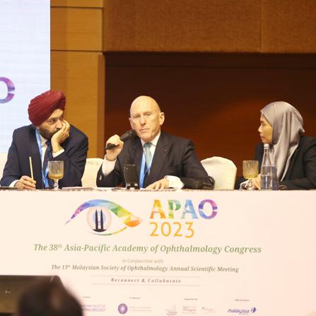
Three illustrious ophthalmologists — this year’s recipients of the APAO and MSO Awards — presented interesting and heartfelt lectures on distinct topics in their fields of interest. The “KOL Video” has been around for a while, but Media MICE’s team uses dynamic production to make the coverage shine like never before at conferences worldwide.

Speaking at Plenary Session II, the 2023 APAO and MSO Named Awards
namely, Prof. Kyoko Ohno-Matsui, professor and chairperson of the Department of Ophthalmology and Visual Science at Tokyo Medical and Dental University (TMDU) — the Jose Rizal International Medal (APAO Award); Prof. A.H.M. Enayet Hussain, director general,


Medical Education of the Directorate General of Health Service — ICO Mark Tso Golden Apple Award (APAO Award); and Prof. Liza-Sharmini Ahmad Tajudin, ophthalmologist and lecturer at the Department of Ophthalmology and Visual Science, Universti Sains Malaysia — Dato’ Dr. S. Selvarajah Award (MSO Named Award), presented inspiring lectures on posterior staphyloma, a lifelong mission to improve eye care in Bangladesh, and making glaucoma patients’ lives better, respectively.
Prof. Dr. Kyoko Ohno-Matsui began her talk by paying tribute to Dr. Jose Rizal, one of the most revered figures in Philippine history, saying she felt great pride for receiving the award after the Filipino doctor.
Prof. Ohno-Matsui presented her lecture on her studies on posterior staphyloma, an eye deformity that is found to be the leading cause of pathologic myopia.

“I will focus on staphyloma because it’s a critical allusion to differentiating pathologic myopia from physiologic myopia,” she shared. “There are three leading causes of blindness
in pathologic myopia — myopic maculopathy, myopic traction maculopathy, and glaucoma or optic myopic optic neuropathy.”
And the cause of developing the above complications is characterized by the eye deformity posterior staphyloma.
A staphyloma, Prof. OhnoMatsui said, is an outpouching of the wall of the eye that has a radius of less than the surrounding curvature of the wall of the eye. To detect staphyloma, besides using wide-field fungus imaging and sweptsource ultra wide-field OCT, 3D MRI can show the entire shape of the eye in a three-dimensional way from any angle. And the tropic eyes are spherical.
Staphyloma formation and axial length elongation are separate phenomena, she said, adding that staphyloma is also found in non-highly myopic eyes (40%) of those with unilateral high myopia.
To find out at what age a staphyloma develops, Prof. Ohno-Matsui said that her team is examining highly myopic children to solve this question.
“Early detection of the presence and degree of staphylomas with wide-field OCT is necessary to establish therapies targeting staphylomas, before it is too late. Therapies targeting staphylomas before developing blinding complications are truly expected to save the eyesight of patients,” she concluded.
Up next, Prof. A.H.M. Enayet Hussain shared his talk, Towards an Equitable and Inclusive vision: Incorporating Social Determinants of Health in Medical Education in Bangladesh, as well as his 30-year vision in dedicating and improving healthcare in Bangladesh.
With the inauguration of the Bangladesh Pediatric Ophthalmology Unit in 2001, Prof. Hussain spearheaded many initiatives in improving eye care, particularly for children.
This includes the Bangladesh Childhood Cataract Campaign, which, since its launch in 2004, has successfully performed 27,000 surgeries (including 9,000 bilateral congenital cataracts); the Sylhet project to strengthen the pediatric eyecare system; integration of child eye health into the Integrated Management of Childhood Illness (IMCI) program in Bangladesh; first national ROP Summit involving national and international participants in February 2020; and a national level advocacy by leadership on childhood blindness prevention in Bangladesh.
“We now have Eye Health Program in
“There are three leading causes of blindness in pathologic myopia — myopic maculopathy, myopic traction maculopathy, and glaucoma or optic myopic optic neuropathy.”Prof. Kyoko Ohno-Matsui Prof. Kyoko Ohno-Matsui
four districts in Bangladesh,” enthused Prof. Hussain. “Trained healthcare service providers work at refugee camps to provide primary eye care services, and we have published an End Line Report with The Fred Hollows Foundation on restoring vision to empower rural and marginalized women and children in Bangladesh,” he continued.
Prof. Hussain also shared his thoughts on access and inequality to eye care, which is greatly affected by social determinants, such as social exclusion, gender inequity, racism, early childhood development, educational opportunities, and employment condition.
“These are the non-medical factors that influence health outcomes. These inequities are due to unfair and avoidable differences in social status. Reducing long-standing inequities in health would require action by all
sectors in society,” he said.
He also stressed on incorporating social determinants of health (SDH) in medical education and posed some challenges for the future.
“There is a lack of relevance of SDH in clinical practice, and we need a change in the medical curriculum and community involvement,” he shared. “The journey has started and hopefully we’ll be able to do this sustainable goal of 2030.”
Lastly, Prof. LizaSharmini Ahmad Tajudin shared her presentation entitled, Understanding the Unmet Needs of Patients with Glaucoma: Lifestyle, Psychological and Genetics.

“Glaucoma is a complex disease and there is strong evidence for genetic predisposition,” she said. “Family history is a risk factor. There are potential genetic markers but no
specific panel for genotype-phenotype testing.”
She continued: “Medicine is personalized and it is difficult to predict the progression of the disease. But how does a patient with glaucoma face their life and what happened to them at home?”
As glaucoma is a progressive disease, patients have to deal with the physiological changes with age, as well as other systemic comorbidities. When they are in their retirement age, they may face financial difficulty.
“Their quality of life will differ because of socio-culture and belief,” Prof. Tajudin said. “And, of course, this quality of life will differ between those in urban, suburban, and rural areas.”
She also shared some of her findings on the various aspects of daily living for a glaucoma patient, such as navigation, mobilization, and reading. She also mentioned ways that may help them improve, such as occupational rehabilitation, Chessington Occupational Therapy Neurological Assessment Battery (COTNAB).
As a chronic disease, glaucoma will have a psychological impact on the patient. Although, it may be difficult to detect in older adults due to age, they may have anxiety and depression.
“Based on our study, we have found patients who suffer helplessness, emptiness, worthlessness, and hopelessness. And this increases as the severity of glaucoma increases. This depression seems to be higher in countries such as Malaysia, as compared to other developed countries. So we should be be more sensitive and empathetic towards them,” Prof. Tajudin noted.
“Patients’ needs and expectations are important. Perhaps in the future, we’ll be able to identify genetic markers [that will let us] know who actually will have aggressive diseases, so we can prepare the patient psychologically to cope better with the disease,” she concluded.
“Based on our study, we have found patients who suffer helplessness, emptiness, worthlessness, and hopelessness. And this increases as the severity of glaucoma increases. This depression seems to be higher in countries such as Malaysia, as compared to other developed countries. So we should be be more sensitive and empathetic towards them.”Prof. Liza-Sharmini Ahmad Tajudin Prof. Liza-Sharmini Ahmad Tajudin
Undeniably, while there have been breakthroughs in imaging, data security is a major concern in digital healthcare. Questions also arise on whether the economic pay-off for using artificial intelligence (AI) is greater than the cost, and whether clinicians can build their own models with no IT skills other than Facebook, Twitter, or Instagram.
In yesterday’s session on Applying Big Data, Artificial Intelligence and Telemedicine in Ophthalmology during the second day of the 38th Asia-Pacific Academy of Ophthalmology Congress (APAO 2023), delegates discussed burning topics such as how to boost trust in the use of AI in ophthalmology, and many more.

While there have been breakthroughs in imaging, including pattern recognition and image classification, data security is a major concern in medical data, said Prof. Anran Ran, scientific officer from the Department of Ophthalmology and Visual Sciences, the Chinese University of Hong Kong.
“Technologies ensuring data security are needed for ophthalmology,” she noted. Retinal images are unique and can be used as biometric identification, like fingerprints.
Federated learning (FL), a type of collaborative strategy, enables mobile phones to collaboratively learn a shared prediction model, but keeps all training data on each device, protecting data, and reducing the risk of data leakage.
“FL has potential in ophthalmology,” continued Prof. Ran. FL can also be applied to different ocular imaging modalities. It also enhances the equity of eye care, as institutions with smaller sample sizes can contribute to and benefit from it. Finally, it serves as an education tool, as it learns knowledge from diverse datasets with different disease prevalences and severities.
Future directions for the use of FL would be to incorporate it with other technologies, such as in smartphonebased ocular imaging, in eye care apps for personalized disease monitoring, and auto machine learning (ML) to involve more institutions without AI experts.
The use of FL also needs to be evaluated in real-world settings, she concluded.
Speaking of auto ML and AI, is it possible to build a model with no prior coding experience?
This is the question that Prof. Javier Zarranz-Ventura from the School of Medicine, University of Barcelona, sought to answer.
While most clinicians have little to no IT skills, Prof. Zarranz-Ventura reassured us that it is possible to use auto ML.
“Basic tools are relatively straightforward,” he argued, adding there is a need for good quality data and specific training on ML applications. Several studies have been carried out, including one on Automated Machine Learning Models Applied on Optical Coherence Tomography Angiography for Detection and Classification of Diabetic Retinopathy in Diabetes Mellitus Type 1,’ by Javier ZarranzVentura et al., and Code-free Deep Learning for Multi-modality Medical Image Classification by Edward Korot et al.
Prof. Dr. Paisan Ruamviboonsuk, clinical professor at the College of Medicine, Rangsit University, Rajavithi Hospital, Bangkok, Thailand, spoke about the health-economic considerations of AI in ophthalmology.
“Economic evaluations can provide ‘value-for-money’ information to those making decisions about the allocations of limited health care resources,” he said.
A cost-utility analysis of deep learning (DL) versus human graders (HG) for diabetic retinopathy (DR) screening was carried out in the Thai national program, which included the cost of treatment of diabetic macular edema (DME) using bevacizumab.
In general, AI appeared to be costeffective compared with human screeners in many economic evaluation models using AI for screening diabetic retinopathy (DR). The cost of AI is a very important parameter to determine the costeffectiveness of AI in health care, he noted.
In the Thai model, DL costs more from a provider perspective but cheaper from a societal perspective. What’s more, adherence to referral is another important factor to determine the cost-effectiveness of DR screening models using AI.
Molteno3® is no stranger to the glaucoma world. This glaucoma drainage device (GDD) has been shown to deliver consistent, long-term intraocular pressure (IOP) control in cases of severe and complex glaucoma. US ophthalmologist Dr. Joseph F. Panarelli, who is an authority in GDD, shared his take on implanting the Molteno3.
In order to ensure optimal outcomes, Dr. Joseph F. Panarelli has fine-tuned his surgical technique for Molteno3 implantation.
He starts the procedure with the placement of a 6 o’clock micro traction stitch that allows for enhanced exposure. “I like to
by Tan Sher Lynnopen up a horizontal meridian and continue by dissection up past the twelve o’clock area and maybe go just an additional clock hour before making a second relaxing incision. The superior temporal quadrant is my preferred quadrant of choice for implantation and if it is not suitable I will move to the inferior nasal quadrant,” he said.
In cases where the patient has more scarring due to prior subconjunctival minimally invasive glaucoma surgery (MIGS), he elects to free up the scar tissue before proceeding to hook both the superior lateral rectus muscles and free them from the Tenon’s attachments, after which he places the Molteno3 in the superior temporal quadrant.
“One of the nice things about the Molteno3 is that it really hugs the globe nicely, so as you tuck it back into the quadrant it really doesn’t move forward that much. This makes it easier when you perform the placement of the temporary ligature, to allow for time for the implant to encapsulate,” he said.
“It’s very important for non-valved GDDs to have the ligature in place and it needs to be water-tight,” he added, noting that there should be no movement of fluid posterior to the ligature upon priming the eye with balance salt solution.
“The GDD needs to be secured approximately eight to ten millimeters posterior to the limbus. The more

posterior you can place the GDD, the better the flow. There’s also a lower likelihood of postoperative strabismus. The knots of the non-absorbable suture need to be rotated into the eyelets, so that we don’t end up with erosion or extrusion of these knots down the road,” he advised.
After the GDD is secured in place, he moves on to implant the tube into the anterior chamber. Firstly, the eye is pulled back into primary position, before trimming the tube with a Westcott scissors, making sure not to over-trim the tube.
He then bends a twenty three gauge needle and makes a tunnel track into the eye to insert the tube, ideally about two millimeters into the anterior chamber. “We want to make sure it’s parallel to the iris plane and far away from the corneal endothelium. Given that this is a non-valved GDD we need some method to control the pressure early on so I’ll pass a ten o’clock vicryl stitch to the occluding ligature. I want to see a nice percolation of fluid through that fenestration,” he said.
First introduced in 1966 by Prof. Anthony Molteno (who invented the world’s first glaucoma drainage device), the Molteno3 has gone through various evolution and refinement until it reaches the form that it is today —the third generation of Molteno.
Thus, far from being an outdated technology, the Molteno3 technology actually reflects a history of extensive research into bleb formation and function, and is considered by many to be the gold standard in GDD technology.
With a contoured plate, the novel
design of the Molteno3 is designed to hug the sclera perfectly. At just 0.4 mm, it is also the slimmest plate on the market. This smooth polypropylene plate reduces the risk of fibroblast attachment, supporting better bleb formation over the longterm. It slides easily between tissue planes and adjacent extraocular muscles, allowing for a shorter, more simplified surgical procedure with less patient discomfort. Additionally, repositioning is relatively easy to achieve the desired outflow when needed.

For more information on Molteno3, visit: https://glaucoma-molteno.com.
Then, a corner patch graft is sutured in place over the tubing to prevent erosion of the tube down the road. Dr. Panerelli notes that he likes to tunnel the tubes closer to twelve o’clock as he is able to obtain better coverage with the upper eyelid and a lower likelihood of tube erosion in the future.
The final step is to close the conjunctiva. “At this point I will often loosen the speculum, bring the eye back up into the primary position and try to advance the conjunctiva up to the limbus. Once it is in place I will close each of the relaxing incisions. I typically try to get deep bites for the first two or three bites so that the tissue is less likely to cheesewire and then take more superficial bites as I am getting further posterior from the limbus,” he said. Once a nice closure is obtained, he then injects the antibiotic and steroid mixture beneath the inferior conjunctiva and palpates the globe to make sure that the desired pressure is achieved.
Dr. Joseph F. Panarelli is a US-based ophthalmologist specializing in the treatment of adult and pediatric glaucoma. He is certified by the American Board of Ophthalmology and is a member of the American glaucoma Society and American Academy of Ophthalmology. He is a glaucoma specialist at NYU Langone Health, where he is also an associate professor of the Department of Ophthalmology at NYU Grossman School of Medicine; program director of the Glaucoma Fellowship Program; and director of the Division of Glaucoma Services, Department of Ophthalmology. He chose to specialize in glaucoma because it allows him to provide ongoing medical care and surgical treatments to patients to improve their quality of life. He has published several new surgical techniques as well as new findings comparing traditional glaucoma procedures to current ones. He has also been part of numerous clinical trials for new surgical devices for eye procedures.
Joseph.Panarelli@nyulangone.org
that uses the VisioLite® Er:Yag laser to create tiny micropores in scleral tissue to tackle ocular rigidity, and uncrosslinking scleral microfibrils to decrease biomechanical stiffness — allowing recovery of a dynamic range of focus (DRoF) inside of the eye.
Presbyopia is not easy to treat, but promising technologies and methods are on the horizon to improve this condition, as presented during the refractive surgery symposium on the second day of the APAO Congress.
It’s inevitable that everybody will develop presbyopia. But treating this condition is difficult. While emmetropic presbyopes face worsening vision, for ametropic presbyopes, gaining distance vision is an attractive proposition. But they need to accept sacrifice in terms of intermediate/near vision, sharpness/quality of vision, photic phenomena, and stereopsis — noted Dr. Robert Ang from the Philippines.
According to Dr. Ang, accommodation is actually a dynamic range of focus mechanism of adjusting your near vision. “Our work began with understanding
accommodation and how age affected presbyopia. We developed a novel therapy called the laser scleral microporation (LSM) technology, which addresses the root cause of presbyopia, not involving changes in refraction or optics of the eye,” he continued.

The LSM technology restores true accommodation by enhancing depth of focus while maintaining contrast sensitivity and stereopsis, and improving binocular vision. It is a minimally invasive, painless, and quick bilateral procedure
“LSM is unique because it can improve intermediate and near visual acuity without touching the visual axis and without compromising distance vision. Early results suggest LSM to be a safe and effective procedure for restoring effective range of focus in presbyopes,” Dr. Ang remarked.
For the past decades, the capsular bag has been the preferred location for the placement of non-accommodating intraocular lenses (IOLs), but it may not be the best location for an accommodating IOL (AIOL).
According to Prof. Dr. Jorge Alio from Spain, the capsular bag seems to be the wrong location for AIOLs due to its unavoidable decadence and fibrosis once the crystalline lens is emptied. He noted that there are various failed approaches in AIOLs caused by poor methodology, commercial bias, and poor monitoring of the investigation.
“The capsular bag is not the place for AIOLs,” he said. “First of all, the capsular bag is a basal membrane of the lens epithelium. Once it is emptied, it has no functions and there are no anatomical reasons for it to exist. Fibrosis and atrophy are unavoidable as it has no function to accomplish and no anatomy to support,” continued Prof. Alio.
He presented an endoscopic primate study which demonstrated that after five years, neither the epithelium nor ciliary body had any atrophy or damage due to the haptics’ location at the sulcus.
“The forces generated at the zonularanterior capsule system are those to be used for AIOLs. Hence, sulcus implantation is the only option for AIOLs,” he concluded.
The use of glaucoma drainage implants to reduce the intraocular pressure (IOP) in glaucoma patients is increasingly popular among surgeons — but they are not without issues.

Also called ‘aqueous shunts’ or ‘glaucoma drainage devices’, glaucoma drainage implants are small devices that are placed in the eye to help lower the IOP and prevent further optic nerve damage in glaucoma patients. Like any surgery, however, glaucoma drainage implants carry some risks of complications.
To avoid the pitfalls, learn from experts in glaucoma surgery as they share tips and experiences during the External Drainage Surgery for Glaucoma symposium, happening today.
Bleb needling is typically performed when a patient’s trabeculectomy is not working properly and scar tissue has formed in an attempt to get the trabeculectomy functioning and lower the eye pressure.
Even though the procedure is generally considered safe and works well in over half of patients, some patients may develop scar tissue which requires a second needling procedure months or years later. Others may experience worse vision due to hypotony, or have blurring of vision due to the antiscarring medication used. There are also possible complications, which include hyphema, a flat anterior
chamber, choroidal effusions, conjunctival buttonholes, and endophthalmitis.1,2,3
Hence, bleb needling should be performed with caution. In his talk, Bleb Needling: Is It Necessary? — Dr. Norhalwani Husain will share his thoughts and experience on whether bleb needling is necessary or not, so that the risks of complications can be minimized.
Glaucoma drainage implants have been indispensable in the fight against blindness, but they are not without complications. In fact, a host of undesirable events can arise. For example, hypotony may develop after tube shunt surgery, along with its associated complications such as corneal edema, choroidal effusions, anterior chamber shallowing, cystoid macular edema, and suprachoroidal hemorrhage.
Other possible complications include diplopia, strabismus, proptosis, tube erosion, failure, corneal decompensation, endophthalmitis, and visual loss.4
In his talk, Tube Complications and Nightmare, Dr. Joseph Anthony Tumbocon will share cases of tube issues and discuss how they can be rectified or prevented. Another session that should not be missed to ensure good surgical outcomes!
The PAUL® Glaucoma Implant (PGI) is a new valveless glaucoma drainage device (GDD) made using implantable medical-grade silicone. This aqueous shunt device is designed by Professor Paul Chew with the intention to reduce postoperative complications seen with current shunts.
However, literature evaluating its efficacy and safety is still limited. It is therefore highly recommended to hear from surgeons who have implanted the device themselves, so that one can make better choices when dealing with it.
In this symposium, Dr. Eng Hui Gan will be sharing his experience of using the PGI in his talk entitled, My Early Experience with the Implantation of PGI. Be sure to catch him for some priceless advice!
References
1. Pederson JE, Smith SG. Surgical management of encapsulated filtering blebs. Ophthalmology. 1985;92(7):955-958.
2. Mardelli PG, Lederer CM Jr, Murray PL, et al. Slit-lamp needle revision of failed filtering blebs using mitomycin C. Ophthalmology. 1996;103(11):1946-1955.
3. Gutierrez-Ortiz C, Cabarga C, Teus MA. Prospective evaluation of preoperative factors associated with successful mitomycin C needling of failed filtration blebs. J Glaucoma. 2006;15(2):98-102.
4. Sarkisian SR Jr. Tube shunt complications and their prevention. Curr Opin Ophthalmol. 2009;20(2):126-130.
Date: February 25, 2023
Time: 16.30 to 18.00 H
Venue: Conference Hall 2 (Level 3), KLCC
The fight against our own immune system is never easy. Autoimmune diseases (AD) affect almost every part of the body, and the eyes are no exception.
Gain a deeper understanding of autoimmune eye diseases and their management in the Autoimmune Eye Diseases: From Pathogenesis to Treatment symposium on Day 3 of the 38th AsiaPacific Academy of Ophthalmology Congress (APAO) 2023.
Here are a few sessions you might wanna catch…
There are various autoimmune diseases that commonly affect the eyes, and one of them is the VogtKoyanagi-Harada (VKH) disease. This rare, granulomatous inflammatory disease of unknown origin affects other body systems as well, including the ears, skin, and the covering of the brain and spinal cord.
However, the most significant manifestation is bilateral, diffuse uveitis, characterized by pain, redness, blurring of vision, and rapid vision loss. The standard treatment for VKH disease is the use of high-dose systemic steroid drugs initially and often followed by immunosuppressive therapy.1
Learn the details of the treatment of this potentially debilitating disease from Dr. Peizeng Yang’s talk, “Treatment of Vogt-Koyanagi-Harada Syndrome.”

The ocular surface microbiota refers to the non-pathogenic microorganisms that colonize the conjunctiva and cornea. There is increasing evidence that this microbiome plays a key role in ocular health and diseases. Changes in the ocular microbiome in patients with dry eyes and in healthy subjects have been reported, and alterations of the ocular microbiome have been thought to contribute to the pathophysiology of dry eye.2
Catch more about microflora and the measures that can be taken to maintain a healthy ocular surface microbiome from Dr. Richard Lee’s talk, “The Intraocular Microbiodata.”
Uveitis often happens in people who have an autoimmune condition. The Ocular Autoimmune Systemic Inflammatory Infectious Study (OASIS) describes the epidemiology of 2,200 patients in a tertiary eye center in Singapore presenting with ocular inflammation and their etiological factors in OASIS - Report 1: Epidemiology and Classification;3 while OASIS - Report 2 focuses on the investigations and management of these patients and discusses the disease-specific complications and outcomes.4
Delve into the details of this condition in Dr. Rupesh Agrawal’s talk, “OASISA Centralized Registry for Autoimmune Disease.”
1. Lavezzo, MM, Sakata VM, Morita C, et al. Vogt-Koyanagi-Harada disease: review of a rare autoimmune disease targeting antigens of melanocytes. Orphanet J Rare Dis. 2016;11:29.
2. Qi Y, Wan Y, Li T, et al. Comparison of the Ocular Microbiomes of Dry Eye Patients With and Without Autoimmune Disease. Front Cell Infect Microbiol. 2021;11:716867.
3. Chen EJ, Bin Ismail MA, Agrawal R, et al. Ocular Autoimmune Systemic Inflammatory Infectious Study (OASIS) - Report 1: Epidemiology and Classification. Ocul Immunol Inflamm. 2018;26(5):732-746.
4. Low R, Chen EJ, Agrawal R, et al. Ocular Autoimmune Systemic Inflammatory Infectious Study (OASIS) - Report 2: Pattern of Uveitis Investigations in Singapore. Ocul Immunol Inflamm. 2020;28(1):92-99.
Date: 25 Feb 2023
Time: 14.30 to 16.00 H
Venue: Meeting Rooms 403-404 (Level 4), KLCC

Proliferative diabetic retinopathy (PDR) is a severe complication of diabetes that could lead to loss of sight. In yesterday’s “Retina (Medical) Symposium on PDR,” researchers discussed the latest studies and treatments for PDR.
People with diabetes can develop diabetic retinopathy (DR), where high blood sugar levels damage the blood vessels in the retina. There are two main stages of DR — non-proliferative diabetic retinopathy (NPDR) and proliferative diabetic retinopathy (PDR). PDR is a more severe type of the disease, where damaged blood vessels close off, causing the growth of new, abnormal blood vessels in the retina.
Dr. Paolo Antonio Silva from the Philippines talked about evaluating whether the detection of predominantly peripheral lesions (PPL) improves the ability to predict rates of retinopathy worsening over time beyond the risks associated with baseline Early Treatment Diabetic Retinopathy Study (ETDRS) Diabetic Retinopathy Severity Scale (DRSS) level.
“Proportion of eyes experiencing DRSS worsened by two or more steps from baseline or receipt of DR treatment through four years. Eyes with fluorescein angiography and pars plana lensectomy (FA-PPL) had a 1.7fold greater risk of disease worsening over four years,” said Dr. Silva.
“The association between disease worsening and retinal nonperfusion
and FA-PPL supports the increased use of ultra-widefield fluorescein angiography (UWF-FA) to complement the color fundus photography in future efforts for DR prognosis, clinical care, and research,” he added.
PDR treatments: What we need to know?
Increased understanding of the biochemical mechanisms underlying PDR is providing new therapeutic approaches with the promise to be both effective and less destructive than current photocoagulation techniques.*
“PRP reduces severe vision loss by 50% or more, while Ranibizumab achieves non-inferior vision outcomes through five years. Meanwhile, aflibercept achieves superior vision outcomes at one year, compared to PRP,” shared Prof. Dr. Susan B. Bressler from the United States.
Meanwhile, Prof. Dr. Jun Kong, also from the US, discussed whether both panretinal photocoagulation (PRP) and antivascular endothelial growth factor (anti-VEGF) should be used in combination in the absence of diabetic macular edema (DME) on all patients with PDR.
“Patients were observed to have faster neovascularization (NV) regression
with anti-VEGF, which may protect against acute incident DME or exacerbation of pre-existing DME,” explained Dr. Kong. “Place PRP can be used for permanent treatment to save visual acuity (VA), which may reduce vitreous hemorrhage (VH) and tractional detachment (TRD) if patients do not return for timely follow-up. Lastly, combination treatment may reduce anti-VEGF treatment burden,” he continued.
“The combined treatment of anti-VEGF and PRP was more effective than PRP alone. Anti-VEGF can be considered to defer PPV in PDR VH eyes with prior completed PRP and no TRD,” said Dr. Onnisa Nanegrungsunk from Thailand.
Prof. Dr. Bressler spoke on lapses in care with anti-VEGF during the management of PDR. She said about 30% of randomized control trials (RCT) anti-VEGF assigned participants dropped out within five years (similar to PRP participants). She also said that 50% of RCT PDR patients were alive five years later after experiencing one long lapse in care (including the ones who dropped out). Lastly, she shared that 75% of participants returned after their first long lapse.
It is important to keep in mind that a comprehensive approach to diabetes care is particularly critical for patients with advanced retinopathy, and only with the coordinated care between eye care providers and many other members of the diabetic patient’s medical care team can optimal ophthalmic outcomes be achieved.* Reference
* Silva PAS, Cavallerano JD, Sun K, et al. Chapter 48 - Proliferative Diabetic Retinopathy. Retina (Fifth Edition, Vol. 2). https://doi.org/10.1016/ B978-1-4557-0737-9.00048-5.


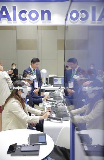
The biggest Asia-Pacific ophthalmological congress is back on track in Kuala Lumpur. It’s the first time the Asia-Pacific Academy of Ophthalmology (APAO) Congress is physical again post-pandemic. The industry is exploding with ongoing eye care innovations, industry news, and educational content that RECONNECT eye care specialists worldwide. And here we are once again COLLABORATING with APAO as the Official Media Partner. So follow us through the conference with event highlight snapshots!


Dr. Loy also noted that studies have shown that injecting anti-VEGF six to 14 days pre-op significantly improved post-op best-corrected visual acuity (BCVA) and reduced recurrent vitreous hemorrhage and intraoperative bleeding.
Retinal experts shared tips for preventing hemorrhage and treating combined traction and rhegmatogenous retinal detachments, as well as the use of silicon oil in diabetic vitrectomy, on the second day of the APAO Congress.
Over the past years, advances in retinal surgery have made many formerly blinding conditions, such as diabetic retinopathy (DR), more manageable. Retinal experts shared their knowledge and experience for better surgical outcomes during the “Retina (Surgical) Symposium on Diabetic Retinopathy and Retinal Vascular Diseases,” held yesterday.
In proliferative diabetic retinopathy (PDR), severe vision loss can occur from complications of retinal neovascularization and fibrovascular proliferation. Even though these are indications for pars plana vitrectomy, intraoperative bleeding is very common, making surgical maneuvers very difficult because of poor visualization.
Postoperative vitreous cavity hemorrhage can occur in 20% to 30% of cases as well, noted Dr. Marie Joan Loy from the Philippines.
“Preoperative anti-vascular endothelial
growth factor (anti-VEGF) injection is effective in regressing retinal neovascularization, thereby decreasing intraoperative and postoperative bleeding, complications, and surgical time, as well as improving surgical outcomes,” shared Dr. Loy.
“Bevacizumab is the most studied anti-VEGF agent. A lower dose of bevacizumab (0.625 mg) appears to be effective and safe compared to higher doses,” she continued.
“Doing surgery within six days of anti-VEGF injection appears better especially in young patients and in those with vitreous hemorrhage. Meanwhile, peri-operative use of antiVEGF agents decreases the risk of early post-vitrectomy bleed, according to the Cochrane Database of Systemic Review,” she concluded.

Meanwhile, Dr. Kian Seng Lim from Malaysia noted that combined traction and rhegmatogenous retinal detachments (TRD/RRD) are probably the most challenging surgical retinal cases.
“These are typically found in young diabetics with minimal PVD, which adds complexity to the surgery. Early surgical intervention is normally required,” he said.
“During surgery, I would attempt to remove as much posterior hyaloid as I could. The use of triamcinolone can be helpful,” Dr. Lim shared, adding that he pays meticulous attention to hemostasis throughout surgery as well.
“During surgery, retinal tears are usually found just adjacent to the fibrovascular stump. I attempt to release all tractions adjacent to retinal tears, which includes the fibrovascular tissue, posterior cortical remnant, and blood clots,” he continued. “My priority is to achieve macular attachment, firstly by releasing traction on the macula and optic disc, and secondly by releasing traction on the retinal tear. I believe that total dissection of
fibrovascular tissue adds considerable risks to the surgery, and I avoid retinotomy and retinectomy at all costs.”
There have been numerous advancements in vitrectomy technology. “The discovery of anti-VEGF has made most of the complications in DR and TRD/RRD surgically treatable and with good outcomes,” he remarked.
The prevalence of DR among people with diabetes in Asia ranges between 30% and 46% higher than in western countries, and Indonesia is one of the countries with the largest number of people with diabetes in the world, noted Dr. Referano Agustiawan from Indonesia.
“Various vitreous substitution with silicone oil and long-acting gas
has been used during pars plana vitrectomy in cases with advanced diabetic complication. However, little clinical data are available regarding which vitreous substitute is best suited for this patient population,” he shared.
Dr. Agustiawan added that silicone oil promotes retinal reattachment by providing extended intraocular tamponade, compartmentalizes the eye, and confines angiogenic substances to the posterior segment, as well as acts as a diffusion barrier to oxygen — thereby preventing a decrease in anterior chamber oxygen tension after vitrectomy.
“Silicone oil tamponade is useful in severely diseased eyes with PDR, rubeosis, and previously failed vitrectomy,” he continued. “Silicone oil’s potential benefit is its stability inside the eye for up to a year, thus providing longer support to the retina and inhibiting the progression of neovascularization compared to other
tamponades,” he said.
Nevertheless, silicone oil also has its disadvantages. Migration of silicone oil droplet into the retina and optic nerve can cause loss of myelinated optic nerve fibrosis, and there’s a risk of secondary glaucoma due to silicone oil droplet in the trabecular meshwork. Besides, there may be a need for a second surgery. In addition, silicone oil is associated with fibrovascular proliferation, has a lower surface tension, and is unable to completely fill the vitreous cavity.
“Although it remains inconclusive whether silicone oil is the best choice for diabetic vitrectomy, in my opinion, silicone oil is handy in complex cases of diabetic vitrectomy and cases of iatrogenic breaks that occur during surgery. Also, it should be used when one is less experienced or just starting with diabetic vitrectomy,” concluded Dr. Agustiawan.
 by Hazlin Hassan
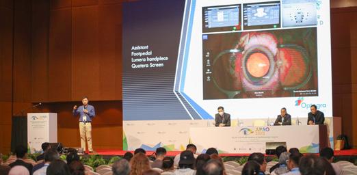
Make your surgical workflow even more efficient while achieving quality outcomes. Gain a completely new experience in phaco surgery with the QUATERA 700, the VISUMAX 800, and the SLT — all from ZEISS.
On Day 2 of the APAO Congress, Carl Zeiss Meditec (ZEISS, Jena, Germany) showcased some of its cutting-edge digital solutions aimed at helping doctors enhance their clinical efficiency and improve patient care — from refractive surgery, glaucoma surgery, and phaco surgery.
From the VISUMAX 800 to the QUATERA 700 and ZEISS SLT, ZEISS’s cutting-edge tools are, indeed, “Seeing Beyond and Shaping Today’s Innovations.” .”
Prof. Colin Chan, a refractive, cataract, and corneal eye surgeon at the Vision
Eye Institute, Sydney, Australia, shared his experiences in using the Visumax 800 — describing himself as a “happy ZEISS user.”
Speed and precision are very important to Prof. Chan, both in his hobby as a bird photographer in his spare time and as a surgeon. And the VISUMAX 800 ticks all the boxes and more, representing the latest generation of ZEISS femtosecond lasers, with reduced laser time in comparison to its predecessors — all while making tissue separation with SMILE pro from ZEISS easier.
“One of the things I do in my spare time is bird photography, and it demands speed, precision, and tracking. I demand the same on my
laser platform,” he shared.
In Prof. Chan’s opinion, SMILE is the refractive procedure for Asian eyes. He said there is a higher prevalence of moderate and high myopia in Asia, and Asian eyes — particularly females, are prone to dry eye, whether before or after laser, particularly LASIK.
“We know that if you treat high myopia, you get more dry eye. And we also know that in high myopia, LASIK becomes more inaccurate. So, therefore, SMILE addresses these ‘Asian’ issues,” he said, adding that several studies have shown that SMILE is more accurate in moderate to high myopes than LASIK.
The key advantages of SMILE
compared to conventional laser (LASIK/PRK) include better accuracy in higher myopia (Asian population), dry eye levels similar to PRK with recovery more similar to LASIK (Asian female population), and ‘truer’ optical zone.
Speed, speed and speed! That is the most impressive upgrade of the VISUMAX 800 – flaps are created in 8 seconds and a lenticule with SMILE pro takes only 10 seconds. The faster speed of the VISUMAX 800 has other benefits: patient comfort, reduced risk of suction loss and reduced surgeon anxiety, said Prof. Chan.
In conclusion, he sums up his experience with these three words: “Faster, better centration, accuracy.”
Dr. Amir Hamid, a consultant ophthalmic surgeon at Optegra Eye Health Care, United Kingdom, also shared his QUATERA 700 experience, and how the digital cataract workflow has changed his practice.
The QUATERA 700 offers chamber stability in all surgical situations and a digitally-integrated surgical workflow. It unites all elements of the ZEISS Cataract Workflow, saving surgeons from having to switch between multiple user interfaces and parameters or different setups.
Dr. Hamid describes his practice as primarily refractive lens exchange and premium cataract surgery.
It is a high-volume model with 16 eyes per four-hour list. Some 80% of cases involve bilateral surgery, with 70% receiving toric intraocular lenses (IOLs).
Safety and accuracy in his practice are improved through the use of technology and a partnership with ZEISS, in particular with the ZEISS FORUM EQ Workplace, which is the implementation of a cloud solution for cataract surgeries.
His practice now uses software-based IOL calculation, selection and ordering, which is an advantage compared to manual marking, which is timeconsuming, messy, and inefficient. One of the most important benefits is eliminating transcript error.
“The key benefit to the ZEISS FORUM EQ Workplace is that it virtually eliminates post-occlusion surge and a digitally-integrated surgical workflow that connects OR devices for more efficiency with one surgical view, one sterile cockpit, and one data access. It also provides automated ultrasound power on demand with efficient followability,” Dr. Hamid explained.
What does it mean in reality? “Greater safety, efficiency, excellent results, a more relaxed surgeon, and happy patients,” he enthused.
In summary, the QUATERA 700 allows for a stable anterior chamber in any setting.
Furthermore, any case complexity and improvements in the workflow allow safe and efficient surgery with less staff in the OR.
The ZEISS SLT: The true lunch break laser?
Dr. Edgar Leuenberger, a glaucoma specialist at the Asian Eye Institute, Philippines, spoke about the state-
of-the-art glaucoma treatment, the ZEISS SLT or Selective Laser Trabeculectomy.
“The ideal glaucoma treatment should be quick, painless, safe, effective, cost-effective, non-invasive, and can be performed during lunch breaks,” Dr. Leuenberger said.
Laser glaucoma treatments have evolved from when Argon Laser Trabeculoplasty (ALT) was first introduced in 1979 to the ZEISS SLT today.
In 2022, the National Institute for Healthcare Excellence (NICE) in the UK recommended the SLT as the first-line therapy for newly-diagnosed primary open-angle glaucoma (POAG).
The selection of laser energy is based on the pigmentation of the patient’s trabecular meshwork. With ZEISS VISULAS green laser technology, no cavitation bubbles are visible and the titration process is simplified. This saves time and reduces unnecessary energy delivery to the patient’s eye.
“The rotating aiming beam enables accurate positioning of the laser and improves visibility of the trabecular meshwork during treatment,” explained Dr. Leuenberger. Every ZEISS SLT laser application with a total diameter of 400 μm consists of 52 squared spots. The squared spot shape ensures a higher homogeneity of the laser energy distribution.
Further, Dr. Leuenberger shared results of an SLT approval study conducted in Germany, on 34 eyes with 16.3% IOP lowering and half of the patients with more than 20% IOP lowering. Cases of juvenile open angle glaucoma saw pressures lowered by 28% at 2 weeks. Overall, 50% of the eyes treated have more than 20% intraocular pressure (IOP) decrease in the second week.
As one of the first users of the ZEISS SLT outside of Europe, Dr. Leuenberger’s patient experience is very similar to the Germany study (IOP lowering: 15.7% vs 16.3%). More long-term, well-designed studies are needed, he added.
On Day 2 of the APAO Congress, a panel of international speakers shared their expertise and views on myopia and artificial intelligence in an information-packed “APAO-AAO-NEI Joint Symposium: Strategic Priorities and Opportunities for Collaboration With the Asia-Pacific Community.”

The symposium featured clustered talks on three topics: myopia by Dr. Michael F. Chiang and Dr. Marcus Ang, data sharing and data models by Dr. Sally Baxter and Dr. Taji Sakamoto, and artificial intelligence by Dr. Aaron Lee and Dr. Haotian Lin.
Dr. Michael Chiang, director of the National Eye Institute (NEI), shared that NEI had 3,500 unique research projects (11% involving non-US collaborators) and over 800 non-US investigators (from 66 countries) participating in NEI-funded research. In March 2022, NEI hosted global societies to discuss areas of mutual research interest and identified several priorities for collaboration.
“Myopia is what I call epidemic within East Asia with 70% to 90% prevalence,” said Dr. Chiang. He added that genome-wide association studies (GWAS) show 200 loci associated with myopia, and there is now a hot debate over various environmental mechanism linked to myopia.
“Current research that involves genetics, animal models, and epidemiology is often limited because these factors involve self-reporting. So overall, the cellular mechanisms of ocular growth in myopia are unclear,” he continued.
Dr. Chiang disclosed that the NEI has engaged with the National Academies
of Sciences, Engineering, and Medicine (NASEM) to commission a two-year study on Myopia: Pathogenesis and Rising Incidence.
Meanwhile, Dr. Marcus Ang from the Singapore National Eye Centre (SNEC) proposed that in looking forward, we should focus on three key areas of research in reducing high myopia.
“Instead of just evaluating myopia in children and adults, we should evaluate who among them will eventually have a visual loss,” he said. “For risk prediction for myopia progression, we can identify the high-risk group so that we can focus our limited resources on those populations. And there should be more efforts around research and clinical translational works to identify and target the nonresponders in studies.”
Dr. Sally Baxter from Viterbi Family Development of Ophthalmology and Shiley Eye Institute in the United States highlighted the importance of data aggregation and harmonization, especially for AI models.
She introduced the Observational Medical Outcomes Partnership (OMOP) common data model that allows harmonization of data from different sources and transforming them into a common format, thus enabling efficient analyses.
Dr. Baxter also brought up concerns on the issue of data privacy versus data sharing. “Everyone’s interested in sharing data and creating better models and doing validation. But there’s also this concern about data privacy, especially in the current environment, not just health data but data in general. How do we do data governance?” she asked.
On the other hand, Dr. Taiji Sakamoto from Kagoshima University, J-CREST, Japan, spoke about issues that are yet to be addressed in the social implementation of digital health, such as ethics, cost-effectiveness, legislation, and security against adversarial attacks.
Meanwhile, Dr. Aaron Y. Lee from the University of Washington introduced a study on AI Ready and Equitable Atlas for Diabetes Insights, which compared data from four ethnic groups.
Dr. Haotian Lin from Zhongshan Ophthalmic Centre, Sun Yat-sen University, Taiwan, concluded the session with his talk on bridging healthcare with privacypreserving technologies. He introduced three models: Federated learning, which shares model updates instead of raw data to protect local data; a 3D reconstruction digital mask that can extract disease-relevant features while hiding sensitive data; and blockchain, which tracks data contribution and ownership.

In 2015, all United Nation Member States committed to achieving 17 sustainable development goals (SDGs) by the year 2030. In the “Ophthalmic Epidemiology and Prevention of Blindness Symposium,” held yesterday, speakers discussed global eye health and the SDGs, as well as improving eye care through ENGINE.
ENGINE stands for “Eyecare Nurtures Good-health, Innovation, driviNg-safety, and Education.” It aims to assess the specific ways in which eye care delivered at crucial moments across the life course can help achieve the SDGs.
According to Zhang et al., efforts to improve eye health contribute to the advancement of several SDGs, including those not exclusively healthrelated.*
Queens University Belfast and LV
Prasad Eye Institute in India (Rohit Khanna PI) are the lead partners of ENGINE. In total, there are 29 partners, including government ministries, universities, NGOs, patient groups, and companies.
The ENGINE Executive Steering Syndicate (ENGINEERS) coordinates Trial Management Groups and Trial Steering Committees for four studies exploring the impact of refractive care.
The four component studies include CLEVER in India: Slowing cognitive decline; STABLE in Vietnam: Improving motorcycle safety; THRIFT in Bangladesh: Promoting mobile banking; and ZEAL in Zimbabwe: Better learning opportunities for children.
“To be practical, trials like these
depend on a very high rate of eligibility. The majority of the target population requires either near or distance glasses in these settings. Carrying out such trials for less common conditions, such as diabetic eye disease, glaucoma, or even cataract, would simply be unwieldly,” shared Prof. Nathan Congdon from Queens University Belfast.
“Refraction interventions are the easiest to scale up in low-middle income countries because they are cheap, and only modest training is required,” he added.
There is strong evidence that vision loss and cognitive decline track together in longitudinal studies. However, trial evidence is needed to determine the cause and effect for large-scale implementation, because none were ever done. To date, India spends 1.5% of its GDP on dementia care, and there are no affordable treatments and prevention strategies for dementia in low- and middleincome countries.
“Vision impairment is a risk factor for cognitive impairment. Possible mechanisms for vision loss and cognition decline include pathophysiological processes (microvascular disease), a decline in social interaction and physical activity, depression as a consequence of vision loss, and loss of cognitively challenging activities such as reading tasks,” shared Dr. Srinivas Marmamula from India.
Traffic crashes are a global cause of death for people aged between five and 29 years, and 90% of these accidents occur in low- and middleincome countries.
Moreover, there is little published evidence that central vision matters for crashes in rich countries because there are effective laws to regulate driving. So far, most studies have been conducted on older drivers, who are hampered by slow reflexes and vision problems.
In Vietnam, the burden of crashes constitutes 2% to 3% of GDP. At least 60% 75% of crashes in Vietnam involve motorcycles. Eighty percent of Vietnamese households own a motorcycle, and 93% of the means of transportation are motorbikes. “It has been observed that the age group with the highest number of accidents are young people aged 20 to 29. Moreover, 40% of accidents happen at night,” revealed Mr. Le Van Dat, who is from the Transport Development and Strategy Institute (TDSI) in Vietnam.
The STABLE project in Vietnam aims to assess the impact of providing glasses to myopic motorcyclists who have been involved in a crash and nearcrash events (CNC). The Vietnamese Ministry of Transport and TDSI are core members of the STABLE team.
Bangladesh has recently moved all social safety net payments to the elderly online, partly in response to the pandemic. However, pilot work by ENGINE’s partners shows 75% of potential recipients identify poor near-vision as a major barrier to the adoption of mobile banking.
“Access to finance has been recognized as a tool for poverty alleviation and enhancing human welfare. However, a large majority of the global population, especially in lower- and middle-income countries, remains unbanked,” said Mr. Atonu Rabbani from Bangladesh in his presentation entitled, Access to

“Mobile-based financial services are helping to close the banking gap for the poor in many countries. However, since the ability to use a mobile device is a prerequisite for accessing DFS, uncorrected presbyopia can be a major barrier against taking full advantage of DFS, especially in lowresource settings. In Bangladesh, 62% of respondents aged 15 to 49 have presbyopia with 3.2% coverage of spectacles, and the prevalence of presbyopia is expected to be higher in those aged 50 and above,” Mr. Rabbani explained.
The THRIFT trial looks into whether presbyopia correction and basic digital financial training can lead to greater adoption and use of digital financial services.
Strong trial evidence shows that the provision of myopic glasses improves children’s educational outcomes. Furthermore, there is growing evidence that uncorrected hyperopia (long-sightedness) may also be associated with impaired learning. So far, no trials have been done in the area, and most school screening programs test only distance vision.
“Several studies show children with uncorrected refractive errors score worse on achievement tests. The findings suggest that improvements in reading may result when their vision is corrected, but randomized control trials (RCTs) are needed to establish a cause and effect relationship between vision care and learning,” shared Ms. Priya Adisesha Reddy from India in
her presentation entitled, Global Eye Health and the SDGs: Vision and Learning (SDG 4).
A study of almost one million children across 30 low- and middle-income countries found that children with vision impairment were two to five times less likely to be included in formal education.
The ENGINE component studies offer hope to many people with uncorrected vision problems in low- to middleincome countries to improve their productivity and learning potential, as well as cognitive abilities. We look forward to better outputs from ENGINE in the coming years.
* Zhang JH, Ramke J, Mwangi N, et al.
Global eye health and the sustainable development goals: protocol for a scoping review. BMJ Open. 2020;10(3):e035789.


