The UNIVERSITY ofWESTE;RN ONTARIO
-An interdisciplinary medical science publication; established 1930Spring 1994
olume 63 Number 2
TAY TAYPER Wl."E344D 94-07-05
"EDICAL JOUR AL$B[TAY1$BUNIYERSITY OF WESTERN
Plastic and Reconstructive Surgery INSIDE: • • • •
Dr. David Lloyd on Curriculum Change McGuffin Award Winner: Sacroiliitis Testicular Torsion Origins of Plastic Surgery
• Tendon Injuries of the Hand • Reconstruction of the Oral Cavity • Class Reports, Stitches In Time, and more ...
Your patients don ,t feel their hypertension. They shouldn ,t feel their medication.
....... ..
Once-a-day
~~~
.,~
v .... .-.~ • .-~•••
Controlled Deli very d il t iazem HC I / NORDIC
111flt PMA C
1"1
~ NORDIC LABO RATO RI ES
- - .. , .........., .............. o...c-o.
t
Incidence ol edema: 26 %. for the lull list ol side effects foncluding first degree AV block). please see the BPI provided in this journal or the product monograph.
® Cardizem
is a registered uademarl< of Marion Merrell Dow Inc.. U.SA
I.,.... I PROCACD
9~00 1 A - E
EDITORIAL
STAFF
1riHI1E
IEU II§§lLJIE
CUNICAL NEUROLOGIC SCIENCES: NEUROLOGY & NEUROSURGERY
Editor-In-Chief Jeffrey Politsky, Meds '94
Layout Jeffrey Politsky, Meds '94
Associate Editors Anne Silas, Meds '94 Ross Mantle, Meds '95
Accounting Anne Silas, Meds '94 Ruth Dippel, Meds '95
Staff Liason Ming-Ka Chan, Meds 95
Mailing & Circulation Glen Hooker, Meds '94v Rose Lai, Meds '96 Ellie Tsai, Meds '97 Lillian Wong, Meds '97
SUBMISSION DEADLINES
Public Relations Adam Enchin, Meds '97 Sanjeev Kaila, Meds '97 Sindu Kanjeekal, Meds '97 Anoosh Sharif, Meds '97 Bao Tang, Meds '97
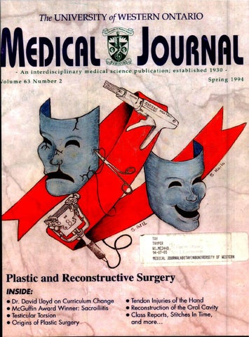
COVER ART: Depic ts the traditional ma s ks of Greek drama representing tragedy and comedy. The tragic mask suffers from sub orbi tal, nasal, mandibular and frontal bone fractures as well as a cleft lip, while the comic ma s k is in tact. Between these are, top to bottom, an Auto Suture Multifi r e Premium disposable skin s tapler, a Gilles eedle Driver, and Brown Dermatome used in plastic surgery.
A ssistant Editors Contributing Editors Elin Ringstrom, Meds '96 Jay athanson, Meds '96 Sonny Bhalla, Meds '97 Susannah Huh, Med s '97 Jaqueline Pradko, Meds '97 Copy Editors Priya Chopra, Meds'95 Cindy Hawkins, Meds '96 Maureen Gottsman, Meds '97 ina Singh, Meds '97
Advertising View An Ad Ming-Ka Chan, Meds '95
A rtwork Joe Kim, Meds '96v Mark Bernstein, Meds '96 Romy Croitoru, Meds '97 Sudeep Gill, Meds '97 Sanjeev Kaila, Meds '97
Class Representatives Justin Amann, Meds '94 James Me abb, Meds '95 Peter Swedko, Meds '96 Sonny Bhalla & Susannah Huh, Meds '97
~
Printer Willow Press Limited
Denotes Director
ARTICLES ........ Sept. 26, 1994
Sudeep Gill, Meds'97 Sanjeev Kaila, Meds'97
••••••••••••••••••••••••••••••••••••••••••••• UWO MEDICAL JOURNAL ADVISORY COUNCIL Jeffrey Politsky, Editor-In-Chief Anne Silas, Associate Editor Ro s Mantle, Associate Editor Jeff Kolbasnik, Exec. V.P. Hippocratic Council Harsh Hundal, President Hippocratic Council2 Dr. J. Silcox, Assistant Dean, Student & Faculty Affairs3 Dr. J. Howard, Faculty Liason Hippocratic Council• Dr. M. Inwood 5 Dr. . Muirhead6 Dr. R. McMurtry, Dean of Medicine2.7 ' Chairperson, Ex Officio Member, 1 Department of Obstetrics and Gynecology, St. Joseph's Health Centre, • Department of Gastroenterology, Victoria Hospital, 5 Department of Haema tology and Oncology, St. Joseph's Health Centre, • Department of ephrology, University Hospital, 7 Department of Orthopaedic Surgery, St. Joseph's Health Centre . 2
ALL CORRESPONDENCE regarding Journa l content MUST be sent to the Editor of the Journa l (N OT to members of th e Advisory Council) . Letters to the Editor will be published and edited at the discretion of the Editor. Th e Advisory Council was created to assist managerial & business aspects of UWO Medical Journa l operations . THE ADVISO RY COUNCIL HAS NO ROLE REGARDING CONTENT. A ll material published in the Journal reflects solely the views and opinions of the authors of the material printed and not necessarily the editorial staff or the Advisory Council of the Journal.
••••••••••••••••••••••••••••••••••••••••••••• U. W.O. Medica/Journal 63 (2) 1994- - - - - - - - - - - - - - - - - - - - - - - - 59
GUIDELINES TO AUTHORS The purpose of the U.W.O . Medical Journal is to provide a single forum for original articles based on clinical or research medicine of topical or historical relevance . Since the readership of the Jo u rnal is interdisciplinary, articles published will attempt to reflect the wide range of medical disc iplines. Informal peer review is required, i.e. non-specialist authors are encouraged to collaborate with or at minimum have their work reviewed by a specialist in the field . This individual, if not a co-author, is to be acknowledged at the end of the paper. Short biographical notes on the authors are to be included at the beginning of each paper. Affiliation with UWO is not a prerequisite for authorship. Submissions are to include three copies double spaced and the full text on 3.5" computer diskette in Microsoft Word (Macintosh) or Word Perfect (IBM) format. Journal staff artists are pleased to assist with pictorials, which are normally printed in black and white. Submissions and disks become the property of the Journal. The Jo urnal reserves the right to correct grammatical and stylistic errors. References are indicated numerically in the textâ&#x20AC;˘ and lis ted as endnotes in order of appea r ance.2 Punctuation comes before reference numbers and sentences are separated by one space only. Examples of Journal reference format follow: J, Thomas 5, jan MA. Clinical value of polysomnography. Lancet 1992; 339(2):347-50. or 2. Dement WC, Ca rskadon MA , Richardson G . Excessive daytime sleepiness in the sleep apnea syndrome. In: Guilleminault C, Dement WC, eds. Sleep apnea syndromes. ew York: Alan R Liss, 1978:23-46. 1. Douglas
Please direct s ubmissions, including re turn ad dress, pho ne and fa x number, to: UWO Medica l journal, Health Sciences Big., University of Western Ontario, London, Ontario N6A SCI. Tel: (519) 661-2076 Fax: (519) 661-3797
or Ross Mantle, Editor, Fax. (519) 434 9199 Tel. (519) 434 2299
The U. W.O. Medical Journal is an interdisciplinary medical science publication, established in 1930. The Journal is published three times each academic year: Fall, Winter, & Spring. Subscription is $17.00 per year. Š All material published in the U. W.O. Medical Journal is copywright protected-no section of the U. W.O. M edical Journal may be reproduced witho u t the expressed written permission of the editorial board of the Journal. 60
Dr. Jolm Mount L. J. Sandy Wetstein . B A.. CA Chief of Psychiatry. Parmer. St. Joseph's Health Cemre KPMG Peat Marwick Thome President. London Academy of Medicine
"Practicing medicine in the '90s require~ that we pay much more attention to practice and personal finances. Sandy and his colleagues at KPMG know our concerns well. In addition to practice accounting and tax matters, they help me with with overall financial lifestyle and retirement planning." - Dr. John Mount
1400 - 130 Dufferin Avenue , London (519) 672-4880
- - - - - - - - - - - - - - - - - - - - - - - - U.W.O. Medica/Journal 63 (2) 1994
CONTENTS EDITORIALS ··· ················· ······· ··· ························ ··········· ·· ········· ······ ··········· 63
FACULTY NEWS Deans' Corner ... ... ...... ..................... ..................... ..... ... ..... ..... ..... ...... 67 Class Reports .... .. ................ ... ................. ............. ................ ...... ...... .... 68
ARTICLES Testicular Torsion (P. 74)
Interview with Dr. Lloyd on the new curriculum proposal Jay Nathanson .......... .... ................................................................ 72 A Brief Review of Testicular Torsion Marc Anthony Fischer and Dr. J.D . Denstedt ................ ............. 74 Is there an Obligation to Treat Patients with HIV? Jay Nathanson ..... ....... ........... .......... ............. ... .............. ...... ........ 77 Detecting Early Sacroiliitis: Babak Raissi and Dr. L. Thain ........................................... ... .. .......79 IFIEA1I1UIRUE §IEC'II'IION g
PLASTIC AND RECONSTRUCTIVE SURGERY 8
Detecting Sacroiliitis (P. 79)
Origins of Plastic Surgery Mark P. Bernstein and Dr. C. Scilley .... ............ ...... .... ...... ...... ....... 85 Breast Implants, Public Dilemma Dr. J. Mohammad and Dr. L.N. Hurst ..........................................87 Pressure Ulcers: A review Sonny Bhalla and Dr. R. W. Teasell ...... .......... ........ ............. .......... 90 Hand Fractures Dr. Rizwan A . Mian and Irfan A. Mian ......... .............................. 94 Tendon Injuries of the Hand Dr. Rizwan A . Mian .................. ...... ............. ... ............................ 97 Reconstruction of the Oral Cavity following Surgery for Squamous Cell Carcinoma Mark Taylor and Dr. T. W. Matthews ............... ............................ 99
Feature Section: Plastic and Recon structive Su rgery (P. 84)
!!MEDICAL HUMOUR!! Stitches in Time ........ ..... ...... .. ............. ... .. ................................ .......... 103 Dr. Jason Bowels ....... ............................... .......................... ............... 109
PROBLEM SOLVING Thinking on your Feet ........... .................. ......................... ................ 106 Medical Vocabulary ... ..... ............................. ..................... .. .............. 107 U. W.O. Medica/Journal 63
(2)1994------------------------
61
Once-a-day
PRESCRIBING INFORMATION •CAADIZE/(CD Onc....-.7 Conuohd DtiYtryc.p..IH 120~~~&o 180 "''o HO "lind JOO "''o
THERAPEUTIC CLASSIFICATION Antihyptmnsi¥t ind Antianpal
""'t
INDICATIONS AND CLINICAL USE AloGuoA I. CARDIZ£/1 CD is indiattd lor lilt...,..._. ol'"'- subltanpno (tflon-assoaattdanpno) -t'lidtnctoi"SSO9""' ill potitnts w!lo ,......,IJII19!DI:Ilti< d«pitt ~tt doses ol bt11-blocitn ondlor orpvc nitntts or w!lo annot U>ltntt mo...,...._ CARDIZ£/1 CD 11117 bt llitd ill combiNtion wi1!l bt!l-blocl.tn ill ct.onic sable anpn. pocitnn wi1!l normal otnerintor function. Whtn sudl concomiant thtnj>y is illtroduct<, potitnu- bt monitored dostly (Sot WARNINGS).
-
!ii:lct lilt ..r.ty ind lffiocy ol CD apsulos ill lilt """"'"'''" ol unsablt or ruosputic """" hu not bttn submntattd, "" o1 dlis formuboon lor tfltst indialiom • not rtCOIIIIIItlldtd.
CARDIZ£/1 CD • rndiattd lor lilt trotmtnt olll1ild to modtrm t11t11ua1 hyptmnsooo. CARDIZ£/1 CD silou~ nonno1y bt ustd ill mo.. patients in whom uutmer:t with diurtoo or btu~m tw bten ineffecti'f't. or has bten wotiattcl with unacctpaMe ad.ent tfftru. CAROfZEH CO an be Wd u an initialqtnt in those ~titnu in whom the use ol diuretics lndlor bfu.bJocien is contl"aannfiated. or in potitnn wi1!l medial concitions ill which tfltst dnip &tqUtlllly """ strious adotnt tlf«U. s.m., ol (()IICIIITtl'< "" ol CARDIZ£/1 CD wi1!l odltrii!Ci!yptrunsm ~ hu "" bttn tmbishtd.
CONTRAINDICATIONS Dilowm HClo contnlldiattd: I. In potitnts wi1!l sidt...,. syndromt ""'~" ill lilt prtstnCt ola functionin& ""tricubr poctnuktr; t In pocitnn with stecnd or thirO dqrtt AV blod:
l. In poOtntS With known hyptntnsi<Mty to lfrlliutm; 4. lnpotitntswil!lsmnhypo"""""(lessthan !O.,.fl&systoic:~ s. In ~ inbmion potitnts. ......... loft - b r blurt llllllifesttd ..,. pulrno<wy CO!IItstion: 6. In prtiiWICJ ind ill ....... ol ~ pottntial WARNINGS Ct.ltuc CC101DtJCT100 Oitimo! prolonp AV nodt rt!nctory ponod> wisilout .p1ic>r>t1y prolonp!& 1111U1 nodt rtC""'J txCI\'C m potitocs With srdt synd,_ This tlftct ""7 l1ltly rtsuft m abnormaly slow hwt rttts (9miarbrly • potitnts wi1!l sidt ..., synd"""') or steon6- or thir<dqrtt AV blod (6 o/1206 potitnts or OJI~ Fint dqrtt AV bloci wu obstmd ill SJI ol potitnn rKfim& c.uotlE11 CD (Itt ADVW£ REACTIONS~ Concomiwlt vse of cfikiu:em wid! btu.blod:m or ditftllis m~y mutt in Jdditi¥t effects on ardi:ac concfuction. CC*GHTM Hwr F.u.UII 8taust hu a otpti<t 1IIOUOpK tlftct • .., ind n a!tca ardiac c~ lilt ""'& shaaW only bt UStd """""""" ond undtr art!lllllltdialsupor-mioo • pocitnn With C<><psti¥t Mile bart (1tt also CONMNDICATlONS).
U.. wmr I<T•-to.oaos 1M combN.tion d diltiuem and btta·b6ocbn WV'TWS aution WICt 11 some pa:tients ldditm effects on hwt rttt, AV conduction, blood prtUUrt"' loft - - b r function ..... b t t n - Clost medial suptrrision is rtCOIIIII!tlldtd. Gtntnly, diltiut111 shaaW ""bt ;.on to poritnn wi1!l ~loft ...aiarbr llnction while thoy rocliot bta-blocl.tn. - · i l l txetpoonal CUtS whtn. •lilt opilion ollllt ~ concomiant"" is considtrtd sudl ust shaaW bt llll<iluttd pluoiJ iltloospiai"""'OikiutmJi'rtsno pro<Ktion apimt llltdJn&tn ol tbnrpt bta-blocl.orwithdrtWJI ind sudl withdrtWJI- bt dont bflllt p!utirtduction ollllt dost ol bt!l-blocl.tr. HYromtsooo Since ditiuem en ptripfttnl YUOibr rHisWKt. dtcruses tn b6ood premrre IN.f ocasionlly rtSUk in S)'l'l9tormtit hypottnsoL tn ~tienu wi111ll1pll or trrl!ythmils M& ~ dnrp.lllt additiontl hypotfii!M tlftct ol diltiutm shaaW bt t>ktn ioto cOIISIIItntion.
......m.
Aarn Hu•nc bQuoT In rtrt iomncts. .pianttlmliom ill...._ phosphttm. CPillDH. SGOT. 5GPT ind .-,mptoms coosisttnt wil!IJCW ht9J!ic qury 11m bttn obstrotd. That rucliom ht,. bttn , _ - disconcinuttion ol d"'l thtnj>y. Althoup 1 Cllllll rtltlionship to dilou"" hu not bttn ts!lblishtd ill aJ asts. 1 dni& ilductd hyptntnsi!mcy ro- is sul9fCltd (1tt ADVW£ REA~ As wi1!1 "'' dnr& &Mn "'" prolonrtd ponods.labontory portmtttn silou~ bt moMortd It "'"br illttmls.
PRECAUTIONS lttrAE> HftATIC OIRaiAI. fvNcnoo 8taust liltiwalo txttnsi'ftJy 01ttlbobtd bflllt or ind t><Citttd bflllt bdnfy ind ill bilt. roonitorio& ollabontory pmmtttn ind all!ious '""'&• ti<rttion '" rK..-ndtd ill poOtnn wi1!l implirtd htplti< or rtnalluncnon (Itt AOVW£ REACTIONS). l'moATIICIJR Tht safHJ oldiciut111 ill dlilc!rtn hu noc ytt bttn tmbislltd.
-KoTMEIS
Ditiuem has- reporud to be txcrtttd 1ft lwman- One rl90't suuem dg[ (OI'(entt1tions .. brtut l'lill'l'll7 approxiNtt wum Wttk. Since dilliwrl..r.ty • newborns hu not bttn tsublisl!td. n ~ not bt I""' to nunin& mothtn. U.. orlltf ElDuLt Administrttion ol dillimm to tidtrty pocitnn ("" "' tqUJI to 65 ol 'l•) roquins aution. Tht inciCtnet oltrlttnt rtXlions is tpprox> mtttly Ill hqtltr ill dlis croup. Those trlttnt rucliom which occur - • fr~ indudt: ptriphtnl tdtmt, brtdyara. ptlpintion. dizmm. mil ind polyurit. Thtnlort. porticubr art ill ti<rttion o t6ristblt(1tt DOSAGEAND ADI1INim.ATION). DluG INTIIIACl10MS Di&iUJU: Dillimm ind diplis &IJcosidts ""7 ht" tlftct ill prolonp!& AV conduction. In <inial triJis. COIIC1Ifrtllt -trttion ol ditiutm ind di&oxil ht" rtSUittd ill incrtuts ill strum di&oxil lt"ls wi1!l prolonption ol AVconduction. This incrtut 1117 rtSUft froao 1 dKIWt ill rtnal dwtnct ollf~o · Potitnts on concorlliant tl>tnj>y, tsptdaly mo.. wi1!l rtllll irnplinntnl. silou~ bt an!uly - t d . Tht dost ol cf&oxio .., nttd downward tdjustllltlll lta-lllod<tn: Tht concOIIiant admioistntion ol di · wi1!l bt!1 adr""'li< blodinz dnrp womms aution ind ~ IIIOIIitorq. 5udl 111 wociation mty ht" lll .Cdici'f tlftct M hwt rttt. M AV ccnd:Ktion or on blood prtssurt. (Sot WARN GS.) Apprvpriltt....,. adjust"""" ""7 bt ntct!WJ. AI1Udy ill fi'f normal subjtcu sllowtd thtt diltimno incrmtd proprtno1o1 biotnilbi!y bf tppt0X>1111tt1y 501. Short ..Olortc-acti•c Nitrates: Oitiu ""7 bt SJftly co-tdmioisttrtd With nitntts. but lfltrt ht" bttn l<w controltd stu<!its to ..W.tt lilt ~inti tlftceittntu ol dlis combiNtion. Other Calcium Antaronists: Umittd dinial uperitnct wzttsa dlaE in ctrUin stYert conditions not~ adequatefy to .,enpamif or to Aiftdipont. """'dikiutm ill wi1!l tichtr ol tfltst ""'" ""1 bt btntfocial.
rws
tn-
c...,.._
ADVERSE REACTIONS AloGuoA Tht safHJ ol CARDIZEH CD. admonisttrtd tt l01ts up to 360 me a dty, wu mlutttd ill 365 pocitnn wi1!l ct.onic sublt • trt1ttd ill controlod ind ~ clnial triJ1s. Adftnt ...- - • rfi'OI'Ud ill 21.1% ol potit<ts, ind rtqtlirtd discontinultion ~ Ut. rJ pocitna. Tht 11101tc..,_ art.tnt tf!tcurtpotttd wttt:fint dqrtt AV blod (SA~ dittrltss (3.111~ hudadlt (lOS). uthtnit (2.71). brtdyatdit (lSI), ind """" ptctoris (1.61). Tht folowr( ptretntllt ol art.tnt tffocts. dMdtd ..,. IJI!tlll. .... rtpO<U¢ Crionsaolv. Fnt dqrtt AV blod (SA~ brtdyarOG (lSI~ anpno ptctoris (1.61~ ptriphtnl tdtmt (1.4~ ptlpinliom (1.11~ llld •tntricullrtxtrlSJ!tolts(OA~
Ctntnl NomMis SJSttm: Oinintu (3.01). httdtdlt (3.01~ uthtnit (2.71~ insotMit(l.ll~ ntrY0U111t1S (0.81~
Gutroiattsliul: N>~~~tt(L41~ lfwr!ltt (OJI). Dt,..tolocial: Rull (0.81). Othtr: Amblyopa (OJI~ Tht folowin& addiciontl art.tnO tlfKn ht" ocO>rTtd wi1!l tn oncidtnct ol ltss than OJI ill <inial trills: bundlt brtndo blod. Ytntricubr tldoyardia. ECG tbnornlllity,suprt•tntricubr txtrlSJ!tolts. chtst pUo. syncope. postllrtl hypostnsion. portsthtsit. """"'· dtprts"""- mtnal conlusioo, impoctn<~ tbdoloinll pUo. constiptaon. Gl . tpiswis.I'IIChtl riplicy. m)'llcQ.
-
AWHJ mlacion wu arritd out ill controltd swdits ill 378 hyptrttnsiYt politnn tmttd wi1!l CARDIZ£/1 CD tt doses to 360 me a clay. Adftnt tlftcts wm rtpottld ill 30.71 ol potitnts ind roquirtd d'sconcinuttion ol thtnj>y ill WI. Tht _, c"'""'"' trlttnt tl!tcn wort: httdtcho (8.71~ tdtmt (4.01~ bndycria (3.71~ ofrnintss (l.l%), ECG abnonntity (2.91~ uthtnit (liS) ind lint dqrtt AVb1oci (liS). Tht folowinc ptretn"'t olarlttnt tlfKts. of!Yidtd by IJI!tlll, wu rtpotttd: Ciltdiorucular: Edtmt ptriphtnl (4.01~ brtdyardit (l.ll). ECG tbnormUtits (2.91~ lint dqrtt AV blod (liS~ arrioytllnul(l.il~ Y2SOdiotion {IM!Irnc) ( 1.61~ lu!dlt brtndo blod (0.81~ criomtply (OJ I~ hypott<sion (OJI). Ct otnl Nmovs Systtm: Httdtcho (8.71~ cinintss (3.41~ ut11tnrt (2.6~ somnoltnct ( 1.11~ "'"""""" (1.1 ~~ GutTOiotosliul: Coroapttion (1.11~ dysptpot (1.11~ diarrtota (0.61). l.obor>tlll)' Tosts: IGPT incrust (0.81~ Otll<r: l.tvkoptnll(l.l ~~ nocnrriJ (OJI). Tht folowin& tdditiontl art.tnO tlfKn ht" ocewrtd wi1!1 tn incidtnct ol ltss than OJI • <inial tntls: systDic: ""'"""· supr>Ytntncubr txtruystolts. rnqr.int. tldoyardia. inctrutd tpptCitt. incrtut • wfllht.lhminunt. bilirvbontmio. hyptnonc....._ thnt irlsomnG. '""~" ...,.... pnrricus. rts11. inctrutd ptnpimion. polyuria. tmblyopit.- ind tlmliom ~ uotint kintst.....,. phosphttast. ind SGOT.
OVERAU CARDIZEM SAFETY PROFILE In <inial trills ol CARDIZEH abita. CARDIZ£/1 SA apwlts ind CARDIZ£11 CD apsu1ts iroYo1vinJ "'" 3JOO poutnts. lilt most common art.tnt mctions wort httcbdlt (4.61~ tdtmt (4.61~ cinintss (JJI~ uthtnit (2.71~ &nt-dtcm AV blod (2.41~ brtdyantia (1.71~ flllhonc (I J~ """' (1 .41~ mil (I.JS~ ind CJs9t9sil (I.~!~). In addition, lilt folowin& ....a -• rfi'OI'Ud wil!lt lroqutnCJ olltss than l.o:l Ciltdionscvlar: ~ trrhythmit.lu!dlt brt.'ldl blod. t>dlyar{ra. ""tricubr txtruysto1ts. c..,..... hwt blur~ syncope. AV bloci (1Kon6- or thir<-dqrtt~ hypottnsion. ECG tbnonnt1iats. Nt"ovs Systtm: AnlntsiJ. dtprtllion. pit abnormality. """""""' somnoltnc~ .....,.tions. ptrtstfotsQ. ptnOIIlity '"""'- tlllllltVI. trtmor. 1bnornW drums. insomnil. GutTOinttsliul: Anoruia. ditrrlota. dfs,....._ mild tlmliom oiSGOT. IGPT.LDH. ind W1int phospllt111t (!tt WARNINGS). """""&· wtqht incrrue., thnt. c~uon. o.....coJocial: Ptttd>ra~ pn>ritus. p~>otostnsrtmt-. Othtr: Amb/yopG. CPI( incrwt. dyspnt.a. tpoStWS. fJt l'rilltion. byptrJiyc"""- ltxUll dilliarlots. lllSll IIOCtllnl. osttoartocubr .,.... iropottne~ drf mouth. polyurit. hJiltnlnC ..... Tht lolowinc postmtrl:ttinc t¥tnn ht" bttn rtpottld infrtqutndy ill pocitnn rtctiYil& c.uDtZEI1 aloptcia. trythtrnl multiformo. txfoliotJYt dtnnttitis. txtrlpJrtlllidtJ IJI'P(OIIII. Cinli'rtl hypt<pluil. htmolycic llltllliJ, dttxhtd rteinJ. incrwtd blttd<1c tint.ltukoptnra. purporrt. rt11110potloy, ind ~In tddition."""' sudlu ~~~~ iobrction ht¥t bttn obstmd which lrt not rodily lfisuncuoshtblt froao lilt lllwnl histotyolllltdiswtill tfltst plt>tll<l. Aruoborolwti-Oocwntnttd CUtS oi,....,UZtd rtsll, chtrtcterutdultukOCJtoCiutx ruarlitis. ht" bttn roporud. Howt¥tr, • dtfioitiYt aust ind tlftct rtblionship bttwttn tfltst t¥tnn llld CARDIZ£11 thtnj>y is ytt to bt tst>blishtd.
"""oons.
C""'""""
SYMPTOMS AND TREATMENT OF OVERDOSAGE ~ wi1!l ..... diltiutm hu bttn oOstntd ill ' ""'- Eicfot (8) politnn rteOYtrtd wisilout stqutbt "'" l .... dtys. Ont pot>tnt wt1o had irltsttd tn rinown liiiOUIIt o1 ciltimno. tolmmidt ind alcollol t>q>tritnetd • &al ardiac trmt Doses inctsttd rtr:ctd froao IJ to IOJ 1'11111. Bndyarofra. AV bloci ind hJpoctnsion wort nottd i> most pocitna. In lilt ..... ol ~ "'watrtttd rtsponst.tpprq>riltt supporti¥t rnruurts- bt tmploftd • addition to pstnC ..,.. Tht folowinc mwurts ""7 bt coosidtrtd: kADY<AaA
Administtr •tropnt. f lfltrt • no._. to npl blodtdt. tdmmttr -ottrtnol auOOII!Iy. HIGHDfGI<E AVIIloa Trt.1C as lor bndycria tbo¥t. FU.td hqh dqrtt AV blod shaaW bt trottd With ardiac pocq. CAiooAc f ALUIIIE Adroiroisttrioo""9i<•ltnnfosoprottrtnol.do9tlllint or~) ind diurttics. HYPO,._ Vasopmson (f.l. cloptnon "' lmrttrtnol biwtrttt~ Aautl erta!llltllt llld ....,. silould dtptnd on lilt lt'ltricy ollllt dirooaJ situttion.
DOSAGE AND ADMINISTRATION ......... DosJ&tsfor lilt trt1tmtnt ol"""' shaaW bt tdjusttd to odl pol>tllt's nttds, Swtin& wil!la dolt o/120 "'to 180 "'1 OIICt daily. lndMdutl pocitnn mlJ rtspcnd to hicfotr doses ol "9 to 360 "'1 OIICt dtily. Whtn IIKt!WJ, titrtrion shaaW bt amtd 011t Oftr l 7 to 14 dty ponod. Politnn controltd ondiltiutlll tlont or il combiNtion with odltr mtdialiom may bt SJftly swnchtd to CARDIZEH CD apsu1os u lilt"'""' oquinltnt toal cbiy dose. 5ubstqutnt ticrttion to hicfotr"' lowtr dolts ..., bt I!Kt!WJ ind shaaW bt inioattd .. dinraly wm>nttd. Thtrt is liroittd t>q>tritnet wi1!l doses Jbcm 360 "''o .......... lilt incidtnct ol art.tn0 mctions incrtuts ulllt dost rncrwtS wi1!l &nt dtzrtt AV
blod. ond HYPEITEMIOII
brtdyardia boom& lilt "'""'"t rtltlionship to dose. Thmf«~ dolts '"'"' than 360 me "' not rtCOIIIII!tlldtd.
O...,t bt iocliYidllllttd dtptndirl on potitnt's toltrtnct ind rtsponliYtntU to CARDIZ£/1 CD apsu1ts Whtn ustd u monothtltpJ. usv>l stJnio& doses lrt 180 to 2401111 OIICt cbiy, tltfooop 10111t pocitnn OllJ rtspcnd to 120"' OIICt cbily. 11uirnuml11Cihypttttnsr tlftct is......, obstmci Wr JWOiimttly l to 4 Wtfb olditrtpy: thtrtfort.....,. ~ shaaW bt sdltdu!td KCordioctJ. Tht usv>l....,. """ studied ill <inial trills ... 240 to 360 me onct cbiy. A........, cbiy dost o/360 "'onct cbiy shaaW no< bt txettdtd. Tht '""'&• ol CAADIZEH CD or concom.-a nt tntihyptrUII!iYt 'l"'U mty nttd to bt J<1usttd whtn tddi:o& ont to lilt odltr. Itt WARNINGS ind PRECAliTIONS ""'""' ... wi1!l bt!l-blocl.tn. Hyptrunsift po<itnn conuohd on CARDIZ£/1 SA t1ont "' ill c....,...tion wi1!l odltrllltil!yptrunsit 'ltnn ""7 bt saftly swnchtd to CARDIZ£/1 CD It lilt """ toa1 cbily dose. s.>stqutnC ticrttion to hiptr or lower dolts 1117 bt ""t!WJ and shaaW bt inicilttd u dinrally WJml\ttd.
CAAOIZEH CD apsulos shaaW not bt chtwtd "' crvshtd.
AVAILABILITY CARDIZ£/1 CD 120 me apsulos '" Rljllllitd ill boa1ts o/100. bdlipt ........ bUt apsult • wi1!1 CAAOIZEH CD 120 "''· CARDIZ£/1 CD 180 me apsulos "' Oljlplitd ill boalts o/100. bdlli&fot blutlli&fot nrr~ bUt ~ • onpmttd With CARDIZ£/1 CD 180 "'· c.uDtZEI1 CD 240 1111 apsulos"' Rljllllitd ill boalts o/100. E>dl t blutlli&fot bUt apoolt is imj>rinttd wi1!1 CARDIZEH CD 240 "''· CAAOIZEH CD apsulos"' suppitd ill boalts o/100. bch li&fot blutlicf>t 1"7 apsult is inpriottd wi1!l CARDIZ£/1 CD JOO "''·
"'""ltd
J00"''
Productl1orqrtplt- .. rtqutst ~~~~is 1 rqistertd trtdtnwlt oll1trion l1trrtl Dow Inc.. UJ.A. O RDlC lABORATORIES ---·~ w, ~.~ .......... o-c....~o
MeMBI'R
I PM8 I I PMAC I C AA 9l0S o4 8 - E
POSTCARD FROM VANCOUVER: THE RECONSTRUCTION OF THE UWO MEDICAL JOURNAL ay 17, 1994: I arrived in Vancouver just two days ago. Six action-packed days have passed since the MCQQE-I's ended. The task I now have at hand is the acquisition of a dwelling (quite a task here). Since my forays began, my feelings for this city have intensified- this of Canada is simply spectacular. I've chosen to write my final editorial, as Ed In-Chief of the UW edical Journal, on the recent ction,' of the Medical ournal. given the featur this issue. I've ch se orne rather gather my thoug},lts-Eng ·sh Bay, Stanley Park. In I've even h time, by means of a complex to predict that the ronto Maple Leafs will inate the Canucks from the playoffs in six gaJ;rf~f" In 1990-91, and Shirley opportunity to oec: m1fe and I had previo altering the Journa One of the first
M
former Ed.-In-Chief) describing his pleasure with the changes. We met with Dr. Inwood several times to discuss various Journal issues of consistency, finance, and faculty involvement. Over the year, we worked at enticing contributors, creating controversy, and proving appeal. More significantly, however, ("I.Q.,...~,.. ans had been laid to improve faculty lv~~~...._e~;pe~ci·c:1lly with the Dean. The change red to be well-timed with the . Shirley, Dr. Inwood/ and I extensively over holw to · two routes ultimately suppor~ 'through th_~_/.....--7 1 ent-faculty ccnincil (witlll1i.e ent being a C9'flsistent base to rapidly overtur . undergraduate a d a strong sub · ·on drive. 1 did not end in the infrastru lt
t.
umerous as the creation department ournal eplete tit was to single Editor-Inmedi -le al that brought to the 'fore a latent, but longa bund n ver the ing, rivalry between the two most powerful persp five , largely cal undergraduate student organizations the cod liver oil fortification). """"'"'"''" ~,l._'"'lrlfedical Journal and the Hippocratic Council. This regular part of the Journal after rivalry had recently been quite repressed since the o, of publication. The often referred-to Journal was more weak than it was powerful. the Journal occurred in the mid-late 19 However, a rapidly developing Journal with lasted until Shirley and I decided to return editorials and articles relating sensitive issues raised Journa l to its former quality. Based on Journal models the hackles of certain Council members (including like Brain Research and the original size of the UWO the pre ident, vice-president, and other outspoken Med. }., Shirley and I chose to change the size of the members). Suggestions such as censorship of my Journal to 7 x 10 and publish each issue with a editor ials, a strong resistance to hierarchical feature section to provide a focus. Soon after restructuring, and even a dispute over the use of the publication of that first "new" issue, Shirley and I term "Editor-In-Chief," became the order of the day received a letter from Martin Inwood (Meds '69, U. W.O. Medica/Journal 63 (2) 1994- - - - - - - - - - - - - - - - - - - - - - - - -
63
Editorials [only one person suggested censorship, although improved Faculty relations and support led both I'm sure other members were anxious, after I had the Journal Editorial Board and the Hippocratic written two consecutive editorials, one on the utility Council to independently strive for Journal-Council of didactic instruction, and the other on the pass-fail separation. grading system and the attempt to blunt In all likelihood, though not definitive at this competition]. Despite the turbulence, however, the time, a recommendation will be made for the Journal carried on: the Council permitted the Journal to establish formal ties with the Faculty of Journal to create an Advisory Council, and to alter Medicine and to relinquish, slowly or suddenly, its the design of the Editorial Board, with the condition ties with the Hippocratic Council. Although of approval from informal peer review every four operating closely with the Faculty rather than with months for one year (of all of my proposa only 'Hippo' will initially be taxing and very much a payment of the Journal staff was rejected). process of touch and feel between the two historic By August of 1992, a UWO M edica l oi:Et:int:rJF-...'-':Nl'r~'"•"nt tions, the ultimate potential of the Journal Advisory Counci was merely 'signatures' munication medium of UWO and the being realize . t about the same t ime, a community will be tapped only guaranteed a e~f ing contract had b arran the _Faculty. of Medigne, and providing e ou funds to p sh wo Hippocratic Council.1 quality publidtions ··year. oducing issues he Journal is ready (and each year, as had been done previous1 , was no a of publicapon. And in it3longer a po 'lity. As e l, mark ting and s post-b?~' the Jo.JJ-¥-nctrls . advertising I had done, suggeste . a change ts it never ha ~ef~re. Three yea rs form the useJof colo¥~ to devote my .~ e to improve the to an 8.5 x 11 covers. On cha ges, ouriial level compjltal>le o the best such acquired eds '95) as .. Canada. Three later, I'm more Editor (it year,•when 'th__~....result d :very grateful for Meds '94, CoI will m1ss the actlion, tp.e .t urmoil, the Journal thrill, but right now I' m · V ~~;oliver and Joe Kim (Me on't permit excessive· s timentality. I have ste f a d· variety to climb. end of Edit rial, have taken the 1 certain people. 'm npt going to hold time-that's for(sur ! ·s issue is once Be to re a interview with nge (a necessar y ntle, Anne Silas, (Meds '94), Glen and Jeff Mount (ptoduction rna excellence), Dr. Martin w'....- "" . . . wood (former editor and great inspiration), Dr. Dr. Howard, Dr. Muirhead, Dr. McMurtry, Despite financial -=--..--·.- ·-· tc~~-- and my fellow Hippocratic Council members. I also thank all the current Journal staff and anyone else Editorial Board managed so who has contributed through a cartoon, drawing, or that not only has its debt been repaid article. Thank you to all of the Journal subscribers, surplus of funds for important equipment patrons, and advertisers. I especiall y thank the created as well. As a direct result of this reader-for appropriately showing interest in a related fiasco, the Hippocratic Council undertook to worthy publication. Best wishes to my classmates; form a task force to make recommend a tions best wishes to the Journal; and to Ross-just do it regarding the governance of the Journal. With an annual operating cost equalling or exceeding the right. C' est complet. Hippocratic Council budget, the Journal became a Jeffrey 'Politsky, .Jvfeds '94 liability that the Council was unable (and perhaps rt.ditor-1 n-Cfiief unprepared) to underwrite . As well, conflicting views of the Journal's future, coupled with greatly
e
64
- - - - - - - - - - - - - - - - - - - - - - - - U. W.O. Medical Journal 63 (2) 1994
Ed i to ri a l s
IT HASN'T SUNK IN YET â&#x20AC;˘..â&#x20AC;˘ I believe most of my Meds '94 colleagues would support the premise that this fourth and final year of med school has flown by. What happened? Electives, ix weeks in the classroom ... whoosh ...and all of a sudden the LMCC' s were a part of history. In retrospect, I feel my greatest achievement this year has been the successful completion of the CIMS ma tch. Although many people were eager to give ad vice and preparatory aid, no one was able to ad equatel y warn against the grueling pressure, stre s, and work involved in the entire process. The fin a lity of the outcome of this ordeal and the premature stage at which we had to make lifelong career choices, formed a solid background of stre tha t never waned. Remember your personal statements? I am proud that I managed to create such open-ended drivel about my future. How many theories exist to explain the manner in which the programs make
their short lists and final decisions? So many of us suffered in some form at the hands of "52-pickup" decision-making. Our pocketbooks felt the brunt of infl e xible programs and conflicting interview schedules. My phone and credit card bills will never again see such glory! Such is my brief review of the pitfalls of the CIMS match. I consider myself luckyI am happy with the outcome. But I am also well aware of the fact that I will never again have to repeat this task- whew!!?! Finally, saying goodbye. This is going to be harder than I thought. I wish good luck to all of my classmates as we embark on the next step of our ca r eers . Jeff-every body knows! Ross-I'm confident that the Journal will continue to flourish in your energetic hands.
Yfnne Silas , M eds '94 associate rrdztor
NAOMI WOLF AND MALE AESTHETIC SURGERY I was surprised to learn of the strong distaste many local plastic surgeons have for a number of ne w er procedures conceived for male cosmesis including pectoral implants and penile lengthening ( ee Letters to the Eaitor) . These, it apfears, are considered "fringe" medicine (a form o "treating people' s neuroses with a scalpel" ) and may cast professional doubt on the practitioners who would perform them. My first reaction to this attitude was to view it as a hold-over of paternalistic traditions wherein a very important asset of the female is her appearance, w hereas males who would worry about such matters are diminished in the eyes of their male and female peers. It seems unfair that the surgical alteration of the female form should be accepted as appropriate and of great psychological value, while developments in analogous procedures for the male are suppressed. Many self-described feminist writers, however, do not support this interpretation. Rather than confer upon the gander what is ostensibly good for the goose, they argue that women are victimized by unhealthy forces in society which drive them to seek unnecessary surgical intervention. According to aomi Wolf, author of The Beauty Myth, "The surgeons are playing on the [beauty] myth's double standard for the function of the body. A man's thigh is for walking, but a woman's is for walking and looking 'beautiful.' If women can walk but believe o ur limbs look wrong ... we feel as genuinel y d eformed and disabled as the unwilling Victorian h y pochondriac felt ill. " Wolf goes on to quote Hippocrates, resolutions arising from the Nuremburg trials, and the Declaration of Geneva in an attempt to show that urgery for aesthetic reasons violates generally recognized tenets of medical ethics. 1 In this
feminist view, aesthetic surgery should be suppressed in both males and females. What of the surgeons? Wolf's assignment of blame to plastic surgeons intent on profit does not appear to be generalizable. Indeed, cynicism on the part of plastic and other surgeons regarding purely aesthetic procedures is widespread. 2 Is this negative attitude justifiable? Schain reports in Cancer that "For years, it was assumed that women who ought breast reconstructive surgery were less able to cope with an altered body image than their mastectomy counterparts who did not seek out this procedure." However, a number of studies reviewed by the author concluded that women seeking breast reconstruction "were exhibiting evidence of positive coping and assertive problem solving, not neurotic compensation for an unresolved narcissistic injury." 3 Perhaps this result is attributable to the fact that these studies looked at procedures done to restore normal appearance rather than surpass it. Nevertheless, I w onder whether a similar study done in normal individuals undergoing purely aesthetic/rocedures would also show positive coping an assertive problem solving, rather than the popularly conceived neuro es. Wolf notwithstanding, I have a feeling that aesthetic surgery is not such a bad thing in many case . Efforts to satisfy the putative high demand for aesthetic procedures in males should not be stifled.
'Ross Mantle, Med '95 a ssociate rtditor 1 Wolf,
. The Beauty Myth. Toronto: Vintage, 1991:228-39,324-5.
2 Goldwyn, RM . Plastic surgeons on the make. Pia tic and Recon tructi ve Su rgery 1985, 75(2):251. 3 Schain, WS. Breast reconstruction. Update of psychosocial and pragmatic concerns. Cancer 1991 , 68(5 su ppl):1170-5.
U. W .O. Medical Journal 63 (2) 1994- - - - - - - - - - - - -- - - - - -- - - - - -
L
65
u@ NO CENSORSHIP HERE. HONEST!
BORDERLINE HUMOUR
I recently met with Ross Mantle, Associate Editor of the UWO Medical Journal, because of several concerns that the Division of Plastic Surgery had. The first [concern] was his plan to carry a lead article about the penile lengthening procedure as carried out by Dr. Robert Stubbs in Toronto. His arguments for carrying the article were that it was newsworthy, controversial, and piqued the public's interest thereby increasing the circulation of the Journal. [... and that it represents a new procedure in the male comparable to current aesthetic procedures in the female, - Ed.] I stated that we felt that it was an inappropriate subject for the Journal for a number of reasons. First, it represents a bizarre fringe of plastic surgery and imply confirms the stereotypical image of the discipline as treating people's neuroses with a scalpel. Certainly it does not seem to coincide with the purpose of the Journal as a " ingle forum for original articles based on clinical or research medicine [of topical or historical relevance, - Ed.]." In addition, it seems to me that thi should be an opportunity to inform the UWO medical community about the strengths of the Division of Plastic Surgery at UWO. Given that Dr. Stubbs did not train at Western and indeed has no University affiliation even in Toronto, [an article about one of his procedures] seems inappropriate. Generally [penile lengthening] is a subject better suited to a tabloid newspaper rather than a medical journal. I took some pains to make it dear to Mr. Mantle that we are not attempting to censor the content of this issue. The final decisions concerning content appropriately rest with [the editorial board]. However, I did state that it was our po ition that if this article formed a part of the issue on plastic and reconstructive surgery that we would be unwilling to act as either authors or resource persons for student contribution to the issue.
I am writing to express my dismay regarding Dr. David Colby's attemp t at medical h umour, entitled "Misadventures in Psychia try" in the Fall 1993 issue of the UWO Medical Journal. I have two objections to this article. Dr. Colby's caricatures of psychiatrists are too ludicrous to be eriously offensive, although I que tion whether it is appropriate that clinical teachers should be portrayed in such a fashion. My more serious concern is about Dr. Colby's depiction of patients that he was privileged to have contact with as a student. I have no he itation in saying that the pejorative way these patients have been described is totally inappropriate and offensive. Dr. Colby's description of patients trivialized their difficulties . Although Dr. Colby attempted to, it is difficult to extract much humour regarding suicide. Dr. Colby' sharpest barbs appeared reserved for patients with a diagno i of borderline personality di order. It is beyond my under tanding what use an otherwise excellent medical journal would have for such an article, and suggest that some further consideration be given to what is con idered "humour" appropriate for publication. Yours truly, Paul Steinberg, MD, FRCP(C). As ist. Chief, Dept. of Psychiatry, UWO.
Q
MILESIJA
Sincerely, Douglas C. Ross, MD, FRCS(C).
IN SEARCH OF THE CLOCK ... Your interesting article in the UWO Medical Journal vol. 63(1) "In Search of the clock" struck a chord in my distant memory in that I recalled a colleague of mine who was one of the pioneers in the studies of chronobiology in humans way back in the early '60's. His name is Prof. H. W. Simpson, now at the Royal Infirmary in Glasgow, Scotland. His wife wrote two excellent narratives of their "explorations" in this area: Home is a Tent and A Greenland Summer. Both make good bed-time reading.
SERVING YOU BEST ... through people, research and innovation. MILES CANADA INC. Pharmaceutical Division 77 Belfield Road, Etobicoke, Ontario M9W 1G6
C. Anderson, MD, FRCPath, FCAP, FRCP(C) Professor of Pathology, UWO.
66
- - - - - - - - - - - - - - - - - - - - - - - - - U. W .O. Medica/Journal 63 (2) 1994
DEAN'S
CORNER
Health Intelligence Unit: The Vision, The Dream Dr. Evelyn Vingilis
s the role of th e univer si ty in oc iety s imply to generate, interpret and transmit n ew knowledge? Or will univers ities come out of the "ivory tower" and join in meaningful partnerships with comm unit y lea ders and gove rnments, to resolve socie ty's present and future problems? These two questions introduced eufelds' and Spasoff's vision for "the potential and organization of Health Intelligen ce Units." The rationa le for H ea lth Intelli gence Units (HI U) is that pl a nn ers, edu ca tors and providers of health services may lack the information with w hich to respond to the needs of their population. In other words, the ed ucation of health professionals and the planning and provision of ervices are often done in a vacuum withou t relevant population data or other health-related information to guide their development. In ad diti o n, as Mustard (1992) and many o th e rs have s ta ted, the health status of a community often ha less to do w ith health care than with other factor , such as economic, socia l, family a nd environmental factor . The broad determinants of health are not well articulated for the health ca r e y tern. As a consequence prevention and heal th promotion is limited within health care because the information i not readily avai lab le on w hat affects population health and what can be done. Fina ll y, th ere is importan t information that educators, planners and providers have on health that is also not being communicated to the general popula tion. ln other words there a re large amounts of healthrelated data currently available that are not being used to affect change in health sta tus. By definition all HIU's will evolve somewhat d ifferently in order to be re s ponsive to local he alt h
I
At present, the Unit has found opportunities to engage in activities within each of the se functions. However, the direction of th e U nit will be to achieve a bal ance between reactive and proactive activities. In the next six months, the Unit will e ngage in consu lta tion throughout Southwestern Ontario to identify health-related priorities that will subseq u e ntl y beco me programme strategies of the Unit. Clearly, the Unit does not have the resources to fulfill a ll the need s of the commun ity. For this reason, it will be very important to determine priorities so that future ac tivities are focused . The Un it will go be yo nd the Academic mandat e of ga therin g data and engaging in research. The Unit wi ll also move one step further and become engaged in the process of working to change the health sta tu s of the populati on through the medic a l sc hool, k ey community s takehold ers a nd th e public. n
services needs. The following five functions have been identified for the University of Western Ontario's HIU: a) Service: provide response, where possible, on vario u s health related issues as identified b y stakeholders and public; b ) Informa tion : gather a nd be aware of local health-related population data andre ources; c) Resea rch : conduct literature reviews, meta-analyses, secondary data analyses, original research and program evaluations on d e terminants of health, population health and health statu is ue ; d ) Education : provide information obtained for u se in s p ecia li ze d education cu rri c ula , notably in the Faculty of Medicine and in th e development of public information programmes; e) Network: provide the opportunities and forums fo r various h ea lth-related stakeholde rs to colla borate on issues related to ed u catio n , programmes a nd research.
MfDIChL~ SCHOOL~ IS~ JUST~ THÂŁ~BÂŁGINNING i
our real education start s once you
lOur jUuurcU./ partnu- ac~YMJ ~ country
raduate. You 'll need to know how to run your practice and manage y our life. hat s where MD Management, the Canadian Medical Associations financial subsidiary, can help. We have the expertise to help you get ahead and stay there. lOu may IN a biJoctor, b ut an you a UJine.;.; per.10nl
------~~ - ~a~~d~tt;ai~bl 0
n't.'
F'or_ I~foo""o -;'L' _
J 8 Ul:.S~~~-r---JO \ 7
MD Managements business education program combines seminars, publications and audio tapes to give you the right mix of information y ou'll need to run a medical practice and mind your business.
"'""' +
MD Manage.m ent staffs branch offices across the country so you 'll have a financial partner in most major centres. Talk to one of our consultants about getting a practice loan, building an investment portfolio and saving tax .
We'll iJo wbatever it fAke., to lliLIWU your qULJtion.;
MD Managements practice management hotline gives you access to one of Canada 's leading consultants. Call our toll free line whenever you like, ask your question and you11 have an an!!'ver within 48 hours.
"'"'""
U. W. O. Medica/Journal 63 (2) 1994- - - - - - - - - - - - - - - - - - - - - - - - -
67
Faculty
News
CLASS REPORTS MEDS
.I
9 4
THE S.A.P. REPORT by Anne Silas, Justin Amann, & Jeffrey Politsky. [April 23 , 1994 , th e famou s trio is walking out of a second floor classroom]
JP: Well, that's that. We' ve just had our last ever class of undergraduate medicine. Whad'ya say about that?! AS: Great. But what was he talking about? I understood the term surfactant, but after that, adios mes amigos. JA: I agreewoaa .. .. what the f .... *速#THUD%&$. JP & AS (both looking down at the fallen giant, and at all of the napkins strewn about): Are you okay? JA (chirp, chirp): I'm okay. (tweet, tweet) I dunno-what happened?. JP (pointing at the floor): You slipped on the remains of this Club Sub速, and plowed into a pile of unsanitary napkins. JA: Club Sub速? Napkins?! What the ... AS (snatching one of the 'kins): Hey look, there's something on it. It's a naked stick-woman ...... why, there' s stick-women on all of these napkins ....I' m enraged. This is some kind of sick joke against thin women everywhere. Look at this, there's no meat on her. Of all things to use-a fine ball point pen!! why not a felttipped pen; someone answer me. JP: Perhaps we should draw graduation gowns on them before this goes any further. JA: U I weren't so equanamous, I'd suggest bringing this napkin fiasco to the attention of the Dean' s office. But what would the office staff do with all of these napkins? AS: I strongly suggest we leave this sensitive domain and move on. JP: Before we do, truth be told, we are poking fun at a recent incident which was rather pathetic, from start to finish. During a physiology class in the 2nd last week of our basic science options, a couple of egg-heads drew a pregnant professor in a sexually compromised position on a napkinbrilliant, eh! Apparently, just to exacerbate the situation, a recent copy
68
of Pla y boy (the one with Elle McPherson) was being passed around. Anyways, someone found the napkin, and rather than throw it in the garbage w here it belonged, the napkin was taken to the UMEC office and then passed on to Dr. Lloyd. Our class received E-mail subsequently about the code of behaviour. Unfortumately, our whole class looked bad because of a few people who are actually going to be doctors in a few weeks. [Jeff pa uses to let his leng thy diatribe take full effec t-and it has-both Anne and Justin look like they desperately need to use the potty.] And, point finale, Dr. Lloyd did in fact deem the napkin unsanitary and unfit for human use, or consumption. JA & AS: Here, Here. Har, Har. Ar, Ar. RRRRRR. Meow. [th e trio have now made their way to "concrete beach" / AS: Regardez! Est-ce que c'est Norm et Ray? Oui, Oui, c'est orm et Ray. [th e tepid trio wa t ch as No rm approaches, alone] JA, AS, JP: Hey Norm! Norm: Hey guys. How are ya? JP: We're fine, thanks. How come you left Ray over yonder? Norm (l aughing ): Th a t gu y ' s unbelievable. He' s got his eyes on that chick over there. AS: I can' t see that far. Is she pretty? Norm: Pretty, shmetty. She's eating. He doesn' t want her body, he wants her fries. [they all laugh as they watch the woman make a shrieking gesture, toss her fries in the air, and run like a bat out of hell; N or m cont inu es as Ray us i ng his primitive grasp reflex, collects the fries] That actually reminds me of a story about me and Ray during our family medicine rotation in Fort Frances. AS: Do tell! Norm: Well, one night, we decided to pay a friendly visit to the local redneck tavern located across the river in Minneso ta . We arrived at The Roadhou e Tavern, and decided to scope the place. We sat down at a strategically placed table close to the dance floor. I promptly downed half of some cheap, watery American beer and ended up draped unconscious over our table. When I came to, Ray
was being solicited by a middle-aged, bleached-blonde, trying-to-lookyoung-again, biker chick who was also missing a few of her front teeth! All that I could do was say "Don' t resist Ray, don' t resist..." The next thing I knew, Ray was in a crushing bear hug on the dance floor. Actually, it looked like he was enjoying it! Needles to say, when the song was over, we left there in a hurry regardless of our level of intoxication. JP: That's excellent . Someone 's going to have to keep tabs on that wildman-perhaps it' ll be y ou Norm. Norm (calling to Ray) : Maybe so. Anyways folks , we gotta get to McD' s. JA: Hungry? Norm: Not really; Ray wanted to get a summer job as the Hamburgler! AS: Too funny. I' m gonna mi ss those stories. [Not long after N orm's departure, the tireless trio fee l th e g rou nd rum ble 'neath their feet. They all turn around to see Paul D. bounding toward them like a springbuck in heat; not far from him is an odd -loo kin g c reature- bip edal , ema ciat ed , pal e, bearded , w ith it s cephalic end covered by a dirty green rag- th ey briefly won d er about its origin .] Paul: Yo!! AS: Yo! JP: Yo. JA: Yo? Paul: Yo! Is there a cleaning lad y behind me? JP: o. Paul, why would a cleaning lady be chasing you? Paul: Well, it goes back two yea r ; she still has it in for me. JP: You mean the Romanian woman who spends most of her time in the Collip Reading room telephoning her many relativ es? Why don' t you tell us about it. Paul: In ovember of second year I was walking into the 24 hour medsci study room when I noticed a sign posted on the doorway. It said the room would be closed from 08000900 h in the future . I remember thinking how rediculous that sig n was because nobody in his right mind would ever study so early. Two weeks later, it wa s the
- - - - - - - - - - - - - - - - - - - - - - - U. W .O. Medical Journal 63 (2) 1994
Facu l ty Sunday night before our neuroanatomy exam. I wa in Medsci until around 3 am with my two comrades, when my eyes were getting too blurry to read . Although I had not yet memorized the critical spinal cord pathways, I figured that I had to get orne sleep before the exam. I left my note in the study room a nd intended on returning at 00 h to leave myself an hour before the exam to memorize the remaining material.. Unfortunately I was late and did not get back to the room until a round 0830 h when I was already beginning to panic. Then the trouble began. While walki ng into the study room I was topped by a haggard looking cleaning lady. She told me in broken english that I could "no come in" because she was cleaning. Mustering all of my patience, I politely explained that I needed only to retrieve my notes and then I would be out of her way. She became irate. She told me that we had ample warning that the room would be closed and that I should come back in an hour. Pointing to my watch, I told her that I had an exam very soon and I had to get my notes. She continued to block my ent ranc e in protest. Finally in desperation I bolted past her. She creamed after me, ''I'll call the police." "Go ahead," I shouted back as I went into the side room to fetch my notes. A I was walking out of the main st ud y room , two campu police officer were walking in. I walked by them nonchalantly and then turned to look ju t as the cleaning lady pointing at me shouted "There he is! Get him!" The police ordered me to stop. I looked at my watch. I had 20 minute to memorize the spinal tract ! Having dealt with the campus police before, I knew it would take at least 30 minutes to explain. So, I had to choose--who did I fear more, the police or UWO Med School? I looked at the police, and took off. They were after me in a fla h. And after a short chase running down the corridors and ducking into doorways, I lost them. On the top of a stairwell, I sat down to look at my note -15 minute to go-spinothalamic pathwa ys, the posterior column -Good Lord, only 7 minutes
and the corticospinal pathway to go. Crap, here they come. I jumped out into the 4th floor hallway just as they reached the landing. Luckily, a herd of dentistry students were making their way to a lab. I was immediately lost in their ranks. Within 4 minutes, I memorized the corticospinal tracts, and then decided to proceed to the exam-the coast seemed dear. I was almost there when they saw me. I barelled into the crowd of my classmates-excuse me, pardon me. The cops were closing in on me; they weren't more than 4 feet from me when I got to the exa m room doorway. I took my exam from the profe or and lunged into the room. Upon seeing the sadistic look on the professor's face, I remembered an old saying from a Walt Disney movie---"a villain's worst enem y is another villain." I sat down and could see the police halted by the professor at the doorway. I imagine the denouement something like ... as a demonic light blazed from the professor's eyes, his gnarled hand pointing at the two shocked policemen, he snarled in a low, sharp whisper: ' othing of this Airth can get a medical student out of an exam at western, othing!"
News
AS: Wow! that's a great story, Paul. But don't look now- here comes the cleaning lady out of the health sciences building; she's screaming at you in Romanian-something about that feather duster she's waiving and your poop-chute. Pau l (frantic and yelling): Oh Jeez! Hey AI, let's get out of here- that thing hurts. JA: I didn't know you spoke Romanian!? AS: I don't! AI was coming too dose to us. [two and a half weeks have passed; it's now May 11, about 8:00 p.m., @ Fanshawe Park] JP (talking between gulps): Well, we really needed this. 14 hours and 637 rediculously difficult questions later-we really need our membranes stabilized . AS: Oui, oui, oui. What a race. That last exa m wasn't a test of my knowledge, it was a test of my bladder control. JA: What an exam-Kawasaki's disease, bad laser copies of children with no bones, pink cats gnawing at scrotums, and female eggs being penetrated by hamsters-talk about pichezing Ia vache [holding a beer high in one hand and a pizza above Ray in the
.~STir.~
..-.--Astra Canada--...
''Research for a Better Tomo" ow"
Astra Pharma Inc. 1004 Middlegate Road Mississauga, Ontario L4Y 1M4
U.W.O. Medical Journal 63 (2) 1994- - - - - - - - - - - - - - - - - - - - - - - -
69
Faculty
News
other]-Cheers Dude! JP: Party on. [jon I. and his soon-to-deliver wife Tanya arrive] AS: Hey There! [Looking at Tanya]. Wow! Almost there? Jon: You bet. We'll have our little pee-wee soon-gee, I hope he doesn't get that as a nickname, with me being a pee-wee specialist and all. Oh well. Listen, I was going to send this letter to you, but now, I might as well read it. "Dearest Jeff: Just writin' to let you know how HHHHockey went this year. The combined efforts of Meds'94 / 95 resulted in the Meds' Hockey Championship. In the semi finals, "the team" defeated the postgrads in an overtime shootout. We then crushed Meds'96 by about 1510 . The score isn't absolutely accurate, but you get the idea that it was a defensive struggle. It' s a shame that the Meds '94 hockey dynasty (with the likes of Baird, Baron, Eschleman, Fischer, Glazman, Hunt, Izawa, Lawson, Lindsay, MacDonald, Mac ay, Marquart, Plaxton, Politsky, Riddell, Schnell, & Zarzour) must come to an end, but alas, ashes to ashes... Oh, and one last note, the Meds' hockey league came close to folding this year due to the poor turnout for the Meds ' 97 team. This time-honoured tradition MUST CO TI UE. It would be a black spot in UWO Meds' history should such a travesty occur. As one staffman said : "they're letting in too many girls!!" Yours forever babe .. .. Oops, I forgot not to readt that. Tonya & AS (staring indignantly at Jeff): Your forever babe?!?! [3 hours have passed. Most of the class is staggering. However, Faisal is bet;ond stagge rin g-he's become ballistic , ballismic, and choeroathetotic.] JP: Wow. That' s either the best example of alcohol dehydrogenase deficiency I've ever seen or he's the new arcan Poster Boy. AS: o kidding! I guess nobody has a tox creen kit handy. [All three laugh, then puke, then laugh some more (and so on ). As the music plays on, the trio and many others, climb up on the same benches that they slurped beers 4 years ago. They dance and recall fo nd memories (and puke once more for the road).]
70
AS,JA,JP: Remember all those great times of inebriation and debauchery at Fanshawe Park. Yes, and remember Jon R. in path-"personally, I have keloid." How about Simone's curiosity about the longevity of a man's "stuff." Don't forget AI's great question about the infectivity of STD's; yeah it depend s on where you "dip your wick." Or Hoover's birthday bash at the Forum in first year-he saw his first naked woman and then threw up! JP: You know, if I got another dog, I would call him "Quat." AS: Very funny, you're a regular George Costanza. JA: Well, you guys are really lucky, both off to Vancouver. Jeff for brains and Anne to stare at polaroids. I'll look at pictures too, but here. I'll just keep peddling and stay away from the lousy drivers. [By midnight, the music has stopped, people say th eir goodb yes until convocation. People disperse, bumping into objects, fallin g down in the wet grass, and yes, many are throwing up.] JP: Rem e ber folks , as you travel through life, whatever be your goal, keep your eye upon the doughnut, and not upon the hole! See ya!
MEDS
I
9 5
by Jamie McNabb t t he time of writing this s egment, our cia had reached the halfway point of our clerkship. So I ask my elf at this juncture, is the glass half empty or half full? Although there are a few lucky people in the class who have known, even prior to clerkship, exactly what discipline of medicine they wanted to pursue, many of us have approached this year as one in which we attempt to find our true calling. I call those few " lucky" because it seems that with the changes that have occurred in the m dical curriculum over the past couple of years, there appears to be a distinct shortage of time in which to make career decisions. Gone are the good old days when you could actually spend more than two weeks on a rotation before making a decision to spend the rest of your life within it. Unfortunately for u , the time has come and gone where we
A
could have made a difference to these specialization path revisions that will have such a profound effect on our lives. As a matter of fact, when the proverbial ball had started to roll in making those changes, we were still arranging transport of our undergraduate transcripts to OMSAS. It's difficult if not foolhardy to rely on your predecessors to obstruct the bureaucratic process when they are still allowed to make a couple of grand on any given weekend in Woodstock Emerg before they complete their residency . I'm not trying to point the finger at any one group for our predicament (there are too many groups for one finger) because we are as much to blame as anyone. Even if we had banded together as a group of 95 , our collective voice as medical students hardly carries the same clout as Babe Ruth's Louisville Slugger. So here we are, left to deal with what we ' ve been given, and assemble orne semblance of the Yellow Brick Road to professional fulfillment. Our clerkship year is therefore a pivotal one. I think in general, the atmosphere of the hospital is a welcome change to the classroom environment. I mean, what a reassuring feeling it is to realize that after two and a half intense years of undergraduate medicine we have a clinical knowledge equivalent to that of the average ho pital security guard . As time passes, I think these feeling s of self-doubt gradually disappear, but it is certainly a new and foreign experience for many of us. In general, I believe that we as a group welcome the challenge, but if you are like me, it's a truly frustrating experience to leave a service just as one is becoming somewhat comfortable with managing the daily routine. This adds to the difficulty in making our decisions for the future . just when we' re fooling ourselves that we' re becoming somewhat adept at the tasks at hand, we ' re forced to move on before experiencing the rotation in a relative comfort zone. (I gue this serves to keep us humble and provides further opportunity to prove ourselves to our seniors - "So, you can hunt down the ultrasound results at St. Joe's... ow let's see if you can sniff out the urine report at U.H.")
- - - - - - - - - - - - - - - - - - - - - - - - U. W.O. Medica/Journal 63 (2) 1994
Faculty I don't want this report to seem negative. I' m just writing down some of the thoughts I myself as well as others I've talked to are having. We' re only half-way through and as a whole (or rather, as a half) I think 3rd year is an incredible experience, but the word incredible has many interpretations when it comes to de cribing clerkship. So is the glass half-full or half empty? Just a minute, I'll get the resident ...
MEDS by jay Nathanson hh, spring is in the air, and so is CLERKSHIP. Yes, we have passed that milestone of medical school: the filling out of the clerkship forms. The halls of the medical sciences building and UH w ere filled with whispers of "What order did you put down?", "I hope I don' t get 'medicine' first", "I better not get 'surgery' during the summer', and so on. Hold on, gang, we' re almost there- whether we like it or not. Although the first term was hell, most of us made it through with flying colours. Most of us turned off and tuned out for so long after the first set of exams that by the time we remembered that we were in med school, it was time for the second set. The highlight of those two "build up your alcohol tolerance" weeks between the end of exams and w inter break was the ever-so-classy Medicine Semi-Formal, organized by our very own Peter and Michelle. We dined to candlelight and then danced the night away at the Sheraton Armouries in anticipation of our upcoming holidays. With the start of the baseball season, Meds '96 (along with a few wanna-be' s from '97) went off to Skydome for a Friday night Jays victory over the Twins. The main event of this term, was, of course , Tachycardia. Our performance of "Psychogenic Fugue in A Minor" left awed audiences of admirers ardently applauding and a bundantly awarding ample accolades. Our very own Gilbert and Sullivan, Lennox and Stevenson, took a rag-tag bunch of unruly students and managed to make a magnificent and marvellously melodic marathon
A
of medicine - mocking music. Our class' sed ucti vel y-staged, sinfully sensual, Saturday strip-show successfully satiated and surprized sex-starved students. Michelle M's tremendously talented and tightly tuned Tachy Band tantalized thoroughly throughout intermission. The first year class, continuing where we left off (actually, quite a bit lower in the gutter), went through the traditional "Gee, I can say '@#$%"&*!' on stage and get away with it so let's say it as many times as grammatically possible so we can get the most laughs" phase. The graduating class reminisced about the good ol' days and wondered about the future in their farewell Tachy performance of 'The Wizard of 02". Did Meds '95 even have a play this year? I can't remember. Good luck to all on exams. I hope everyone enjoys their LAST summer!
MEDS
~
9 7
by Susanna Huh and Sonny Bhalla y the time you read this, most of you will probably be studying fast and furiously for the impending doom of final exams . However, we have faith that you will have the time to peruse another fine edition of this Journal in its entirety (unless you're as anal as the Class of . .. ). As your devoted writers, we' ve gone to the trouble of compiling a list of the many memorable (and some not-so-memorable) class events since the last Journal. Most stressful ev e nts since opening our acceptance letters: Exams I and ll. ' uff said. Most justifiable excuse to party: OMSW Weekend , McMaster University. Rarely does an event come along where you can drink with 150 other crazed med students under the guise of a legitimate educational endeavour. Character-building in more ways than one ... Kudos to the medical students from Mac; despite their academics (or lack thereof), their organizational skill deserve praise. Most hospitable classmate: Steve "Hair" Herr.
B
News
Part I: Papa and Mama Herr's fine food and drink provided a backdrop for an excellent day of skiing (a nd skinny dipping???). Part II: Med's '97 in "God's Country" , June 24-26. The saga continues: be there for another fine performance as some of your classmates strut their "stuff' (or lack thereof). Best female bonding experience: Laura's baby shower. Some venerable members of our class were dismayed to find that they weren ' t invited. However, the surprise homemade WonTon soup was enjoyed by all ... Most likely to succeed Timothy Dalton: Mike Crouzat. The savage nature of our classmates originally became apparent during War Games in the fall. This term during The Assassin Game, stealth and duplicity reigned supreme as the Class of '97 wreaked havoc on them elves and the facilities. The fierce controversy surrounding the win should ensure plenty of action in the sequel. Best P.R. for Meds '97: members of Med Outreach, M.S.S.R. and the Responsible Sexuality Group. Certain members of Meds '97 have been working diligently with upper year medical students to contribute in different ways to the London community and far beyond . Their hard work reminds us of the difference we can all make. Successful projects this year included the Share the Warmth Drive, Street Kids in London, and fundraising for Tanzania. The Most: Tachycardia 1994. "Two thumbs up" - Siskel and Ebert In true Tacky tradition, our skit was vulgar, crass and, therefore, hilarious. The combined talents that surfaced this year ensure many fine performances to come and a reputation that will outlast our stay here. (At the very least, we'll always be funnier than Meds '96.) A few final words: Congratulations to the new class exec; we expect three years of living up to your campaign promi es. Good luck on exams. Enjoy your summer. And remember, September holds much promise: a chance for us to corrupt the unsuspecting mind of an entire generation of medical students .. . the Class of '98 awaits! n
U. W .O. Medica/Journal 63 (2)1994- - - - - - - - - - - - - - - - - - - - - - - -
71
INTERVIEW WITH THE ASSOCIATE OF MEDICAL EDUCATION: Dr.
LI o y d
on
the
new
curriculum
DEAN
proposal. btJ Jay Nathanson , Med'96
r. Lloyd is the Associate Dean , Medical Education in the Faculty of Medicine. His responsibilities in this role . incl~d e the administration of th e underg raduate med1cal cumculum, the admissions process, and the Learning Resource Centre. H e is involved in teaching in Clinical Methods , Introduction to Clinical Clerkship (ICC ), and Proble_m-bas~ Learning; as well he is a member of the clinical teachmg umt at St. Joseph' s H ealth Centre, Divis ion of Gastroenterology, Department of Medicine.
D
What prompted you to write your paper on the renewal of the medical curriculum? The medical curriculum at The University of Western Ontario has always been very heavily weighted toward delivery of information through lectures. The curriculum is rigid and unfortunately restricts the introduction of new ideas. If one wanted to bring in more teaching, for example, on medical ethics, there would b e little opportunity to introduce ethics into courses. Generally, it would have to be taught didactically rather than allowing students to meet and explore an ethical issue through discussion. What attracted you to a PBL-type curriculum? . I believe that Problem-based Learning (PBL) is rmportant because students are given the opportunity to explore the literature themselves, discuss their findings, recognize their own shortcomings, and learn to communicate with each other within groups. I do not think it is the only method of teaching studen ts . It is certainly sensible, where necessary, to give lectures and seminars. But, much of the information should be learned through discovery because that is the way that students are going to have to continue to develop their own knowledge after graduation. Is anything being done to help alleviate the information overload felt by students? Information overload has always been the criticism of our curriculum and probably of many curricula . The _only way of controlling what is being delivered to students is to bring about some degree of ce ntral control to the curriculum. I'm not saying that we should take control away from the teachers. Rath e r, the curriculum should be designed in such a way that everyone knows what everyone else is teaching, that they communicate this to each other. I believe that if we
72
moved toward systems-based education this could be achieved. My proposal is that within a system -based cours~, the various departments involved in teaching a certam area of a system would get together in a committee setting, decide on what the content will be, who should teach it and how the information should be transferred . What was the response to your proposals from the administration, faculty, and students? I think it is honest to say that the response I've had Some have rejected change outright but, my proposal, have not offered a ny alternative . Others have made the assumption that because a proposal was written down and circulated, it was no longer negotiable. This is not the case . Thi s document was a discu ssion paper that was produced after review of a variety of medical curricula, drawing from them what I believe to be the best components. Some individuals have unfortunately mixed the curriculum model with the method of education and therefore have rejected it based on the fact that there is either too little time given to lectures or too much time given to Problem-based Learning. My response to those individuals is to ask, "If two hours of lectures per day is not enough, and six hours in a day is too much, what i the right mix?" I've had a number of very supportive responses both from individuals and from some of the departments. In some cases I have been almost give a blanket approval for change. As the author of the document I obviously find this encouraging but at the same time I recognize that the 路document is not perfect ~as b~en mixed. m faun~ss to
- - - - - - - - - - - - - - - - - - - - - - - - U. W .O. Medica/Journal 63 (2) 1994
Art i cles What chan ges h ave you recom men ded with respect to the clin ical clerkship? We changed the tim e tabl e and improved th e evaluation method this year. We recognize that th e evalua tion method need some work and the Phase IV commi ttee is studying thi s . In ad dition , when th e clerks hip was last revised , rehab m edicine, geriatric and oncology we re given les time, which ha s bee n identified as a problem . The Phase IV committee has a ta k force th a t is working on ways of re-introducing those aspects of medicine into the clerkship. We have been co nce rn ed about th e timin g of the ta rt of th e clerkship . The proposal is to reduce Phase III from 10 to 7 weeks, and modify the Problem-based Learning component by integrating it with Clinical Methods. ot only w ill tudents be able to start their elective ooner but we a nti cipa te it will be possible to put a March break into the clerkship so that stud ents will have orne extra vaca tion time. What ch anges are being consid ered for Phases I and II? In the model that I prop osed, Ph ase I of th e curricu lum w ill have a Cell Biology course w hich will be an integrated cou rse that deals with both normal and abnorma l cell structure a nd function. There would be an introducto r y co ur e on Biostatistic a nd Epidemiology, an introduction to communica ti on (even using imulated patients) and an introduction to selfstudy methods. There is a sub-committee of the Phase I 路 cour e committee that is currently looking at models for a Cell Biology course. How w ould PBL fit into the curriculu m? Problem-based Learning a we now have it in the cu rri culum is a cou rse. By that I mean it ha a cour e commi ttee, cour e conten t a nd an eva lua tion method . We are explo rin g method of changi ng that. Facu lty Council ha s ag reed that Problem-based learning for tudent tarting in 1995 w ill no longer be a cou rse but a method of lea rning within the Pha e I a nd Phase II courses. We anticipate that thi s wi ll b e a posi tive change in Problem-based learning that will no t alter the course's e mphasis on p syc h osocia l and population health issues which are exceptionally important for the knowledgeable physician. What about the argum ent that important k nowledge migh t be missed in a PBL system? One of the ques tions I am consta ntly asked is how I can be o ure that people would know the information if they are not given it by lecture. The answer to this is we don't know . We can lecture to you, we ca n tes t you , and then generally s tudents go into a clinica l etting and don ' t re membe r th e informatio n . The compla in t from clinicians is that we don' t seem to teach tudents anythi ng during those first two years. Clearly, however, we do. In fac t, a considerable amount of information is
imparted and the students generally pass the exams and achieve very high mark s. Unfortunately, the information is delivered in packages that are completely out of contex t, are memori zed for examinations and then don't promote r eca ll of information at the appropriate time; the appropriate time obviously being at the bedside. One bit of evidence that I can quote that suppor ts PBL is th a t in two groups of students receiving their education through Problem-based Learning or didacti c teach in g, the groups receiving didactic teaching achieve higher marks with the initial test. However, if they are retested six months later, the PBL groups achieve higher marks. W e'll always rememb er Billy Hunter and Zebulon Kincaid . Exac tl y, yo u ' ll a lways re m e mber tho se "cases," even the unu sua l nam es. Somehow it will trigger something and will fit into place when you next meet that kind of ituation. This w ill happen when you go on the ward s a nd you ' ll always remember a patient and p ossibly eve n the bed tha t the patient was in . You'll remember the first case of a heart murmur as well as the per on a nd orne of the situa tions that were associated with that person's problem. The informa tion i lea rned in the contex t of the clinical practice of medicine. How can s tudents p lay a part in the renewal process? There has been a fair amount of student input into this change process already. I would hope that tudents will continue to be involved . Students know that we a re about to pilot a new method of eva luatin g teaching which will be eventually automated thr ough th e Learning Reso urce Centre . Thi teacher evaluation I anticipa te wi ll be a much better m et hod th an we' re curre ntly using. In return for participating and coopera ting with this method of teacher eva luation we expect to be able to give feedback much more rapidly to our teachers. Where those ind ividuals a re performing well we can praise them and awa rd them prize , and where there are problems we a nticipate that we can bring about change. By watchi ng the eva luations come through, we've already managed to make som e changes in the clerkship in the middle of the course. Up to now our evalua tion proces of courses and teachers has been very cumbersome and it is m y hope that thi s ca n be corrected. Student participation in this pilot project will be very helpful. A we m ove to cha nge within the curriculum, we will consult the s tud ent body to ensure that the things that we do are not simply going to place additional stresses o n th e m but that r a ther the c han ges w ill ultimately mak e the Western pro g ram effective, innova ti ve, and sufficiently re ponsive so that change can be achieved without ha ving to cons tantly revamp the cu rriculum . W e recognize that educational programs are not completely in tune w ith the needs of medical practitioners a nd change will be necessary in the medical curriculum to address that.
U. W .O. Medical Journal 63 (2) 19 9 4 - - - - - - - - - - - - - - - - - - - - - - - - -
73
Art i cles
A
BRIEF
REVIEW OF TESTICULAR TORSION Marc Anthony Fischer, HBSc , Meds '94, and fohn D. Denstedt MD FRCS (C)
A
lthough testicular torsion is probably the most common pediatric genitourinary emergency (1 in 160 by age 25 years), it often has a poor prognosis. Gonadal survival is strongly timedependent, so rapid diagnosis and management are necessary. The incidence of torsion is highest immediately postnatally, and between the ages of 12 and 18 years. Loss of a testicle and possible compromise of fertility are preventable outcomes. Hormonal status may also be jeopardized if function of the contralateral testicle is or becomes abnormal. This article will describe Figure I . Extravagi11al Testicular Torsio11 Fi~orure 2. l11trava~ori11al Testicular Torsio11 the types of torsion, associated pathophysiology, clinical findings and management of this disorder. The testicle obtains its sole arterial supply and venous been demonstrated in rats but remain controversial in drainage from the testicular vessels that run in the humans. Studies have demonstrated the presence of spermatic cord . The blood supply to the testicle can be antisperm and antitestis antibodies in humans with compromised if the vascular pedicle becomes twisted. previous torsion. 3 Endocrine function of the torsed gonad Obstruction of outflow leads to venous congestion and does not seem to be affected but may decrease if damage swelling which, if prolonged, progresses to venous progresses to chronic atrophy. thrombosis. Arterial thrombosis, secondary haemorAnatomically, torsion can occur outside or inside the rhaging and infarction follow if the cord remains tor ed. tunica vaginalis, which envelopes the testicle . Testicular damage depends upon the tightness and Extravaginal torsion occurs proximal to the merging of the duration of the vascular occlusion. Experiments in dogs tunica vaginalis with the spermatic cord and therefore the have shown that 4 complete turns (1440 degrees) of the entire testis, epididymis and tunica vaginalis twists about spermatic cord produces irreversible damage within 2 the vertical axis on the spermatic cord above (Figure 1). In hours whereas 1 turn (360 degrees) produces no damage extravaginal torsion, incomplete or absent attachment of for up to 12 hours. ' Testis survival is also time dependent. the gubernaculum to the scrotal wall following testicular Salvage rates are maximized when treatment is descent allows free rotation of the entire gonad within the undertaken within 4-6 hours of onset of symptoms. The crotum. Extravaginal torsion usually occurs postnatally incidence of testicular atrophy increases after 8 hours of (72 %) but can occur in utero (28 %) and therefore may be ischemia time, until, after 24 hours, no testicle can be present at birth. It accounts for 6-12 % of all cases of saved. A direct correlation between testis volume and torsion.' Such ca es are often missed because physiologic torsion-induced ischemia time has been demonstrated . benign scrotal enlargement is common postnatally. It Functionally, decreased semen volume in addition to occurs more commonly on the left side but is bilateral in qualitative and quantitative sperm deficiencies can occur 2% of cases. Intraabdorninal and cryptorchid testicles may not only in the affected testis but in the contralateral one present as torsion due to lack of normal fixation within the as well. Such change are noted only in cases of testicular scrotum. Infrequent report of cases of postpubertal atrophy and not in complete infarction. Deficiencies in extravaginal torsion have been made. germinal function are prevented in the contralateral Intravaginal torsion occurs within the tunica testicle when the affected gonad is excised. Splenectomy vaginalis (Figure 2). It is more common than extravaginal with antilymphocyte globulin pre-treatment has been torsion and affects peripubertal adolescents (age 12-18 hown to abort the appearance of spermatologic years) but 20% of cases occur in men over 20 years of age. abnormalities in rats. 2 An autoimmunologic re ponse Males developing intravaginal torsion have an (sympathetic orchidopathia) is thought to occur and has abnormally high investment of the tunica vagina lis on the spermatic cord, which allows the testicle to freely strangulate within the tunica vaginalis . ormally, ABOUT TilE A UTHORS: posterior fixation of the te ticle within the tunica Marc Anthony Fischer is a fourth year medical student at University of vaginalis via incomplete epididymal investment is Western Ontario. In July 1994, Dr. Fischer will begin a family medicine present. Failure of this fixation, com bined with an residency. abnormal tunica vaginalis inve trnent, results in the "bell Dr. J.D. Denstedt is a urologist at St . Joseph's Health Centre. clapper" deformity, which predisposes to torsion.
74
- - - - - - - - - - - - - - - - - - - - - - - - - U. W .O. Medica/Journal 63 (2) 1994
1
i
Art i cles Infrequently, torsion may occur upon any one of the four nonfunctioning, inconstant, testicular appendices: appendix te tis, appendix epididymis, paraepididymis and vas aberan (Figure 3). These are prone to torsion because of their pedunculated shape . The appendix testis is present in about 90 % of males and is the most frequently twisted of the four appendages. The rarest form of torsion occurs between the epididymis and the testis (Figure 4). This re ults from an elongated mesorchium between the two structures . Cases of append icia I and testicu Ia r-epid id ymal tor ion usually involve adolescent males. Figure 3. Appendicial Testicu lar Torsion Figure 4 . Testicular-epididy mal Torsiou a. puradidynris The cause of torsion is often unknown. b. â&#x20AC;˘PP"'â&#x20AC;˘dix tpidulymrs Certain congenital anatomical abnormalities c. vas 11btnlns d. a,_.,utix I.SIIS predispose to torsion, specifically bell clapper deformity, intraabdominal testicles, torsion may be of a lower intensity. Testicular-epididymal polyorchidism and cryptorchidism. Horizontal torsion i indistinguishable from the more common types (t ransverse) lie of the testis may also predispose to of torsion based on history. torsion. 5 Disparity in testicular size as compared to the Phy ical examination may be difficult because of size of the supporting mesentery may explain common patient discomfort or exces ive scrotal edema. Infants occurrences of torsion in puberty. Medial attachment of often have a firm erythematous unilateral scrotal swelling the cremasteric muscle distally may be an initiating cause. that does not transilluminate. Adole cents present with Dartos or cremasteric mu cle spasm may be involved . more distress; the te ticle is invariably tender with scrotal Cases of familial intravaginal torsion have been reported.6 erythema extending into the groin. Early in the course of Torsion of the left gonad is more common becau e the left tor ion, a transverse testicular lie may be detected within spermatic cord is longer than the right. Trauma to the the scrotum and if so is diagnostic of torsion. A ' knot' crotum or te ticle is often causative, even if apparently may be felt in the spermatic cord above the testicle. in ignificant. Torsion is more common in winter months Ipsilateral loss of the cremasteric reflex may also be particularly when ambient temperatures are less than 2 noticed. 9 An empty scrotum with a painful groin mass, or degrees Cel ius. 7 Testicular tumours and Henoch with abdominal tenderness on palpation, should alert the Schonlein Purpura with gonadal involvement may lead to clinician to the possi bility of cryptorchidism or eco ndary intravaginal torsion . Elevated levels of intraabdominal torsion, respectively. Although reduced te to terone are considered important given the symptomatology is typical in cases of appendicial torsion, preponderance of torsions in the peripubertal and progre sive induration and swelling may make neonatal periods when testosterone levels are at their differentiation from more common types of torsion peak. Finally, elevation and rotation of the testis during difficult. Occasionally a small peanut sized mass can be the nocturnal sexual response may be the rea on for palpated on the upper pole of the testicle. The blue dot torsion commonly occurring upon awakening. 8 sign (infarcted tissue showing through the superior The clinical presentation of torsion varies with the crotal skin) may be noticed . Distinction of te ticulartype of tor ion and the age of the patient . Infant epididy mal torsion from other more common types of (ext ravaginal torsion) are often urprisingl y torsion is difficult on physical exam. a y mptomatic but may be irritable or feed poorly . The differential diagnosis of the acute scrotum Adole cents (intravaginal tor ion) usually complain of an include torsion, as well as epididymo-orchitis, te ticular acute onset of relentle sly progressive intense unilateral trauma in all its forms, acute hydrocele, incarcerated crotal pain that may radiate to the groin, suprapubic area inguinal hernia, Henoch-Schonlein Purpura, te ticular or contralateral gonad. Uncommonly, a more insidious tumour, mumps orchitis, varicocele, spermatocele and onset of discomfort progres ing to pain is een. Inguinal haematocele . Pseudotor ion (scrotal edema and or uprapubic abdominal pain may be due to torsion of erythe ma) can be due to tracking of fluid into the an ectopic testicle in the inguinal canal, or of an scrotum from an intraabdominal disorder. 10 A te ticular intraabdominal testicle. The severity of the pain can vary tumour is often a hard nonpainful mass that rarely considerably and is not proportional to the degree or presents acutely. Mumps orchitis is relatively rare before duration of the torsion. Scrotal welling and erythema puberty, and almost alway associated with parotitis. may develop rapidly but can be more gradual in onset. A Henoch-Schonlein purpura patients often have other low g rade fever with nausea and vomiting is typical, associated vasculitis. Inguinal herniae can be while dysuria or other urinary symptoms are unusual. A distinguished from torsion by physical exam. Varicocele, pa t history of intermittent re olving scrotal pain haematocele, hydrocele and spermatocele can also be (possibly spontaneous torsion and detorsion) is often detected with carefully conducted physical present. Appendicial torsion mimics the ymptoms of exami nations. other torsion except that the symptoms of an appendicial U. W.O. Medica/Journal 63 (2) 1994- - - - - - - - - - - - - - - - - - - - - - - - -
75
A rti cles The problem in the differential diagnosis of torsion is distinction of testicular torsion from epididymo-orchitis. Torsion is a surgical emergency whereas epididymoorchitis is treated medically. A number of clues may be helpful. Age is important as epididymo-orchitis is rare before the third decade and torsion is most common postnatally and peripubertally. Onset of pain is usually gradual in epididymo-orchitis and acute in torsion but this is not always the case. Fever, and dysuric symptoms with or without a urethral discharge, are common with orchitis and urinalysis will often reveal pyuria with bacteriuria. Again, a past history of resolving pain is typical of torsion. Usually history and physical examination are sufficient to make the diagnosis but investigations may be helpful. Imaging techniques such as scrotal ultrasound and Cf Scan are of limited use in the diagnosis of testicular torsion." However, blood flow studies are quite valuable. First, radionucleotide scanning using technetium 99m has been used to assess testicular blood flow.' 2 While theoretically appealing, radionucleotide scanning has limited availability and false negatives are known to occur. It has thus recently fallen out of favour. Currently, colour-coded Doppler ultrasound is being used to demonstrate blood flow patterns superimposed on images of anatomical pathology. This modality has been shown to be superior to radionucleotide scanning for demonstrating testicular torsion. Cases have been reported, however, of failure of colour-coded Doppler to show decreased blood flow in surgically demonstrable cases of testicular torsion. '3 Limited availability and userdependent accuracy are limiting factors. Some recent evidence has shown that scrotal exploration may no longer be necessary if blood flow is shown to be abnormal by Doppler ultrasound . Current recommendation are to operate on any boy with a probable case of torsion based on history and physical examination. In equivocal cases, an emergency Doppler ultrasound study should be undertaken and intervention based upon its result. '• The successful management of testicular torsion requires restoration of vascular integrity of the spermatic cord in the shortest possible time. This begins with the primary care physician. Icing the testicle has been shown to prolong gonadal survival.'5 Manual detorsion may be accomplished after injection of 5-10 cc of Xylocaine around the external inguinal ring, to reduce pain during manipula tion . As rotation in torsion occurs externally when viewed from the feet up, detorsion counterclockwise of the left testicle, and clockwise of the right testicle, by one or two full turns, may serve to detorse the cord. A knot or twist in the cord disappears after a successful manipulation, and pain is relieved. Elective orchidopexy can then be scheduled. Principles of surgical management of testicular torsion involve restoration of blood flow, removal of infarcted tissue, and testicular fixation to protect against the possibility of future torsion of the ipsilateral and contralateral testicle. Orchidopexy following torsion has recently been called into question by certain authors. 7 Perinatal and adolescent torsion are m a naged differently. Testicular salvage rates perinatally are poor because the torsion is often present for more than 24 hours, after which time no testicle can be saved. As a result, these boys can be scheduled for elective orchidectomy and contralateral orchidopexy unless a frank pyogenic process in
76
the infarcted tissue, which requires immediate attention, is present. The usual surgical approach in perinatal patients is via an inguinal incision as the spermatic cord is often torsed at the level of the external inguinal ring. Any concurrent pathology, often an associated inguinal hernia, can be repaired at the time of surgery. Adolescent testicular torsion is a surgical emergency. A diagnosis made based on history and physical examination takes precedence over laboratory and imaging studies. Surgical intervention via a scrotal incision should be undertaken as soon as possible since testis survival is timedependent. Viability after detorsion is assessed by a return of normal colour or by injection of fluoroscein dye and observation under ultraviolet light. If the testicle is obviously infarcted or necrotic, or if viability is in ques tion, it is removed to reduce the risk of development of immunologic damage to the contralateral testicle. Prosthesis can be fitted at the time of surgery or electively at a future date. Often the diagnosis and treatment of testicular torsion may be delayed leading to loss of the gonad. Patients may not realize the seriousne s of their symptoms, or may be loathe to see k medical attention because of embarrassment. Physicians may omit key points in the history or fail to examine the genitals of patients. Loss of a gonad has been grounds for successful litigation in some cases of testicular torsion.•• Torsion of the testicle is an uncommon emergency prese ntation that can be effectively treated. Rapid diagnosis and intervention are required for salvage of the testicle, as survival is ex quis itely time-dependent. A degree of suspicion for testicular torsion s hould be maintained in any boy with scrotal or abdominal pain. REFERENCES 1. 2. 3. 4. 5. 6. 7.
9. 10. 11. 12. 13. 14. 15. 16.
Sonda, L.P. and Lapides, J. Experimental torsion of the spermatic cord . Surg. Forum 12:502, {1961). agler, H.M. and White, R. The effect of testicular torsion on the contralateral testis. f. Urol. 128:343, {1982). Williamson, R.C. . and Thomas W . E.G . Sy mpath e ti c orchidopathia. Ann. R. Col/. Surg. Eng. 66:264, (1984). Leape, L.L. Torsion of the testis-invitation to error. fAM.A 200:669, (1967). Corriere, J. ., Jr. Horizontal lie of the testicles: A diagno tic sign in torsion of the testis. f. Urol. 1707:616, (1972). Stewart, J.O.R. and Maiti, A.K. Familial torsion of the testicle. Br. J. Urol. 57:190, {1985). Mizrachi, S. and Shtamler, B. Surgical approach and outcome in torsion of testis. Urology 39(1 ):52, {1992). Longo, U.J. Torsion of the testis: A new twist. Urology 12:743, (197 ). Rabinowitz, R. Cremasteric reflex in acute crotal swelling. Soc. Ped. Urol. Newsletter 4:103, (19 2). Lynch, D.F., Jr. et al. Pseudotorsion of testis. Urology 21:68, (1983). Lea pe, L.L. Testicular tor ion. In: Pediatric Urology, Ashcraft, K.W. (Ed): Toronto, W.B. Saunders Co, p.432, (1990). Stage, K.H. et al. Testicular canning: clinical experience with 2 patients. f. Urol. 125:334, {19 1). Steinhardt, G.F. et al Testicular Torsion: pitfalls of color Doppler ultrasound . f. Urol. 150:461 , (1993). Ka s, E.J. et al. Do all children with an acute scrotum require exploration? f. Urol. 150:667, (1993). Boyarsky, S. Scrotal cooling and testicular torsion. f. Urol. 141:960, (1989). Boyarsky, Setal. Medicolegal aspects of testicular torsion. Missouri Med. 87:359, (1990).
- - - - - - - - - - - - - - - - - - - - - - - - U.W .O. Medica/Journal 63 (2) 1994
A r t i cles
IS THERE AN OBLIGATION TO TREAT PATIENTS WITH HIV? Jay Nathanson , Meds '96 hroughout history, the practice of medicine ha s been a characteristically dangerous profession. Earlier physicians lived in a world of plagues and epidemics which very often claimed their own lives as well as those of their patients. However, for the past thirty years, the use of antibiotics and antiseptic techniques has allowed the current generation of physicians to practice relatively free from serious occupational risk. With the emergence of AIDS during the last decade, physicians once again face the risk of deadly infection. The modem myth of hazard-free medicine that wa so readily accepted in the past is now in que tion. We must once again ponder the duties of the medical profession and the risks that accompany them. The Canadian Medical Association (CMA) recognizes that a physician (except in an emergency) has the right to refu e a patient.' However, along with the American Medical Association (AMA) the CMA believes that a physician may not ethically refuse to treat a patient whose condition is within the physician's current realm of competence solely because the patient is seropositive (for HN) ... 2J Although this may appear to be a paradox, it may be understood that although a physician can refuse to treat a patient for no reason, he or she cannot refuse to treat for any reason . Obviously, both the CMA and the AMA believe that physicians do have a duty to treat HIV po itive patients and that refusing to do o is both di criminatory and unethical.
T
Therefore it is difficult to base a duty to treat on how physicians have behaved in the past. The historical basis also fails in that it does not account for the changing definitions of acceptable risks . The risks that accompanied being a physician thirty or more years ago may not be considered reasonable by physicians and society in general today. In the First World, the average life expectancy for most people, including physicians, is much higher, as is the average quality of life. Modem day physicians have safely practiced medicine in a hazardfree environment where ris k of death from infectious disease has been long forgotten. Consequently, for these physicians, treating patients where risk is involved may be considered superogatory. Based on this argument, students currently entering medical school may have an obligation to treat, based on the fact that they, for the mo t part, are aware of the dangers of HN. U they do not want to treat people with HN, they have the choice of not pur uing a medical career. This resolution is clearly not acceptable if we wish to establish an obligation to treat that is applicable to the entire medical profession. We must, therefore, look elsewhere for a basis for an obligation to treat HN infected patients.
PHYSICIANS, PLAGUES, AND THE PAST I it possible to derive the obligation to treat from history? In order to do so, we must examine how the medical profession, both individually and collectively, re ponded to infectious disea ses, and what influenced that behaviour. The AMA also uses a historical basis for their sta tement on the treatment of HN positive patients: The tradition of the American Medical Association, since its organiza tion in 1847, is that when an epidemic prevails, a physician must continue his labours without regard to the risk to his own health. 2 However, in reality th e hi s torical record is inconsistent} In many cases, physicians have remained w ith the sick, rather than flee , w hen faced with widesp read infectious disease in their cities. These phy icians were motivated by their compassion, charity, and en e of duty. Many other physicians fled the city during times of epidemic. And many of tho e w ho stayed behind did o only out of fear of public out rage. As Daniel Defoe wrote in his account of the plague: Great was the reproach thrown on those physicians who left their patients during the sickness, and now [when] the-tj came to town again, nobody cared to employ them; the-tj were called deserters, and frequently bills were set up upon their doors, and written, here is a doctor to be let!4
U. W.O. Medical Journal 63 (2) 1994- - - - - - - - - - - - - - - - - - - - - - - - -
77
A r t i cles THE SOCIAL CONTRACT, OR COLLECTIVE COVENANT The ocial contract, or collective covenant, provides a more valid justification for establishing an obligation to treat based on the existence of a contract between society and the medical profession. Society invests heavily in educating and training physicians. Those who are fortunate enough to be chosen are granted the opportunity to study and practice medicine along with the accompanying prestige, privileges, and financial rewards. However, in return for all tills, a social contract is established that obligates physicians to se rve the ociety, who will respond to the health care needs of its citizens, including those with HIV.
THE MEDICAL PROFESSION AND RISKS Ordinary citizens are under no obligation to treat or to care for people with HIV. Physicians, however, have chosen medicine as a vocation and have thus established for themselves a role of caring for and healing the ick. The e profe sional responsibilities convey a certain amount of baseline risk, similar to the e entia! and everpresent occupational risk of a fire-fighter or police officer. Consequently entering into this profe sion, physicians have implicitly agreed to take certain risks. everthele , police officers are not ordered into suicide missions and fire-fighters are not expected to enter a blaze tha t would assuredly result in bodily harm or death. Subsequently, there may be some case in which the risk of physician infection is so high or the benefits of the medical intervention to the patient i o low, that the obligation to treat is limited . Three factors contribute to the cumulative risk for HIV infection among phy icians: the risk of becoming HIV positive from a single contaminated needle-stick, the frequency of needle-stick exposures, and the proportion HIV positive patients in the total patient population. 5 Studies from the late 1980s used thls equation to calculate that, for an internist, the risk of seroconversion from mucous membrane exposure or parenteral inoculation of HIV -infected blood has a high upper limit of 0.4 % annually (incidently, the comparable risk of Hepatitis B is 12% to 17%). 6 Comparatively, operating room surgeon have a risk of 3%. Finally, risks for a surgeon operating in the San Francisco area, with it high population of HIV positive patients, have been estimated to be 49 % annually. 6 We must determine what the medical profession and society consider to be a reasonable risk and establish a justifiable threshold between duty and upererogation. Subsequently, if the risk of a certain procedure is found to be excessively hlgh, the physician would not be obligated to treat the patient. The obligation to treat should also be limited to medical interventions that are expected to significantly extend the life of the patient. However, if the treatment would result in little or no lifesaving benefit to the patient, or if the procedure is medically unessential (such as elective or cosmetic surgery) the physician may morally refuse to treat tills patient.
78
REAFFIRMING THE DUTY TO TREAT This reaffirmation of the obligation to treat must begin early, even before the prospective physicians enter medical schools. Applicants to medical school, should be explicitly informed of their duties to society and that risks may have to be faced as both studen ts or physicians (although it should be understood that students would not be expected to perform exposure-prone procedures on HIY-positive patients until they have the necessary skills) . Those who are at odds with this arrangement simply need not apply. Both physicians and tudent must be fully educated with regards to the actual no ocomial risks of HIY tran mi sion . Instruction on the u e of universal precautions should be mandatory. Po t and Botkin go as far as recommending that schools and hospitals offer a network whlch would provide "free access to standard infection control protocol - hepatitis B check, tetanus, zidovudine (AZT) prophylaxis - and disabilit y insurance to those exposed to infectious agents.6 Finally, s ince many of the victims of AIDS are homosexuals or intravenous drug users, a prevailing attitude may be that of ''I'm not risking my life for them". These beliefs may play a strong part in a physician' s refusal to treat HIV positive patients. Through sensitivity training and meetings with actual victims of HIY and AIDS these discriminatory attitudes might be mitigated. A of last year, 5647 cases of AIDS have been reported in Canada. 7 That number is surely to rise in the future, with little hope of a cure in sight. Physicians and medical students mu t be well prepared to deal with patients with HIV with increasing frequency.
REFERENCES
2 3 4
5 6
7
Canadian Medical Associa tion. Guid e to the Ethical Behaviour of Physicians. Apri11 990. Daniels, . Du ty to Trea t or Rig ht to Refuse? Hastings Center Report. (1991). Kluge, E. Ethical Issues onceming the H!V Status of Physicia ns and Patients. Canadia 11 Medical Associatio11 Jourual. 145:518-9 (1991). Arras, J.D. The Fragi le Web of Responsibili ty: AIDS and the Duty to Treat. Has tings Cen ter Report. 18, (Special Supplement ):10-19 (19 ). Ezekiel, j.E. Do Phy ician Have an Obligation to Trea t Pa tient with AJDS? New England Journal of Medici11e. 318:1686-1690 (19 ). Post S.C . and Botkin, J.R. AIDS and the Medical Student: The Ri k of Contagion and the Duty to Treat. Journal of the American Medical Association. 268:11 9-1193 (1992). Sulliva n, P. Ten Yea rs of AIDS. Ca nadian Medical Associa tio11 Journal. 146:377 (1 992).
n
- - - - - - - - - - - - - - - - - - - - - - - - U. W .O. Medical Journal 63 (2) 1994
~--------------------------------------------------------------------------------------
---
A r t i cles
DETECTING EARLY SACROILIITIS: COMPARISON OF FOUR DIAGNOSTIC IMAGING MODALITIES by: Babak Raissi, Meds'95 and L. Thain, MD, FRCP(C)
he eronegative spondyloarthropa thies (S SP) are characterized by involvement of the sacroiliac (SI) joints, periphera l in flamm a to ry arthropa thy, and absence of rheuma toid factor. Other features may include inflammation of the eye, aorta, lung parenchyma, mucocutaneous areas, entheses, and the high incidence of the ti ue antigen HLA-B27. One of the unifying fea tures of a ll S SP is sacroiliitis. ' Since the clinical diagnosis of early sacroiliitis is often difficult to make, confirmation o ften d e p e nd s on im ag in g eva lu a tion . Ye t th e rad iographic assessment of sacroiliitis is difficult in its ea rly stages. The focus of thi s paper is to compare the utility of plain radiography, quantita tive bone scanning (Q BS) , co mput e d tomo g r a ph y (CT), a nd m ag n e ti c re onance imaging (MRI) in confirming the diagno is of early active sacroiliitis.
T
Table 1: Extent of Radiographic SI Joint Findings Grade 0 - normal Grade I
- equivocal
Grade II - irregular widening of the joint spaces with ad jacent bone sclerosis Grade ill - irregularity of joint spaces, marked ad jacent bone sclerosis & partial ankylosis Grade IV - complete bony ankylosis (fusion)
IMA GING TECHNIQUES ROENTGENOGRAMS Pla in ra diog ra phs o f th e Sl joints a re diffi cult to interpret due to overlying soft tissues and the curved and obliq u e o ri e nta ti o n o f th e join ts .2 In addit io n, pla in radiograph are frequ ently normal in ea rly sacroil iiti , 1 and the d evelopment of radiographic abnormalitie may not become evid ent until 3-12 yea rs after the o nset of infla mm a to r y lowe r bac k p ain .â&#x20AC;˘ T hi s m a kes pl ain radiographs of limited usefulness in the diagno i of ea rly sacroiliitis. The radiographic appea rance of the SI joints was g rad e d 0-4 acco rdin g to th e Atla s of Sta nd a rd Rad iogra phs of Arthritis6 as shown in Table 1.
BONE SCANS Since infla mmation of the SI joints i a ocia ted with o teoblastic activity, the bone can will reveal increased upta ke of radionuclid e in the regions of the acral alae and th e ili a .; A fte r intrave n o u s a dmini s tra ti o n of 99mTec hn e tium m e th y le n e d iph os ph o n a te, th e radionuclide concentra tes a t ites of new bone formation di p laying "ho t spot " on the ca n. Throu gh a process called QBS, the os teoblastic activi ty of the join t ca n be ana lyzed and quanti fied , a llowing comparison of the SI joints' uptake to the peak uptake over the sacrum (Figure 1). ABOUT THE A UTHORS: Babak Rai si is a 3rd year medical student at the UWO. Mr. Raissi is intersted in specializing in radiology. Dr. L. Thain is a radiolodist at University Hospital specializing in musculoskeletal radiology.
Figure 1 Quantitative Bone Scan (QBS) (posterior image reversed). A, Normal QBS in a heathy individua l. B, Abnormal QBS of the left sacroiliac joint, with a SI]/S uptake ratio of 2.4 (arrow) . The right side shows a normal uptake ratio of 1.0. (Courtesy of Battafarano et. a/. '5) .
U. W.O. Medical Journal 63 {2) 1994- - - - - - -- - - - -- - - - - - - - - - - - -
79
Articles Fig ure 2 A , A plain radiog raph report ed as equivocal beca use of mild sclerosis on the right iliac side. As shown, the joint spaces are well preserved and th ere are no definite eros ions. B, CT im age shows a near co mplete obliteration of the articular space bilaterally. Marked sclerosis and irregularity on th e left s ide (arrow) indicating sacroiliitis . (Courtesy of Kozin et. al.'9) .
A
This sacroiliac joint to sacrum (SIJ / S) uptake ratio gives a relative indication of the inflammatory activity in the SI joints: 6•7 SIJ / S uptake ratio= right SI jointpeak + left SI jointpeak sacralpeak X 2 Intuitively, a high SIJ/S uptake ratio should suggest sacroiliitis. However, the utility of QBS has been evaluated by many investigators and results have been conflicting.~>-" There is such a wide range of variation in the normal population that quantitative evaluation has proved useless in detecting early sacroiliitis.12 Therefore, a set uptake ratio limit indicating pathology could not be accurately determined. In one study', 4 out of 34 patients with chronic inflammatory back pain had normal SIJ /S uptake ratios on QBS. All of these 4 patients were HLA-B27 positive with
INFORMATION BUREAU
Please call our toll free number:
1-800-563-5574 • Dietary Fats & Your Heart Booklet • Nutrition Information • Patient Education Materials
e Regiolorod T.- loW!< al Thcmu J. liplon
80
B
clinically diminished lumbar movements. Also, two of the four had diminished chest expansion and one developed iritis. These are typical findings suggestive of ankylosing spondylitis (AS), one of the S SP. These results were confirmed by several other studies which found that 2057% of patients considered to have early clinical sacroiliitis had normal SIJ/S uptake ratios. 6•9•13 Although most of the QBS studies were from the late 70's and early 80's, a recent study' 6 has reproduced comparable results showing that there is not a significant difference between the SIJ / S uptake ratio in controls and patients with sacroiliitis. Furthermore, abnormal SIJ / S ratios were also found in other diseases like hyperparathyroidism, salpingitis, rheumatoid arthritis, systemic lupus erythematosus, and mechanical lower back pain. 14 Russell et. al. 10 found that 7 out of 10 patients in their study with Grade I sacroiliitis on plain radiography had normal QBS results. The same team later reported on the poor specificity of the method ." In addition, other studies revealed that patients with Grade IV radiographic changes of the SI joints frequently had normal scans. 6.s-' 0 •14 This is in keeping with the fact that once the SI joints have fused, there is no active inflammation and one would expect the SIJ / S ratio to return to normal or less. A test which fails to detect up to 57% of the population at risk and has a high number of false positive results is not a useful diagnostic procedure. Although a few studies show QBS to be sensitive, 14. 16 most data support the relatively low ensitivity and specificity for detecting early sacroiliitis, thereby limiting its acceptance in the routine clinical work-up by rheuma tologists. COMPUTERIZED AXIAL TOMOGRAPHY
Examination of the SI joints for th e diagnosis of inflammatory sacroiliitis by CT has also revealed conflicting results by several investigators. 1 ~>-20 Although it is generally accepted that CT provides superior bone and surrounding soft tissue detail when compared to plain radiography, a consensus has not been reached regarding the sensitivity and specificity of CT in the diagnosis of early sacroiliitis. 16•2 1 Studies from the U.S. 18•19 and the U.K.16.20 demonstrated that CT was far more sensitive in recognizing early sacroiliitis (see Table 2) th a n plain radiography, as it was able to identify sacroiliitis was identified in cases where the plain films of the SI joints
- - - - - - - - - - - - - - - - - - - - - - - - U. W.O. Medical Journal 63 (2) 1994
A rti cles
A
8
were either normal or equivocal (Figure 2). Kozin et. aJ. •• showed that in a sample of 16 patients w ho were HLA-B27 positive an d had clinical evide nce of sac roi lii tis, th e sensitivity of Cf was 81 %, compared to plain rad iography at 50% (Grad e II or more). In contrast, Cf failed to show an improved detection of sacroiliitis compared with plain radiographs in a double-blind study of 20 patien ts w ho had radiographically and clinica ll y documented sacroiliitis.'7 This study, however, focused on patients w ho had moderate to severe disease (Grade III or IV) which explains why plain radiographs were as good as CT in detecting sacroiliitis. In advanced stages, the p lain radiographs clearly illustrated pathology in the 51 joints (Figure 3). It was also noted that Cf exposed patients to negligible radiation to the testes compared to plain radiographs of the 51 joints, but at the same time, ovarian exposure may have been increased, depending on the exact position of the gonads. Interestingly, a new study has looked at a three-scan "low dose" cr series of the 51 joints and has shown it to be as accurate as a complete Cf series with a 20-30 fold reduction in total radiation exposure. 5 In this protocol, fewer slices are obtained and angulation is directed away from the ovaries. To examine the specificity of Cf, Volger et. aJ.2' conducted a prospective study of 45 asymptomatic subjects who underwent pelvic CT scanning. They concluded that the CT appearance of normal 51 joints vary with age and characteristic CT findings (See Table 2) alone are not reliable indicators of sacroiliitis in the older age groups. With advancing age, subtle changes associated with osteoarthritis may falsely suggest pathology. 2• Kozin et. ai. •• claimed that the specificity of Cf and plain radiography were comparable. When faced with the " difficult case" of suspec ted sacroiliitis, the following guideline may be helpful 20 : (1) Cf scanning of the SI joints is unnecessary in patients with definite plain radiographic abnormalities or when plain films are normal and clinical suspicion is low. (2) cr scanning is quite valuable for patients with signs and symptoms consistent with the diagnosis of sacroiliac disease, but in whom plain radiographs are equivocal. Overall, CT has been useful for early detection of sacroiliitis when there is high clinical suspicion of ankylosing spondylitis and normal or equivocal plain film findings.••.•s.••.21.n.2•
MA GNETIC RESONANCE IMAGING
c Figure 3 Advanced sacroiliitis with ankylosis. A, Plain radiograph showing ankylosis of both Sf joints (Grade IV). Residual myelographic contrast media in spinal canal also seen. B, CT through synovial part of the Sf joints, showing complete obliteration of joint space. Irreg ular areas of increased absorption and distinct areas of decreased absorption represent abnormal bony sclerosis and regions of prior erosion. C, CT through ligam entou s compartment of Sl joints showing complete bony ankt;losis. (Courtesy of Carrera et. a/.").
MR1 of the 51 joints has provided new insight into the pathological changes in sacroiliitis. A Swedish Study2.'
Table 2: Characteristic CT Findings in Sacroiliitis* 1 - Sacral subchondral sclerosis 2 -Joint space loss 3 -Joint space erosions 4 - Intraarticular osseous ankylosis *any two of these were considered indicative of sacroiliitis30
U. W .O. Medical Journal 63 (2) 1994- - - - - - - - - - - - - - - - - - - - - - - -
81
Articles looked at 27 patients with symptoms compatible with sacroiliitis who had MR images of their 51 joints taken. They found unique information about the disease process in sacroiliitis by showing abnormalities in the subchondral bone and periarticular bone marrow. They described two types of lesions in the periarticular region w hich were differentiated by MRI on the basis of their wa ter content. Type I lesions (high water content) were presumed to be due to inflammatory edema and type II lesions (reduced water content) were presumed to be due to invasive fibrotic tissue or sclerosis. These presumptions were based on a study of patients with early sacroiliitis who underwent open biopsy indicating tha t the earliest subchondral bone lesions appeared to be inflammatory, followed by fibrous tissue proliferation. 26 Type I lesions were characterized by a low signal intensity in the T-1 weighted image and a high signal intensity in the T-2 weighted images (Figure 4). Type I lesions were localized in patchy areas close to the joint space and more or less diffusely distributed in the periarticular bone marrow. They were associated wi th erosions seen by CT. Type II lesions were characterized by
8
Figure 5 Axial MR and CT images of the sacroiliac joints of a 17-year-old patient with suspected sacroiliitis, but normal finding on CT and plain radiography (not shown). Symmetric bands of type I lesions (arrows) in A, the Tl-weighted image (TR/TE 500/22), showing low signal intensih;. B, A normal CT image of the same patient at an equivalen t level. (Courtesy of Ahlstrom et. al. 15 ).
D Figure 4 A , Axial T1-weighted (TR/TE 500/22) MR image of the Sf joints of a patient with AS, showing subchondral type II lesions and fat accumulation in the juxtaarticular bone marrow on the right side. There is irregular joint space in the right joint close to the lesions (right arrow). On the left side, the signal intensity of the joint space is more homogeneous, and type I lesions localized to patchy areas are seen dorsally (left arrow). B, The correspponding CT image shows erosions associated with the lesions in the MRI. (Courtesy of Ahlstrom et. al. 15 ).
82
a low signal intensity in T-1 and T-2 weighted sequences. Type II lesions were associated with erosions and more or less extensive sclerotic changes seen on CT. MRI is not significantly more specific than CT for distinction betwee_n inflammatory and infectious sacroiliitis, because MDI IS unable to distinguish edema from pus.= The prominent advantage of MRI is its ability to allow direct visualization of articular cartilage and its focal destruction, secondary to inflammatory pannus.30 In 7 normal volunteers and 17 patients with clinical and radiological evidence of sacroiliitis (all had plain films, 16 had CT, 1 had SI joint tomography), MR images showed changes in all the cases where CT findings were abnormal and in 2 patients whose CT findings were negative for sacroiliitis. 28 MRI also allowed characterization of three patients with abnormal CT findings as having SI joint osteoarthritis, rather than sacroiliitis. It is important to note that the cause of the
- - - - - - - - - - - - - - - - - - - - - - - - U. W.O. Medical Journal 63 (2) 1994
A r t i cles sacroiliiti in five out of the seventeen patients was AS, two were septic arthritis and the rest were other S SP. In the study by Ahlstrom et. al., 258 14 of the 27 patients were diagnosed as having abnormalities of the SI joints when evaluated by MRI. cr imaging showed comparable results with the exception of one patient in whom the CT was normal, but the MRI depicted type I lesion changes. This patient was 17 years old, HLA-B27 po itive and had a po itive first degree family history of AS. In summary, several studies ';¡28 â&#x20AC;˘29 found that MR appears to allow detection of synovitis and cartilage destruction before any bone changes occur. evertheless, a prospective randomized study with larger sample ize comparing cr to MR in early sacroiliitis is needed to accurately determine the ensitivity and specificity of each diagnostic tool. In addition to its diagnostic purposes, MRI may play a significant role in explaining the pathogenesis of sacroiliitis. Pr ently, however, the role of MRI in the SI joint diseases has not been fully defined.
SU MMARY Sacroiliitis is a significant cause of disability particularly in men under the age of 40. An accurate and early diagnosis of sacroiliitis is very important in order to in titute proper medical treatment. Despite its limitations, plain radiography remains the most widely accepted and available initial tool to evaluate sacroiliac joint diseases. QBS has an uncertain role in the diagnosis of sacroiliitis as it is a method with variable sensitivity and low specificity. CT, on the other hand, has shown orne utility when compared to plain radiography, particularly in cases with equivocal plain film findings. Lastly, although MRI appears to be safe and reliable, due to time and cost con traint , it is probably unnecessary in patients with definitive abnormalities on plain film or CT. MDI i uperior to cr in detecting early sacroiliitis becau e of the ability to demonstrate bone marrow and cartilage abnormalities before subchondral bone becomes involved . Its unique diagnostic abilities, however, may be reserved for the following special circumstances: (1) a clinical u picion of early sacroiliitis in a patient with other imaging tests normal, (2) to confirm a clinical diagnosis of active sacroiliitis in situations of disability determination, and (3) to document response to medical treatment and di ease progression.
6.
7.
8.
9.
10.
11. 12.
13.
14.
15.
16.
17. 17. 18. 19.
20.
21. 22. 23.
24.
REFERENCES 1. 2.
3.
4.
5.
Arnett FC. Seronegative Spondyloarthropathies. Bulletin on Rheumatic Diseases, 37:1-12, (19 7). Taylor HG, Wardle T, Beswick EJ, et. al. The Relationship of Clinical and Laboratory Measurements to Radiological Change in Ankylosing Spondyliti . British journal of Rheumatology, 30:330-335, (1991). Moll JMH, Wright V. ew York Clinical Criteria for Ankylosing Spondylitis. A Stati tical Evaluation. Amznls of The Rheumatic Diseases, 32:354-363, (1973). Mau W, Zeidler H, Mau R, et. al. Clinical Features and Progno i of Patients With Possible Ankylosing Spondylitis. Results of a 10Year Follow-Up. journal of Rheumatology, 15:1109-1114, (19 ). Friedman L, Silberberg PJ, et. al. A Limited, Low-Dose Computed Tomography Protocol to Examine The Sacroiliac Joints. Canadian Association of Radiologists Journal, 44:267-272, (1993).
25.
26.
27. 28. 29.
Goldberg RP, Genanat HK, Shimshak R, et. al. Applications and Limitations of Quantitative Sacroiliac Joint Scintigraphy. Radiology, 128:683-686, (197 ). Esdaile JM, Rosenthall L, Terkeltaub R, et. al. Prospective Evaluation of Sacroiliac Scintigraphy in Chronic Inflammatory Back Pain. Arthritis and Rheumatism, 23:998-1003, (1980). Berghs H, Remans J, Drieskins L, et al. Diagno tic Value of Sacroiliac Joint Scintigraphy With 99mTechnetium Pyropho phate in Sacroiliiti . Annals of The Rheumatic Diseases, 37:190-194, (1978). Dequeker J, Goddeeris T, Walravens M, et. a!. Evaluation of Sacroiliitis: Compari on of Radiological and Radionuclide Techniques. Radiology, 128:687-689, (197 ). Ru sell AS, Lentle BC, Percy JS. Investigation of Sacroiliac Disease: Comparative Evaluation of Radiological and Radionuclide Techniques. journal of Rheumatology, 2:45-51, (1975). Verlooy H, Mortelmans L, et. al. Quantitative Scintigraphy of The Sacroiliac Joints. Clinical Imaging, 16:230-233, (1992). Kjallman M, ylen 0, Hansen M. Evaluation of Quantitative Sacro-iliac Scintigraphy in The Early Diagnosis of Ankylosing Spondyliti . Scandinavian Journal of Rheumatology, 15:265-271 , (1986). Lentle BC, Russell AS, Percy JS, Jackson Fl. The Scintigraphic Jnve tigation of Sacroiliac Di ea e. Journal of Nuclear Medicine , 18:529-533, (1977). Lantto T. The Scintigraphy of Sacroiliac Joints: A Comparison of 99mTc-DPD and 99mTc-MDP. European journal of Nuclear Medicine, 16:677-681, (1990). Battafarano DF, West SG , et. al. Comparison of Bone Scan, Computed Tomography, and Magnetic Resonance Imaging in The Diagnosis of Active Sacroiliitis . Seminars in Arthritis and Rheumatism , 23:161-176, (1993). Taggart AJ, Desa i SM, l veso n JM , et. al. Computerized Tomography of The Sacro-iliac Joints in The Diagno is of Sacroiliitis. British journal of Rheumatology, 23:258-266, (1984). Slavin JD, Epstein , Jordan A , et al. The "Warm" Sacroiliac Joint. Clinical Nuclear Medicine, 15:644-646, (1990). Borlaza GS, Seigel R, Kuhns LR, et al. Computed Tomography in The Evaluation of Sacroiliac Arthritis. Radiology, 139:437-440, (1981). Carrera GF, Foley WD, Kozin F, et. al. CT of Sacroiliitis. American journal of Roentgenology, 136:41-46, (19 1). Kozin F, Carrera GF, Ryan LM, et al. Computed Tomography in The Diagnosis of Sacroiliitis. Arthritis and Rheumatism , 24:14791485, (1981). Fewins HE, Whitehouse GH, Bucknall RC. Role of Computed Tomography in The Evaluation of Suspected Sacroiliac Joint Disease. joumal of The Royal Society of Medicine, 83:430-432, (1990). Vogler JB, Brown WH , et. al. The ormal Sacroiliac Joint: A CT Study of A ymptomatic Patients. Radiology, 151:433-437, (1984). Forrester OM . Imaging of The Sacroiliac Joints. Radiologic Clinics of ort/1 America, 28:1055-1072, {1990). Olivieri I, Gemignani G, et. al. Differential Diagnosis Between Osteitis Condensans Ilii and Sacroiliitis. journal of Rheumatology, 17:1504-1512, (1990). Winalski CS, Shapiro AW . Computed Tomography in The Evaluation of Arthritis. Rheumatic Disease Clinics of North America, 17:543-557, (1991). Ahlstrom H, Feltelius yman R, et al. Magnetic Re onance Imaging of Sacroiliac Joint Inflammation. Arthritis and Rheumatism, 33:1763-1769, (1990). Shichikawa K, Tsujimoto M , et. al. Histopathology of Early Sacroiliitis and Enthesis in Ankylo ing Spondylitis. Advances in Lnflammatory Research. Vol. 9. The Spondyloarthropathies, ew York, Raven Press, (1985). Wetzel LH, Levine E. MR Imaging of Sacral and Presacral Lesions. American journal of Roentgenology, 154:771-775, (1991). Murphy MD, Wetzel LH , Bramble JM , et. al. Sacroiliitis: MR Imaging Finding . Radiology, 180:239-244, (1991). Docherty P, Mitchell MJ , MacMillan L, et. al. Magnetic Resonance Imaging in The Detection of Sacroiliitis. journal of Rheumatology, 19:393-401' (1992).
U. W.O. Medica/Journal 63 (2) 19 9 4 - - - - - - - - - - - - - - - - - - - - - - - -
83
BERLEX~ INC CH 3 I
C:O -D-C-CH
~---'
II
3
0
Research is our tradition and our future Major product categories: Antiandrogen for prostate cancer, antineoplastic for chronic lyrr(:lhocytic leukaenia, oral contraceptive, intrauterine device, contrast
enhancement agent forMAl. Ber1exCMadanc. 2200 32nd All9rue L.achne. Quebec
H8T3H4
FEATURE SECTION This is ue feature pla tic and reconstructive urgery with a trong complement of six articles on the hi tory of plastic surgery, aspects of hand injury and repair, breast surgery, and pressure ulcer . The hand is the mo t commonly injured body part; pia tic surgery of the brea t the mo t commonly performed procedure in pia tic urgery路 and pre ure ulcer the mo t expensive health care problem in North America.
Ultralab"' sees into the future. Even though we 've been designing , building and installing laboratory furniture for 40 years. Norlab starts at the beginning with every project. That means listening. in order to thoroughly understand your lab needs so that we can design your lab for maximum efficiency - not only for today, but for tomorrow as well .as your OC. R&D and analyses needs change. The Ultralab Flexible System is designed to be redesigned . Tops are modular . cabinetry is moveable . counter height is adjustable. access to service lines is easy, and service fixtures are elavated. In addition. cabinetry is available in stainless steel for efficiency in wash-up areas and in environmental/clean rooms
Nortab Inc., 2180 Dunwin Drive, Unit 3 Mississauga, Onl LSL 1C7 Tel: (416) 569-8017 Fax: (416) 569-1570
84
- - - - - - - - - - - - - - - - - - - - - - - - U. W .O. Medical Journal 63 (2) 1994
Feature
THE
ORIGINS
OF
PLASTIC
Section
SURGERY
Mark P. Bernstein , Meds'96 and C. Sci/ley, MD, FRCS(C) "Art, indeed, consists in the conception of the result to be produced before its realization in the material." -Aristotle
P
lastic surgery is the specialized branch of surgery concerned with deformities and defects of the integument and underlying musculoskeletal framework. Two subdivisions of the specialty have evolved: reconstructive, which is concerned with attempts to restore the individual to normal, and cosmetic, which attempts to surpass what is normal. The term plastic comes from the Greek plastikos meaning to shape or mold. The plastic surgeon combines creative art in the Aristotelian sense with surgical principles to achieve these goals in a wide variety of anatomic areas. A brief survey of the origins of twentieth century plastic surgery follows. The earliest reconstructive surgery began with eanderthal man. ' Fire was a hazard. Consequently, evidence of treatment for burn injuries dates back as early as 60,000 BC and documentation as early as 1600 BC in the Ebers Payrus of Egypt. 2 Moreover, Sushruta, the great Indian surgeon often termed the Hippocrates of sixth century BC outlined burn therapy with debridement in the Samhita. 3 In addition, Sushruta described operations for facial reconstruction including detailed lists of 125 surgical instruments, 14 types of dressings, and various sutures and splints. At that time, facial reconstruction was largely devoted to repair of pierced earlobes that had ripped (and become infected) and for amputa tions of the nose. These injuries were frequent occurrences as earlobe holes were stretched to hold large amulets to ward off evil and nasal dismemberment was the punishment for criminals, adulterers and inhabitants of conquered cities. To reconstruct the nose, the Koomas clan used skin from the forehead as a flap turned down and once over on a pedicle containing sufficient vascular supply to maintain viability of the transposed skin and allow regrowth of new vessels and healing into the new site (Figure 1). This is the first documented rhinoplasty. The technique and its success s pread through the Persian , Greeks, Arabs and Jews, and into communities of Iraq and Rome. Facial reconstruction was further vaulted with the advent of the advancement flap created by Celsus shortly after Christ. Later, Galen pioneered the treatment of nasal and jaw fractures . By 600 AD the Roman, Paulus Aegineta, described a battery of facial procedures and an ope ration for hypospadias, until his death halted the progression of Roman surgery. Knowledge then refiltered into Greece and Italy by the 15th century via ABOUT THE AUTHORS: Mark P. Bernstein is a 2nd year medical shtde11t at UWO. Mr. Bernstein is i11terested in persuing a career in plastic surgery. Dr. C. Sci/ley is a plastic surgeon, and trauma team leader at Victoria Hospital.
Figure 1 Author's rendition of Plate Vlll illustrating bandaging for Tagliacozzi's nasal reconstruction using an upper arm skin flap . (After Tagliacozzi, A. De Curtorum Chirurgia per lnstitionem. Venice, Gaspare Bindoni, 1597.)
Persia and Arabia, and Italian plastic surgery was reborn together with art and music in the cradle of the Renaissance. The Branca family of Sicily practiced the Indian method of nasal reconstruction as described by Sushruta. The youngest Branca, Antonio extended his practice by applying the pedicled-flap technique to restore lips and ears. The demand for reconstructive procedures in the sixteen th century grew greatly as a consequence of war trauma, and syphilis. Thus, interest in plastic surgery grew and many pupils of the Brancas continued to practice and pass down the knowledge of their methods. Modern plastic surgery has been built on the stepping stone of a second generation follower of Branca, Gaspare Tagliacozzi of Bologna. He published his treatise entitled De Curtorum Chirurgia Per Institionem (Surgical Tayloring of the Skin) in 1597 and became celebrated throughout Europe. Outlining his modified procedures in precise detail with accompanying illustrations, Tagliacozzi led the way in nasal reconstruction with an upper a rm skin pedicle anchored to the nose and fixed with leather bandaging (Fig ure 2). Regarding his delayed flap he wrote: It is not well to implant the flap in its age of infancy because then it is not strong enough, it has suffered the v iolence of the first operation, and is subject to inflammation or hemorrhage. Nor is its youth the proper time as it is then still not firm enough and is subject to various evils. Nor yet should one await its old age, for by then it has become too wrinkled, blanched, pallid and juiceless. But when it has reached the age of manhood and has entirely hardened, and now begins to be turned, strong enough and fortified to sustain the force of the operation, it is necessary to take the flap, and join it with the missing parts in the union of grafting, for it cannot be done better or more safely. Thus it will satisft; the hopes of ourselves and of the patient abundantly. Between the taking up of the flap and the insertion [about fourteen days] the patient is not limited as to diet and may go about freely.
U. W .O. Medical Journal 63 (2) 1994- - - - - - - - - - - - - - - - - - - - - - - - -
85
Feature
Sect i on
As Tagliacozzi's reputation spread, the University of Berlin, who went on to Catholic church mounted protests against publish Rhinoplastik in 1818. the surgeon for blasphemy claiming that Skin grafting began with Sir Astley deformities by birth or accident were Cooper's closure of a stump with a fullwilled by God. With his death came the thickness graft from the patient's death of his work. amputated thumb. Grafting from the It was not for another two hundred thigh to the nose was then carried out by a colleague of von Graefe's, Biinger, in 1823. years that plastic surgery resurfaced. An article in a London publication, The By the turn of the century, the technique Gentle-man' s Magazineâ&#x20AC;˘, in 1794 was perfected. describing the Indian method of The great diversity and growth of rhinoplasty caught the attention of the plastic surgery has come in the twentieth English Surgeon Joseph Constantine century with its two great wars and the Carpue. The article included an problems presented as a result of gunshot, blast, and burn injuries. Specialized units illu s tration of the patient (Figure 2), Cowasjee, and the details of the forehead grew up in Europe and the United States flap construction recounted as follows: Figure 2. Early In d ia n na sal for the management of these patients . ... a thin plate of wax is fitted to the stump recons tru ction . ( Gentlem en 's These units were pioneered by Gilles in of the nose, so as to make a nose of good Magazine, vol. 64, 1794, p. 891 ; Europe and Blair in the USA. ln the jeremy Norman & Co. , Inc.) periods of relative prosperity following appearance; it is then flattened, and laid on these conflicts, the new branch of aesthetic the forehead . A line is dra w n ro und the wax, which is then of no f urther use; and the operator or cosmetic surgery grew, applying techniques used in treating war injuries to improve facial and body structure. then dissects off as mu ch skin as it covered , leaving Plastic surgery gained respectability as an independent undiv ided a s mall sli p bet wee n th e ey es. Thi s slip preserves the circulation, till a union has taken place specialty in orth America with the establishment of the American Board of Plastic Surgery in 1937. beh veen the new and the old parts ...an incision is made through the skin , which passes round both alae, and goes REFERENCES along the upper lip. The skin is now brought down from the fo rehead; and, being twisted half round, its edge is Solecki , R.S. a nd Sha nid a r, .: A ea nd e rtha l fl ower buria l in 1. inserted into this incision; so that a nose is formed with a orthem lraq. Science 190:880, (1975). double hold, above, and w ith its alae and septum below , 2. Bryan, C. P.: And ent Egyptia n Med id ne: the Payrus Ebers, London fixed in the incision . Ares, (1930). Carpue himself then successfully performed the 3. Bhishagratna, K.K.: An English transla tion of the Sushruta Sa mhita technique and promoted the method, reestablishing based on origin a l San skrit text. 3 Vols. Ca lcutta, Bose, (1916). rhinoplasty across Europe . The Sushrutia n and 4. fro m B.L. : Letter to Editor. The Gentleman 's Magazine, Jerem y Tagliacotian procedures were again executed but further orman & Co., lnc, 64:891, (1794). modified by von Graefe, Professor of Surgery at the n
MOVING TOWARDS A BRIGHTER FUTURE.
ComplimentJ
of l&f
WYETH-AYERST
,.. ~~ CANADA I NC.
86
- - - - - - - - - - - - - - - - - - - - - - - - U.W.O. Medical Journal 63 (2) 1994
Feature
Sect i on
BREAST IMPLANTS, PUBLIC DILEMMA by Jamal M ohammad MD , and Larn; N . Hurst MD, FRCS(C)
ugmentation mammoplasty using prosthetic implant and lipo uction are the two mo t common operations in ae thetic plastic urgery in orth America. It is e timated that two million women have had breast implants in the United State , and about 200,000 women in Canada . Prosthetic brea s t augm entation and reconstruction has been practi ed for seve ra l d eca des a nd has been co ns idered a safe and accepted urgical procedure. Rece ntl y, major public concern have arisen about the safety of the e implants. If brea t implan ts pro ve to ha ve deleteriou lon g- term effect on the health of women, we could face a major public hea lth problem . Thi article di cu se aspects of brea t implant re g ulation , breast s urge r y and it's complica ti ons, and the current litera ture o n long term safety of breast implant .
A
HISTORY OF BREAST IMPLANT REGULATION Surgically placed implants appeared in 1958, the fir t being the lvalon sponge m ade from polyvinyl alco hol. However, fibro us tis ue inva d ed these sponge implants, causing them to shrink and become hard. In 1963, Cronin and Gerow reported the use of silicone gel prostheses in the Un ited States. Their firs t u se in Canada ma y have been in 1969. These implant have va ried in size, hape a nd type of s urface (smooth, textured or coated with polyurethane foam). Foam-covered implants were first u ed in 1972, but did not become popular until 19 1. By the late 1980's, they had generated a major controversy revo lvi ng a round the fact tha t one of the brea kdown p r od uct s of a biodegradabl e foam u sed in Me m e impla nts, 2, 4- toluen e diamine (TDA), wa know n to cau sa rcomas in rat . Meme implants were voluntarily withdrawn from the market by the manufacturer in April 1991, though an expert panel of the Canadian Medical A ocia ti on concluded that " ur gica l re moval of polyu reth a ne foam covered breast implants olely for reason of potential ri k of ca ncer does not appear to be indica ted ." '-2-'-" Althou g h regulation s for pros th etic devices have been in force in Canada since 1975, brea t implant were no t cove red until 1982. At that time, an amendment ex tend ed a premarketing requirement for evidence of afety and effectivene to all devices manufactured after October 19 2 fo r implantation in ti ssues o r bodie for more than 30 days. Other events subsequently affecting the distribution of brea t implant in Canada include the declara tion by the De partment of a tional Health and ABO UT THE AUTHORS: Dr. Jamal Mohamumd is a senior resident in Plastic Surgery at UWO. He will return to Kuwait at the end of his resideucy. Dr. Larry Hurst is Chief of Plastic Surgery at Uuiver ity Hospital, UWO.
Welfare (D HW) in January 1992 of a moratorium on the distribution of the silicone gel-filled implants in Canada following a similar moratorium by the FDA two da ys earlier.
TYPES OF BREAST IMPLANTS The ideal breast implant is chemically inert, non-irritant (no inflammatory reaction), non-carcinogenic, non-allergenic and resistant to mechanical trains. Silicone polymers are used to envelop implants since they cause less tissue reaction than almost any other non-autogenous material. The predominant monomeric building block in ilicone for medica l u e is dimethyl iJoxanase. These polymers are resistant to change with time, remaining permanently soft. Hig h molecular weight ilicone fom15 a tough rubber used in cathet rs, drains and the outer shell of all breast implants in common use. Lower molecular weight polymers form ilicone oil for lubrication and a gel with which implants can be filled to give the augmented breast a more natural consistency than saline.' The most common types of implants are: 1. Silicone gel-filled: The e implant are the focal point of recent public safety co n cerns and the recent moratorium on distribution. Dow-Corning is one of the manufacturers which ha been under sig nificant pressure from the med ia and the public. 2. Sa lin e fill e d: These ca n be filled with va ryin g amount of aline at the time of surgery to ma tch the size of the other brea t exactly. 3. Double lumen: containi ng ei ther sa line or ilicone gel. Th double lumen may offer added protection against implant rupture, but these implants till suffer from the problem of diffusion of silicone gel throug h the outer layer . 4. Polyurethane foam covered implants: The foam was intend ed to decrease cap ular contracture. The popular Meme implant is one of this group that i no longer on the market.
WHO RECEIVES BREASTS IMPLANTS? There are three main indications for women seeking brea t implant surgery: 1. h y popla tic brea t (e ither de ve lopmental , involutional, or with ptosis), 2. asymmetrical brea t , and 3. as part of recon tru ction after urgery for brea t ca ncer. Althoug h national statis tics a re una vailable in the United States and Canada, in 1990 the American Society of Plastic and Reconstructi ve Surgery (ASPRS) noted that 65 % of wo m en reco rd ed as ha v in g undergone augmentation mammoplasty without reconstruction were und er 35 yea rs of age. Patients who had breast implant urge ry as part of brea t recon tru ction we re older, corr ponding to the average age of onset of breast cancer.
U. W.O. Medica/Journal 63 (2) 1994- - - - - - - - - - - - - - - - - - - - - - - -
87
Feature
Section
BREAST SURGERY & IT'S COMPLICATIONS The inframammary incision, which is frequently used, gives the best exposure with the easiest insertion and adjustment of the implant. Areolar incisions (either transareolar or periareolar) are associated with a less visible scar and transaxillary incision (posterior to the anterior axillary fold) leaves the patient with a scar in a very inconspicuous location. Position of the breast implant is either ubglandular (above the pectoralis major) or submu cular (beneath the pectoralis major). Subglandular positioning means an easier and faster dissection, but the submuscular position of the implant beneath the pectoralis major muscle is thought to significantly reduce the incidence of capsular contracture. Complications secondary to breast surgery may occur in the early post-operative period, or later on. Early complications: In addition to the anesthetic risk, there is a 1-3% chance of hematoma giving significant breast pain and swelling. These are managed by immediate evacuation of the hematoma. The incidence of infection requiring immediate removal of the implant is 1-2 %. Other early complications include wound dehiscence, pneumothorax and seroma . Late complication s: There is a 1 % occurre nce of diminished sensation and nipple erection. Other late complications are related to the implant: 1. Capsular contracture: This is the single most challenging and confusing complication of augmentation mammoplasty with incidence varying from 5-50%. Capsule formation around the implant causes variable degrees of breast hardening. The cause of capsule formation and contracture is still unknown and may include a silicone gel bleed, hematoma or seroma, or subclinical infection. To reduce the incidence of contracture, submuscular in ertion, use of steroids in the implant and use of
Required Reading throughout your career For subscription information call: (416) 596-5981 88
saline-filled implants are used with variable success. Capsulotomy, either by an open or closed technique, is performed to correct an established co ntractu re. There is a high r ecurrence rate with th e closed method. 2. Implant deflations: 1-2% of sa line-filled implants deflate, requiring reoperation. 3. Diffusion and Rupture: gel implant bleeding refers to the slow diffusion of silicone components through the intact envelope. Rupture of the implant occurs when the envelope is breached and gel escapes. The spilled gel may be contained within the fibrous capsule around the implant, but if this capsule is fragile, can escape from the implant site through tissue planes causing a change in breast shape. Diffusion is known to occur in all gel implants, while the incidence of implant rupture is thought to be 4.6% over the lifetime of the implant. 5
SCREENING WOMEN WITH BREAST IMPLANTS Methods available for detecting implant rupture include examination by the woman, physical and visual examination by the physician, mammography, ultra onography, CT and MRI. Screening mammography guidelines for the general population should also be applied to women with implants. There is a pecial mammographic displacement technique which displaces the implant away fo r full visualization of the breast ti sues. â&#x20AC;˘
SAFETY ISSUES RELATIONSHIP TO BREAST CANCER . The possibility of a causal relationship between saline gel implants and breast cancer is a very topical issue. This has been extensively studied in basic science labs as part of implant safety testing. o relationship was found. Two clinical largescale cohort studies by Deapen et al. (1986)7 and Berkel et al.(1992) 8 revealed no excess risk of breast cancer when compared with the general population . Several studies in the United States are under way to address this issue and in Canada the D HW (Department of ational Health and Welfare) is planning a large-scale study in Ontario and Quebec. This study is endorsed by both plastic surgeons' and women ' s test groups. Silverstein suggested that breast cancer in women with implants may be more difficult to diagnose and thus be detected at a later stage, which would result in a lower survival rate. On the other hand, some studies suggest that breast cancer is detected at an earlier stage in women with implants, and the s ur vival rate among women with implants and cancer is not lower than expected.
- - - - - - - - - - - - - - - - - - - - - - - - - U. W .O. Medica/Journal 63 (2) 1994
Feature RELA TIONSHIP TO AUTO-IMMUNE DISORDERS
The second major concern is the potential for increased risk of connective tissue disorders (rheumatoid a rthritis , h e molytic anemia , thrombocytopenia , cle roderm a, systemic lupus ery thematosus, human adjuvant disease). Suspicion of an association between silicone gel-filled implants and these disorders has arisen from over 100 case reports. However, to suggest a causal a ociation, the e diseases must be shown to occur more fr equently in women with implants than in women w ithout implants. A study presented at the annual meeting of the American College of Rheumatologists in ovember of 1993 by researchers from the Mayo Clinic concluded that women who received breast implants were not at increased risk for connective tissue diseases based on a sample of 824 implant recipients and 1634 controls studied over a 30 year period.â&#x20AC;˘ In addi tion , reported cases of so-ca lled auto immune disease in women with implants are far le numerous than would be expected in women without implants. The reported number, even if multiplied ten-fold to account for under reporting, would not equal the expected incidence. 10
has arguably overshadowed the importance of clinical evidence. Indeed, the British Ministry of Health reviewed the orth American data on silicone gel filled implants six months ago and disagreed completely with the stand taken by regulatory bodies here. These implants are available today in the U.K., Italy, Japan, and many other countries. Wh a tever the future of implant regulation, empirical s upport with respect to th e physical and mechanical behaviours and biologic consequences of ilicone gel-filled implants is mandatory. Also, pecific guidelines should be developed for informing women about the risks and benefits of breast implants and on the optimal guidelines for surgical procedures and management.
REFERENCES 1.
2. 3. 4. 5.
WHAT IS N EXT? Attitude toward breast implants are polarized and feelings are intense. At the FDA hearings, some women testified that they would rather be dead than not have their silicone gel implants, others felt that their lives had been destroyed by their implants. Still others demand an end to the FDA moratorium so that they may have access to the gel-filled implant. The recent announcements of a global monetary settlement for some implant patients in the United State focused attention intensely on the issue. Emotional and sen ational dialogue in the public media
Sect i on
6.
7.
8.
9. 10.
Brand , K.G. Foa m-covered implants. Clinical Plastic Surgery, 15: 533 (1988). Herman, S. The Meme implant. Plastic and Reconstructive Su rgery, 73: 411 , (1 984). Kerrigan, C.L. Report on the Meme breast implant, prepared for the Department of ational Health and Welfare, Ottawa, (1989). Brod y, G.S.: Silicone technology fo r the plastic surgeo n. Clinical Plastic Surgery , 15: 517, (19 ). DeC h o ln o ky, T. Au g m e nt a ti o n m a mm o pl as t y, sur vey o f com plica ti o ns in 10,94 1 pa ti e nts by 265 surgeo ns: Plas tic and Reconstructive Surgery, 45:573-577,(1970). Silvers tein, M.j. Mammographic measurements before and after a ug menta tio n mammo plasty. Plast ic and Reconstru ctive S urgery, 86:1126, (1 990). Deapen, D.M . Au gm entatio n mammoplasty and breast ca ncer. A 05-year upd a te of Los Angeles stud y. Pla stic and Reconstructive Surgery, 89:660, (1 992). Berkel, j., Birdsell, D. and jenkins, H., Breast augmentation: A risk factor for breast cancer? New England foumal of Medicine, 326:16491653 (1992). Special bulletin: ASPRS, Dec. (1993). Angell. Breast implants, protectio n or paternalism? New England Jouma l of Medicine 326:1695-1696, (1992). n
SEARLE While providing quality health care products to Canadians for more than forty years, Searle Canada Inc. remains committed to education, medical research and corporate social responsibility.
ARTHROTEC'
CYTOTEC
U. W.O. Medical Journal 63 (2) 19 9 4 - - - - - - - - - - - - - - - - - - - - - - - - -
89
Feature
Section
PRESSURE ULCERS: A REVIEW by Sonny Bhalla, Meds '97 and Robert W. Tease/1, MD., FRCP(C)
INTRODUCTION
P
ressure ulcers are differentiated from venous stasis and ischemic ulcers by their pathophysiology and clinical presentation. 95% of pressure ulcers occur at five sites: sacrum, ischial tuberosities, greater trochanters, lateral malleoli, and heels (Fig. 1).' They are characterized by deep tissue necrosis disproportionately greater than the overlying skin necrosis. The wound is often coneshaped with its base directed toward the underlying bone, making it difficult to judge wound size based on surface presentation alone.2 The initial presentation is an area of erythema overly ing a pressure point. Venous stasis ulcers are most common over the inner aspect of the leg and ankle along the long saphenous vein. The e ulcers are shallow, tender, and movable with the skin. 3 Venous stasis ulcers generally occur as a result of long-standing venous insufficiency due to faulty valves, deep vein occlu s ion, mu scle dysfunction, or pump d a mage. 2 Ischemic ulcers are associated with arterial insufficiency. Other common signs of arterial insufficiency may be present, such as weak or absent peripheral pulse, pale skin, hair loss, and nail dystrophy. The ulcers themselves usually occur on the lateral ankle or the ball or heel of the foot, and often have irregular borders and o verlying eschar. 3 Diabetics often suffer from ischemic ulcer due to the peripheral vascular di sea s e and peri pheral neuropathy associated with the condition. Chronic skin ulcers affect approximately 1o/o of the population of industrialized nations.< The prevalence of pressure ulcers in particular ranges from 3% to 14% in acute care settings, and from 15% to 25% in long-term care settings.5.. Two populations are at particularly high risk: spinal cord injury (SCI) patients and elderly patients. 2580% of all SCI patients develop pressure ulcers, and the ensuing complications account for some 8% of the deaths in this group.' With regard to elderly patients, it has been estimated that patients over the age of 70 acco unt for nearly two-thirds of all pressure ulcers.7 The occurrence of pressure ulcers in this group rapidly follows the onset of acute illness; one stud y found that 70 % of ulcers that developed in elderly patients occurred within the first two
ABOUT THE AUTHORS
Figure 1. The five sites at which 95% of pressure ulcers occur: sacrum, ischial tuberosities, greater trochanters, lateral malleoli, and heels.
weeks of hospitalization .3 Among geriatric patients and nursing home residents, pressure ulcers are associated with a four-fold increase in risk of death.8 The development of a pressure ulcer can increase nursing costs by as much as 50%. The total cost of treating an ulcer has been conservatively estimated at $15-20,000 in Canada/ 3 and at $30,000 in the United States.8 In the Unite d States, direct annual costs for treatment of press ure ulcers are approximately $1 billion, whil e indirect annual costs are approaching $5 billion.â&#x20AC;˘
PATHOPHYSIOLOGY There are many external and intrinsic factors that can contribute to the formation of a pressure ulcer (see Table 1). The three primary external forces associated with the development of pressure ulcers are pressure, shearing, and friction . 10 Pressure is the most important of these three factors . When external pressure is greater than mean capillary pressure (32 mrnHg), capillary circulation is impeded and tissues become hypoxemic. If pressure is not alleviated, cells will die primarily due to lack of oxygen. The ulceration can extend to deep fascia, muscle, and bone. Other factors contributing to cell death include osmotic stress from the resultant edema, and increased concentrations of toxic wa s tes due to the s ubseque nt occlusion of lymphatic flow .
Sonny Bhalla is a student in the first year of the Doctor of Medicine program at The University of Western Ontario. Mr. Bhalla holds a HBSc degree in Life Science from The Pennsylvania State University and has an interest in the central nervous system. Dr. Robert W. Teasel/ is an Associate Professor in the Deparhnent of Physical M edicin e & Rehabilita tion and the Chief of the Departmen t of Physical Medicine & Rehabilitation at University Hospital. H e earned his MD at The University of Westem Ontario and completed his post-graduate training at the University of Ottawa. Dr. Teasel/ is the editor of two books and has published extensively in journals and contributed book chapters. Dr. Teasel/'s interests indude stroke rehabilitation, whiplash injuries, conversion reactions, and chronic pain.
90
Table 1.
Factors Contributing to Pressure Ulcer Formation Extrinsic pressure shearing friction moisture
Intrinsic immobility edema sensory loss metabolic disease anemia old age poor nu trition
- - - - - - - - - - - - - - - - - - - - - - - - - U. W .O. Medical Journal 63 (2) 1994
Feature The duration and intensity of applied pre s ure are inversely related; the greater the pressure, the less time needed to cause a g iven amount of tissue damage . Further, low pressures maintained over long periods of time lead to more damage than higher pressures over short periods of time.3 When lying supine on a firm bed the pressure on bony prominences at the contact surface averages between 40 and 70 mmHg. 10 It has been shown that 70 mmHg of pressure applied for 2 hours leads to irreversible tissue damage.11 Shearing forces are created from the s lidin g of adjacen t surfaces over one another. Shearing forces are created over the sacrum, for example, when a patient slowly slides downwards over the surface of the bed from a semi-reclined position. These forces lead to stretching of the subcuta neous vasculature, resulting in thrombosis and undermining of the d ermi s . 12 Local tiss ue s uffers damage second ary to the ensuing hypoxemia. Frictional forces, crea ted, for example, when a patient is dragged across the bed, ca n lead to superficial tissue damage. Whereas pressure and shearing forces exert their damaging effects primarily on the deeper tissues, friction often results in s uperficia l erosions. 12 Moisture from perspiration or urinary or fecal incontinence contributes to skin breakdown by causing maceration of the skin. 13 While pressure, shearing, and friction are the primary con tributin g ex ternal factors, there a re m a n y factors intrinsic to patients that place them a t particular risk of developing pressure sores. Perhaps most important is the level of ph ys ica l activity of th e patient. Immobile or debili ta ted patients may not be able to shift po ition to relieve pressure. Also of importance is loss of pain and pressure sensa tion which would normally cause a patient to shift position to alleviate painful pressure, even while asleep. Emaciated patients who have a very thin layer of fat and mu scle padding over bony prominences are a t higher ri sk of pressure ulcer formation. Malnourished patients are at increased risk: adequate protein, vitamins, and minerals are necessary to main tain tissue integrity. Decreased levels of plasma proteins, on the other hand, can ca u se ed e ma which d ec reases nutrient d e livery. Anemia con tributes to ulcer format io n by decreasing oxygen availability to stressed tissues.
Table 2. Shea Criteria for Ulcer Classification Grade 1: Persistent erythema of intact skin, reversible with intervention. Grade 2: Partial thickness skin loss including epidermis and parts of the dermis. Wound is moist, pink and painful, but free of necrotic tissue. Grade 3: Full thickness skin loss and partial involvement of subcutaneous tissue. Wound may present as a shallow cra ter covered by necrotic tissue, undermining, exudate, and / or infection. Grade 4: Deeper tissue destruction th at may include muscle and/or bone. The wound may present as a deep crater with necro tic tissue, undermining, exudate, and / or infection.
Section
CLASSIFICATION O F PRESSURE ULCERS Classification of an ulcer can be clinically useful when trying to select an appropriate therapy. Ulcers are usually classified by the Shea Criteria, a four-stage description of ulcer progression that grades ulcers by depth of tissue damage (see Table 2). Accurate classification becomes difficult if necrotic tissue is covering the wound. 14
COM PLICATIONS Pressure ulcers frequently produce complications that, in the case of Grade 3 and 4 ulce rs, may prove lifethreatening. Generally, it is best to assume that all pressure ulcers a re infected with polymicrobial flora, including aerobic a nd anaerobic microorganisms. 3 Th e major complica tions of infected pressure ulcers are osteomyelitis when the ulcer involves bone, and septicemia.8 Transient bacteremia frequently occu rs during debrid em e nt of pressu re ulce rs . 15 This is of p artic ular importance to patients with vascular implants or other prosthetic devices because of the risk of secondary infection of the prosthesis. 8 Other common compli ca tions include periostitis, sinus formatio n, sep tic arthritis, amyloidosis, and a nemi a secondary to protein loss. 1
PREVENTION In the case of pressure ulcers, prevention is far easier and much less expensive than treatment of an established ulcer. The goa l of the health care team s hould be to mitigate the effects of any contributing factors (see Table 1). Patients who are bedridden or who cannot participate in physical activity must be repositioned at least every two hours unless a tolerance for longer periods can be demons trated b y a la ck of erythema. 14 Dail y skin inspec ti o n ca n be ca rri ed o ut b y pati e nt s and their families at hom e, and is of particular importance in insensate areas. A variety of pressure-equalizing devices can be used to reduce pressure over bony prominences. Mattresses and wheelchair cushions with air-and-foam, egg-cra te, gel, and water consistencies attempt to evenly redistribute local pressure. Wheelchair-bound patients are taught to regularly push up with their arms every few minutes to raise their torso off the wheelchair and reduce pressure. Patients w ith casts should have windows cut out at pressure points, and in the case of leg casts, heel protectors to help diffuse pressure. Shearing forces can be prevented by ei ther avoiding the semi-reclining posi tion or providing a well-padded footboa rd to prevent sliding down the bed . A sheepskin coveri ng over the mattress has been shown to reduce shear on the skin and also to keep skin dry by improving vapour loss.3 Pillows or other paddings may also be used to reduce shearing forces. Friction can be reduced by simple measures such as providing patients with loose bedclothes, and by lifting patients instead of dragging them across the bed . It is important to ensure that patients remain clean and dry; poor hygiene and moisture contribute to skin breakdown and subsequent infection. Bed sheets should be changed frequently, and skin should be sponged and
U. W. O. Medica/Journal 63 (2) 199 4 - - - - - - - - - - - - - - - - - - - - - - - -
91
Feature
Section
dried thoroughly afterwards. Uri n ary and fecal incontinence must be managed to reduce moisture. General health measures to reduce the risk of pressure ulcer development include prevention of anemia and malnutrition.
TREATMENT There are five basic principles that must be followed in the treatment of a pressure ulcer once it has developed; only the latter three will be discussed in this section: 1) improve the patient's general health and eliminate factors contributing to the development of the ulcer; 2) restore blood supply to the area by relieving pressure; 3) decrease necrotic tissue and eschar at the wound site; 4) cleanse and disinfect the wound site; 5) stimulate granulation of epithelium.
NECROTIC TISSUE REMOVAL Necrotic tissue and debris impede healing of the wound . If the wound is a low grade ulcer involving mainly the epidermis and dermis, necrotic tissue can be removed by simple excision. If there is a large amount of necrotic tissue, or if the ulcer involves underlying fascia, muscle, or bone, surgical debridement in the operating room is indicated. 3
IRRIGATION AND DISINFECTION All wounds should be irrigated and cleansed, and antimicrobial therapy should be instituted for infected wounds. Saline irriga tion of the wound is effective in removing bacteria, debris, and exudate from the ulcer. '6 Wounds are commonly cleansed by common disinfectants such as hydrogen peroxide, acetic acid, and povidone-iodine, but such solutions are toxic to growing fibroblasts and may actually impede wound healing. 17 If there is evidence of septicemia, cellulitis, or osteomyelitis, systemic antimicrobial therapy is indicated. The antibiotics chosen should have broad activity to cover both aerobic and anaerobic pathogens. Specific antibiotics can be later implemented depending upon the results of cultures taken from soft tissue, blood, or bone biopsy. Topical antimicrobials may be used for surface infections, but their efficacy remains unproven. 8
DRESSINGS Wounds should be covered with dressings that allow air flow and are conducive to granulation. Saline-soaked dressings and synthetic occlusive dresssings do provide the physiological environment for fibroblasts to grow and form granulation tisssue.'• After debridement, cleansing, and irrigation, the wound should be covered with a traditional wet-to-dry layered dressing. The first layer is a fine-meshed gauze placed over the entire surface of the wound. As the wound dries, tissue and debris become
Bioniche A new emerging Biopharmaceutical company requires a research nurse to be part of its clinical research team.
Basic requirements: • • • • • • • •
Nurse (BSc preferred) Experience in clinical trials Experience in Urology/Oncology Excellent commmunication skills Dynamic, self motivated, organized Required to travel within Canada Bilingual (preferred) Computer experience an asset
Send resume to: Dr. Ingrid Van der Linden Bioniche Inc. 27-69 Bessemer Rd. London, Ontario N6E 2V6
92
- - - - - - - - - - - - - - - - - - - - - - - - U. W.O. Medical Journal 63 (2) 1994
Feature
Section
embedded in the gauze and the wound is effectively debrided on removing the gauze. The next layer is a saline soaked dressing to absorb exudate. Ths wet-to-dry dressing should be changed 2-3 times daily and continued for several days until exudate is reduced and granula tion tissue appears. Patients and their families can be instructed on changing this dressing at horne. After the wound has been debrided, exudate has been reduced, and infection has cleared, synthetic occlusive dressings (SOD's) should be considered. Of the four groups of SOD's available, hydrocolloids are the most effective,' 8 while biodressings and gels are effective only on very superficial lesions. 3 SOD's have been shown to increase healing of pressure ulcers,' 9 to decrease wound pain, to increase speed of epithelialization/ 0 and to improve the cosmetic appearance of surgical wounds.21 Howeve r, there is concern that SOD's ma y actually increase infection potential; the effec t of SOD's on microbial growth remains to be established. 20.n
directly above the ulcer site. Amputation may be required in cases in which extensive osteomyelitis is present, or the defect is too large to be covered with a flap .8 Many options, such as enzymatic debridement and h ydrophilic polymer absorption from dressings, and surgery are available, but few have been proven to be effective. 23
SURGERY
6.
Surgery is indicated when ulcers fail to heal properly with conservative treatment such a dressings. These are ge nerally late-stage grade 3-4 ulcers in which bone, artery, or nerve may be exposed . 1 Surgical objectives include removal of necrotic and chronic scar tissue to promote re-epithelialization, and closure of the ulcer.8 Closure is accomplished via rnyocu taneous flaps, skin hornografts, or primary closure such that the scar is not
7.
REFERENCES 1.
2. 3.
4. 5. 5.
8.
9. 10.
11.
/
12.
12.
13.
tv1ARK YOUR DIARIES 14.
ADVANCED SEMINAR FOR PHYSICIAN MANAGERS OCTOBER 6, 1994
The Ontario Hospital Association is planning a one day program specially suited for Medical Department Heads or Directors and Chiefs of Medical Staff. Program brochures will be available approximately 6 to 8 weeks prior to the program date. For further information regarding this progran:', please. phone ~he Education & Conventaon Servaces Offace at (416) 429-2661, Ext. 5591/3302 or fax (41 6) 429-5651 .
15.
16.
17. 18. 19. 20. 21 .
22.
OHA Convention & Exhibition November 7- 9, 1994
23.
Teasell R., Dittmer D.K. Complications of immobilization and bed rest. Part 2: other complications. Ca n Fam Phys 39:1440 (1993). Falanga V. Chronic wounds: pathophysiologic and experimental considerations. I lnves Derm 100:721 (1993). Trott A. Chronic skin ulcers. Emerg M ed Clin North Amer 10:823 (1992). Callam M.j., Harper D.R., Dale J.J., et al. Chronic leg ulceration: socioeconomic aspects. Scott Med I 33:358 (1988). Cassell B.L. Treating pressure sores stage by stage. 49:36 (1986). Allma n R.M. Pressure ulcers a mo ng th e elderly. N Eng / I Med 320:850 (1989). Moss R.j., La Puma j. The ethics of pressure sore prevention in the elderly: a practical approach. I Am Geriatr Soc 39:906 (1991). orman D.C., Castle S.C., Cantrell M. Infecti ons in the nursing home. I Am Geriatr Soc 35:796 (19 7). Michocki R.J., Lamy P.P. The problem o f press ure so res in a nursing home population: stati tical data. I Am Geriatr Soc 24:323 (1976). Goode P.S., Allman R.M. The prevention and management of pressure ulcers. Med Clin North Am 73:1511 (1989). Kertesz D., Chow A.W. Infected pressure and diabetic ulcers. C/in Geriatr M ed 8:835 (1992). Kosiak M. Eti ology of decubitus ulce rs . Arch Phys M ed Rehab 42:19 (1961). Hunter J.A., McVittie E., Comais h j.S. Lig ht a nd e lec tr on microscopic tudies of physical injury to the skin. II. Friction. Br I Dermato/ 90:491 (1974). Levine J.M ., Simpson M., McDonald R.J ., et al. Pressure ores: a plan for primary care prevention. Geriatr 44:75 (1989). Sulzberger M.B., Cortese T.A., Fisman L., et al. Studies on blisters produced by friction . 1. Results of linear rubbing a nd twisting techniques. I In vest Dermato/ 47:456 (1966). Spoelhof G.D., Ide K. Pressure ulcers in nu rsing ho me patients. Am Fam Phys 47:1207 (1993). Glen hur H ., Patel B.S., Pathma raja h C. Trans ie nt bacteremia associa ted with debridement of decubitu ulcers. Milit Med 146:432 (1981 ). Davis j.C. Local management of problem wound s. Ln Davis j.C., Hunt T.K., (eds): Problem wounds: the role of oxygen. ew York, Elsevier, 1988, p 211. Lineweaver W., Howard R., Soucy D., et al. Topical antimicrobial toxicity. Arch Surg 120:267 (1985). Mulde r G .D. , LaPan M. Decubitus ulcers : update on new approaches to treatment. Geriatr 43:37 (1988). Eag lestein W.H. Experiences with biosynthetic dressings. f Am Acad Dermato/ 12:434 (1985). Eaglestei n W.H., Mertz P.M., Falanga V. Occlusive Dressings. Am Fam Pract 35:211 (1987). Eaton A.C. A controlled trial to evaluate and compare a sutureless skin closure tec hn ique (OP-SITE Closure) wi th con ventional skin suturin g a nd clippin g in abd om inal s urge r y . Br I Surg 67: 57 (1980). Barnett A., Berkowitz R.L., Mills R., et al. Comparison of synthetic adhesive m ois ture vapour pe rm eable a nd fin e m es h gauze dressings fo r split-thickness skin graft donor si tes. Am 1 Surg 145:379 (1983). Knight A.L. Medical management of pressure sores. I Fam Pract 27:96 (1988>. n
U.W.O. Medical Journal 63 (2)1 994- - - - - - - - - - - - - - - - - - - - - - - -
93
Feature
Sect i on
HAND FRACTURES by Rizwan A Mian , MD and Irfan A Mian he involvement of the Plastic Surgeon in ha~d surgery is often overlooked by the pubhc. Treatment of the hand in fact occupies a large proportion of most plastic surgeons' tim~ . The hand is a wonderful combination of strength and finesse that, as a result of its constant exposure to the environment and its extreme position is the most commonly injured area of the body. Hand complaints account for approximately 10% of all emergency room visits. ' The approach to a patient wi~ a hand injury. s~o~d be a systematic one in orde~ .to avmd overloo~g IDJW:Ies. The general approach modified from Overton IS effective:
T
Initial assessment and resuscitation; Hemorrhage control: pressure and elevation; History: this should include the mechanism of injury, the degree of contamination and tetanus status; 4. Evaluation of neurovascular status and tendon function: an open tendon injury should be examined through a full range of motion, as the tendon injury can move in relation to the skin injury; 5. Anaesthesia; 6. Direct wound inspection; 7. Cleansing the hand; 8. Radiographs; 9. Definitive treatment; 10. Follow up. 1.
2. 3.
To fully assess the injured hand, the anatomy of the hand should be thoroughly understood. However, within the scope of this article, it is not possible to review this area adequately. The bones of the hand are the most commonly fractured bones of the human body.2 The hand bones can be divided into two groups: the immobile center of the hand made up of the index and long metacarpaJ:> and ~e mobile remaining hand bonesY In general, the ~o~ile index and long metacarpals can tolerate less deviatiOn from the anatomic position than can the mobile elements, where movement can compensate for a less than perfect position. An assessment must be made for the presence of rotation of the fracture components and th1s must be recognized and corrected if present. Essentially no rotation is acceptable in phalangeal and metacarpal fractures.~ . Hand fractures should be initially managed With Ice, elevation, analgesia, reduction if necessary and referral to a hand surgeon if required. Stable, undisplaced, phalangeal fractures may be treated by dynamic splinting ~r "buddy taping." Distal phalangeal fractures are treated With dorsal,
ABOUT THE A UTHORS: Rizwan Mian is a PGY1 resident in Plastic Surgery at the UWO. lrfan Mian is a medical sh1dent at Dalhousie University.
94
Middle
FDS Figure 1. Apex volar angulation of a proximal phalanx fracture. volar, or Stack splints. Radial or ulnar gu tter splints are usually acceptable for short term immobilization of middle or proximal phalanges and metacarpals. Also, a dorsal and volar slab may be combined to immobilize hand fractures. The hand should be immobilized in the position of safety, which entails wrist extension of 20 to 30 degrees, MC flexion of 70 to 90 degrees and fu ll extension of the interphalangeal joints. Operative correction is usually reserved for irreducible, unstable, significant intra-articular or open fractures . Intra-articular fractures may result in joint stiffness, pain and chronic degenerative arthritis even with appropriate care.2
DISTAL PHALANGEAL FRACTURES Extra-articular fractures by and large require only splinting for five to seven weeks . Intra-articular fractures are typically on the dorsal surface and they pr~sent as a mallet finger. These are usually treated by splmting. If the intra-articular fragment is large there is controver~~ as ~o whether internal fixation is required. The deCisiO n IS based on the surgeon's preference. Fractures involving the volar surface are rare.2
PROXIMAL AND MIDDLE PHALANGEAL FRACTURES Proximal phalangeal fractures have a typical apex volar angulation pattern due to the actions of the extensors and interosseous muscles (Figure 1 ). Proximal phalangeal fractures are more common than middle phalangeal fractures because of the transmission of the majority of axial forces on the finger to the proximal phalanx. The middle phalanx has two main deforming forces acting on fractm:es in this region: the FDS and the central extensor tendon slip. Fractures at the base of the middle phalanx tend to have an apex dorsal angulation because of the flexor being distal to the fracture and the extensor being proximal. Fractures at the neck angulate in an apex volar direction .because of the predominant action of the FDS.2 Extra-articular fractures result from two general mechanisms: a direct blow or a twisting force applied along the longitudinal axis of the phalanx. The latter type of force often results in an unstable spiral fracture more often seen in the proximal phalanx because twisting forces applied to
- - - - - - - - - - - - - - - - - - - - - - - U. W.O. Medical Journal 63 (2)1 994
Fea t ure the middle phalanx are transmitted to the PIP and result in a dislocation. Undisplaced, stable fractures may be treated by buddy taping or a gutter splint for ten to fourteen days, with follow-up by a hand surgeon. Clo ed reduction should be attempted for displaced or unstable fractures. These should then be referred to a hand surgeon and may require opera tive fixation if the reduction fails, is unstable or the fragments move at a later date. 2 Intra-articular fractures of the proximal phalanx may be displaced or undisplaced. Undisplaced fractures are not common but may be treated with buddy taping and early range of motion. Displaced fractures will require an operative repair if an adequate reduction cannot be obtained and maintained. Intra-articular fractures of the middle phalanx result in one of four types: undisplaced fractures, which are treated with buddy taping and early motion; displaced condylar fractures; comminuted basilar fractures; and avulsion fractures . The second and third patterns are unstable and require an anatomic reduction. They need an operative repair and should be referred on. Avulsion fractures involve the extensor tendon, collateral ligaments or volar plate. They are usually small fractures and can be missed. Often they can be treated by splinting with appropriate follow up. If the fragment is large or the collateral ligaments are involved, an operative repair is more likely to be required. Avulsion fractures have several potential complications of their own, including boutonniere deformity, loss of extensor function and joint instability.
Figure 2. Apex dorsal angulation of a metacarpal neck fracture.
METACARPAL FRACTURES Metacarpal fractures may be divided into two broad groups: those of the thumb metacarpal, which are distinct because of its high degree of mobility and tho e of the remaining metacarpals. Fractures of the thumb metacarpal are uncommon whereas the small metacarpal is the most commonly fractured bone in the hand. 2 Each of the index through to small carpometacarpal joints (CMC) has a varying degree of mobility, increasing from the index to small CMC. The index and long CMC's have minimal motion in the antero-posterior plane while the ring and small CMC's have about 15 and 25 degrees of motion respectively.' The more mobile the CMC is, the greater the residual angulation of the fracture that is permitted. Because of the shape of the head of the MC bones, the colla teral ligaments of the MCP are lax in extension and taut in flexion. This may be important when attempting to reduce a metacarpal fracture. Extending the MCP may facilitate reduction of metacarpal shaft fractures, while MCP flexion allows reduction of metacarpal neck fractures . Complications of metacarpal fracture include persistent rotation, interosseous muscle fibrosis, decreased exten or function and a painful grip, along with complications that
Section
are common to all fractures. Fractures of the index through small metacarpals are divided into four groups: head, neck, shaft and base. Thumb metacarpal fractures can be divided into intra and extra-articular fractures.
METACARPAL HEAD FRACTURES Metacarpal head fractures are usually due to a direct blow or crush. Open reduction and internal fixation is often required to restore normal joint anatomy. Comminuted fractures of the head tend to do poorly, even with optimal management. Early range of motion exercises are important to avoid MCP stiffness.
METACARPAL NECK FRACTURES These fractures tend to be unstable and exhibit apex dorsal angulation (Figure 2). They tend to settle back to the original position after reduction. The neces ity for accurate reduction depends on the metacarpal involved and the degree of angulation. Up to 10 degrees of angulation is considered acceptable for fractures of the index and long metacarpals. The extent of residual angulation that is accep table with ring and sma ll metacarpal fractures is controversial, but 35 degrees in the ring metacarpal and 45 degrees in the small metacarpal are rough guidelines that may be used.' The most common etiology of this fracture type is striking a solid object with a clenched fist. Fractures of the small metacarpal neck are referred to as boxer's fractures. Lacerations associated with these fractures should be considered to be due to striking the mouth of another person and precautions taken, i.e., the wound should be irrigated and debrided, antibiotics should be prescribed and a delayed primary closure should be carried out. If there is significant angu lation, rotation or displacement a closed reduction hould be attempted using the method of Jahss: 4 anesthesia is provided; the PIP, DIP and MCP are each flexed to 90 degrees; and a dorsal force is applied along the axis of the shaft of the proximal phalanx and a simultaneous volar force is placed proximal to the fracture site on the metacarpal haft. Rotation can also be corrected at this point. Once reduction has been obtained, the fracture should be stabilized with a gutter splint in the position of safety. If the reduction is not adeq uate , especially with index and long metacarpal fracture , referral to a hand surgeon is required . If the fracture fragments are in good position, splinting is adequate.
METACARPAL SHAFT FRACTURES There are three types of shaft fractures: transverse, spira l or oblique, and comminuted. Longer spiral fractures are usually due to an indirect blow or a rotational torque, while the other fracture patterns are most often the result of a direct blow. Spiral fractures more commonly result in rotation or shortening of the metacarpal as opposed to angulation which is seen with the other patterns.' Undisplaced transverse fractures are treated with splinting. Displaced fractures should have a closed reduction attempted, followed by splinting. Spiral and comminuted fractures will require referral to a hand surgeon for a possible operative fixation to obtain and maintain an adequate reduction.2
U. W .O. Medical Journal 63 (2) 1994- - - - - - - - - - - - - - - - - - - - - - - - -
95
Feature
Sect i on
METACARPAL BASE FRACTURES This fracture pattern is usually stable but it may still demonstra te significant rotation. The etiology is usually a direct blow or a longitudinal torque applied along the finger. These are treated by splinting or operative repair depending on the fragment pos ition after closed reduction and the degree of intra-articular involvement.
THUMB METACARPAL FRACTURES Fractures of the thumb metacarpal are rare because of the high degree of mobility of this bone. They are either intra or extra-articular. Extra-articular fractures are more co mmon than intra-articular fractures. Extra-articular fractures may be either transverse, oblique or epiphyseal in children. Direct trauma is the most common etiology of fracture. The mobility of the thumb metacarpal allows 20 to 30 degrees of angulation to be acceptable. Fractures w ith g reater than this level of angulation s h o uld be reduced. Immobilization s h o uld be ac hi eved with a thumb s pi ca cast. Oblique fractures often req uire operative fixation because of their inherent instability. 2 Intra-articular base fractures a re of two types: the Bennett fracture and the Rolando fracture (Figure 3). A Benne tt fracture is a n intra-articular base fract ure combined wi th a dorsoradial CMC dislocation. The main distracting force is th e abd ucto r pollicis lon g u s. A Rolando fracture is aT or Y shaped fracture involving the joint surface . Both of these fractures require an operative reduction to reduce the fragments to an anatomic position. Rolando fractures have a poor prognosis even with excellent treatment. The most common problems after the e fractures is post-traumatic osteoa rthritis and joint stiffness. 3
A
B
Figure 3. a) a Bennett fracture and b) a Rolando fracture
CONCLUSION The nature and severity of hand trauma is diverse. The treating physician sho uld have a sou nd understanding of the anatomy of the hand and a systematic approach to the examination and treatment of the traumatized hand . Hand injuries may require referral to a hand surgeon for definitive treatment or follow up care but the initial management of the injury has a great bearing on the final outcome.
A LITTlE SooNER
REFERENCES 1.
2. 3. 4. 5.
PARKE D AVIS STRMNG To MAKE MIRACLES HAPPEN
Overton DT and Ue hara DT: Eva lu ation of The Injured Hand. Emerg Med Cli11 North Am 11: 595-601 , (1993). Bowman SH and Simon RR: Metacarpal and Phalangeal Fractures. Emerg Med Cli11 North Am 11: 671-701 , (1993). Ashkenaze OM a nd Rub y LK : Metacarpal Fractures and Dislocations. Orthop Clin North Am: 23 19-33, (1992). jahss 5 : Fractures of the Metacarpals- a ew Method of Reduction and Immobilization. J Bo11e joint Surg. 20:178-186, (1938). Burkhalter WE: Hand Fractures. /11st Course Lect. 39:249-253, (1990).
Q
Miracles can happen. But behind every miracle is hard work and determination. The determination to make our lives a little better, the hard work necessary to get closer to a cure. It doesn't happen overnight; it often takes years of dedicated research. But when that research culminates in a breakthrough or a new pharmaceutical, miracles become possible.
Committed to hard work, determination and caring. The qualities that can make miracles happen.
PAIIKE路DIWIS Scarborough. Onlario M l L2 3
96
- - - - - - - - - - - - - - - - - - - - - - - - U. W .O. Medical Journal 63 (2) 1994
Feature
TENDON
INJURIES
OF
THE
Sect i on
HAND by Rizwan A Mian, MD
of the PIP joint with a Kirschner wire or splint. Loss of the function of the central tendon causes a boutonniere deformity. This deformity exhibits flexion of the PIP and extension of the MP and DIP. This occurs because the flexor digi torum superficialis (FDS) is now unopposed and flexes the PIP, forcing the head of the proximal phalanx between the lateral bands. The lateral bands displace palmar ligaments to the axis of motion of the PIP and become flexors of the joint. In addition, the EXTENSOR TENDON INJURY extensor hood retracts proximally ca using extension of the MP and DIP. Usually the boutonniere deformity is As a result of the superficial location on the dorsum not an acute presentation but develops over a two week of the hand extensor tendons are frequently injured. The period. ' tendons may be easily seen and palpated with extension of the fingers. Injuries to the extensor Zone IV is the area over the proximal tendons may be divided into eight zone phalanx. Injuries here are usually open and (Figure 1).' are treated with suturing, as outlined Zone I injuries are located over the above, and sp linting with the PIP in distal phalanx. They may be open or extension for six weeks. 2 Zo ae I closed injuries. Most typically they are Zone V is over the MP. Open injuries Zo ne II Zone II I the result of a closed disruption of the are most common in this region. The Zone IV - - . Zone Y ex tensor by a sudden, direct, axial force examining physician must have a high causing flexion of an extended distal degree of suspicion for a human tooth bite Zone VI interphalangeal joint (DIP). The vast injury in this location. Radiographs and majority of these types of injuries can be examination of the tendon through the full treated by splinting the DIP in extension Zone VII range of motion are important. Human bite for six or more weeks, while the proximal wounds hould be irrigated and debrided. Zone VIII As well, antibiotics must be given, and a interphalageal joint (PIP) is free. A dorsal or volar padded, aluminum splint or the delayed primary closure路should be carried commercially available Stack splint may Figure 1: Extensor zones 011 out. If the joint capsule is injured, it should be used. Open injuries require suturing of the dorsum of the hand. be repaired with a 4.0 or 5.0 absorbable, the tendon with a figure of eight s uture braided suture (e.g. Vicryl) . Splinting of using a synthetic , braided, nonab orbable material (e.g. the hand is required, with the wrist in 35 degrees of extension, the MP's in 15 degrees of flexion and the Ticron or Mersilene). The finger should be splinted as interphalangeal joints in full extension.' above. Acutely, zone I injuries result in a mallet finger, Zone VI injuries on the dorsum of the hand are due which is a flexion deformity at the DIP. If left untreated they can develop into a swan neck deformity that i to lacerations. They a re treated with suturing, as discussed in zone I injuries, and splinting as in zone V cha racterized by flexion at the DIP, hyperextension at the injuries. The e injuries heal well. PIP and flexion at the metacarpophalangeal joint (MP). ' Zone II is located over the middle phalanx. Injuries Zone VII is located under the extensor retinaculum. here are most often due to lacerations and are treated like Repair here is difficult because of the presence of the thick, fibrous retinaculum. Also, retraction of the zone I injuries. It is sometimes necessary to extend the laceration in order to achieve adequate exposure for proximal end of the ex ten so r tendon interferes with repair of the tendon. repair attempts . Most of ten the wound must be Zone III injuries occur over the PIP. The central extended in order to improve exposure. Part of the tendon is most commonly traumatized. Closed injuries retinaculum may need to be opened but a portion should be preserved in order to prevent bow tringing. Repair is here are often due to a direct blow to the dorsum of the PIP or forceful flexion of an extended PIP. Lacerations in carried out with an intratendinous stitch, such as the zone III may penetrate the joint itself. If this is the case modified Kesler stitch and a circumferential running surgica l exploration, irrigation , debridement and stitch using a fine, syn thetic, nonabsorbable, prophylactic antibiotics are required. Closed injuries are monofilament suture (e.g. Prolene). The hand is splinted as for zone V injuries. treated with a PIP extension splint for six weeks. Open injuries require suturing, as outlined above, and fixation Zone VIII is proximal to the extensor retinaculum . Wounds in this region must be carefully explored in an effort to identify all possible injurie . Generally, extensor ABOUT THE AUTHORS: tendon injuries heal well. They are repaired using a Rizwan Mian is a PGYJ resident in Plastic Surgery at the UWO. "figure eight" stitch and splinting as outlined for zone V Dr. Mian has a special interest in hand & upper limb surgery. injuries. endon injuries of the hand are frequently encountered in the emergency room. The primary care physician is often responsible for the initial care of the e injuries. Careful and thorough assessment of the injured hand is critical for a successful ou tcome. Tendon injurie may be initially grouped into extensor and flexor tendon trauma.
T
U. W .O.
Medical Journal 63 (2) 1994- - - - - - - - - - - - - - - - - - - - - - - - -
97
Feature
Section
FLEXOR TENDON INJURY Flexor tendons of the hand are frequently injured, especially by laceration . During the Zone I assessment of flexor tendon injury one should Zone II make the point of observing the position of the fingers at rest to see if Zone II I there is a loss of the normal "casca de." Zone IV Normally each of the fingers exhibits flexion at Zone V the MP, PIP and DIP joints, increasing in degree from the index to Fig ure 2: Flexor tendon zones on the small fingers. This is the volar aspect of the hand. lost when the flexor tendons are cut. Flexor tendon injuries should be referred to a hand surgeon for definitive repair in the operating room. A primary repair is often performed within twenty-four hours of injury but a delayed primary repair, with comparable results, may be carried out up to ten days after injury. The preferred suture material is the same as that described for extensor tendons. A number of tenorrhaphies have been described. Most surgeons u se a form of the Kesler intratendinous stitch with a circumferential stitch around the tenorrhaphy site. Dorsal splinting is carried out pre and post-operatively with the wrist flexed 20-30 degrees, the MPs flexed 45-70 degrees and the interphalangeal joints extended . Post-operatively a dynamic splint is used to encourage early motion. Rubber bands are attached to the fingernails of the splinted hand in order to promote passive flexion of the fingers against which the patient actively extends. Flexor tendon injuries require careful repair, a period of splinting and aggressive rehabilitation therapy. Flexor tendon injuries are classified into five zones of injury (Figure 2).3 Zone I is distal to the insertion of the FDS. Injuries Fle sor FleJ[or Dtsitorua Pi&itoruâ&#x20AC;˘ here present Superficial is Profundus Tendon Tendon with a loss of the ability to flex the DIP. Avulsion Fig ure 3: A lateral view of the relationship of the flexor between the FDS and the FOP . digitorum profundus (FOP) can also present in this zone. It will produce an avulsion fracture at the base of th e distal phalanx. This injury also requires operative repair. Zone II is the area of the fibro-osseous digital canal occupied by both the FDS and the FOP. The relationship of the FDS to the FOP is illustrated in Figure 3. It extends from the MP to the insertion of the FDS. Exacting technique is required during repairs in this area. The surgeon should minimize the likelihood of adhesions forming between the tendons in this zone, which would interfere with the
function of the finger flexors. The pulley system on the palmar aspect of the fingers, that holds the tendons in place, further complicates repairs. Unfortunately, zone II are the most commonly seen flexor tendon injuries.3 Zone ill encompasses the area between the MP and the Distal carpal tunnel. This is the region of the lumbrical origins. Injuries in this region heal well. The complicating factors of the pulley system and the tendon sheath are not present. Zone IV is the carpal tunnel. Injuries in this region often require division of part of the transverse carpal ligament. Effort should be made to return a portion of the ligament to its original position to avoid bowstringing. Care must be taken to examine for additional injury because of the close proximity of other structures. Zone V is proximal to the carpal tunnel. Injuries in this zone are often severe and may be compounded by nerve injury. The priorities of tendon repair are the FOP to all fingers, the FDS to the index finger and the flexor pollicus longus (FPL) to the thumb. The FPL is not included in the above classification. Lacerations of the FPL generally heal well because it has its own tendon sheath and spans only two joints.3
CONCLUSION Ex tensor tendon InJUries can often be treated successfully by the primary care physician. The referral of patients to a hand surgeon for follow-up care or for difficult cases is appropriate. Flexor tendon injuries, on the other hand, need the care of a specialist early on.
REFERENCES 1. 2.
3.
Ha rt RG, Uehara DT a nd Ku tz JE: Extensor Tendon Injuries of the Hand. Emerg Med C/in North Am 11: 637-649, (1993). Doy le JR: Extensor Te nd o n s - acute Injuries.Operati ve H and Surgery. In G reen, DP (ed ): ew Yo rk, Churchill Livingstone, pp 1925-1954, (1993). Hart RG a nd Ku tz JE: Flexor Tend on Injuries of the Hand . Emerg Med C/in North Am ll: 621-635, (1993).
=!1
98
Ell
c:?cav
CANADA INC.
- - - - - - - - - - - - - - - - - - - - - - - - - U. W .O. Medical Journal 63 (2) 1994
Feature
Section
RECONSTRUCTION OF THE ORAL CAVITY FOLLOWING SURGERY FOR SQUAMOUS CELL CARCINOMA btj Mark Taylor, Meds '95, and T. Wayne Matthews, MD, FRCS(C)
quamous cell carcinoma i the most common malignancy of the oral cav ity . There are a ppro xi mately sixty new cases a year per one hundred thousand people. This accounts for three percent of all malignant tumours diagno ed in orth America . The ma jo r risk factors for deve loping squamous cell ca rcinoma of the oral cavity are cigarette, pipe, and cigar moking, tobacco chewing, and alcohol consumption. The most common symptom of oral quamous cell ca rci noma is awareness of a visible oral growth. These lesions ulcerate a their growth progresses and become painful. Otalgia (ear pain) and cervical node metastases are other pre enting complaints which are frequently seen. Clinical s taging is essential a nd directs th e physician's management of the ca e. The conventional treatment modalitie for squamous cell carcinoma include urgery and radiotherapy used alone or in combination. The most common deficits following oral cancer surgery include problems with s p eec h , mastication , and swallowing. Oral malignancy si tes consist either of soft ti sue, bone, or both.
S
SOFT TISSUE RECONSTRUCTION There are everal options available for reconstructing soft tissue defects of the oral cavity. The simplest method is excision with primary wound closure. Almost any soft tissue lesion of the oral cavity can be repaired by primary closure. In reality, howeve r, only small s uperfi cia l defects a re managed in this way because exce ive tension can be created whid1 may compromise the functions of the mouth primarily by restricting the mobility of the remaining tongue.
SPLIT THICKNESS SKIN GRAFTIN G Split- kin grafting is a widely used method of recon tru cti ng th e oral cavity. The graft is usuall y harvested from the anterior thigh of the patient. If this ite i unacceptable due either to cosmetic or anatomic reasons, then the buttock may be used for kin acquisition. A graft 0.08-0.12 inche in thickness is used as it is received well in the oral cavity and the donor site heals rapidly. The graft is placed over the defect and sutured in place by a skin-tomuco a approximation. A number of tie-over sutures are ABOUT THE AUTHORS: Mark Taylor i a 3rd year medical student at UWO with an interest in ENT & Plastic Surgery. Dr. Matthews is a11 Otolaryngologi tat St. joseph's Health Centre, London, Ontario.
~
~ FIGURE 1. Steps in the creation of bilateral nasolabial flap s and their use in closing n defect at the tongue base. then ad ded to anchor the centre of the graft. Split-skin grafting may be useful in superficial defects of the buccal muco a, tongue, palate, and floor of the mouth but does not provide the bulk necessary to reconstruct deep defects, uch as at the base of the tongue. Common problems with these grafts are inappropriate contracture and graft failure in previously irradiated tissue.
LOCAL FLAPS Less commonly used methods of oral reconstruction involve the u se of local and regional cutaneous flaps. Regional cutaneous flaps s u ch as the forehead and deltopectoral are now of historical interest only as they have been largely replaced by myocutaneous flaps. Local flaps, by comparison, such as the nasolabial flap, are still useful in recon tructing the floor of the mouth and the ventral aspect of the tongue (Figure 1). The nasolabial flap is reserved for the edentulou s mouth where the loss of the teeth and alveolar resorption have left a shallow floor and there is little chance of the patient biting through the flap pedicle. Large lateral defects are unammenable to this technique as the flap is limited in length and width due to the presence of the eye superiorly and the need to avoid ectropion (eversion of the eyelid) when the secondary defect is dosed.
MYOCUTANEOUS FLAPS In the pa t decade, pedicled myocutaneous flap have emerged as a standard method of reconstruction for large through-and-through defects of the oral cavity. Their popularity can be attributed to their technical simplicity, reliability, and minimal donor site morbidity. The bulkiness of the flap allows for large defect closure but also camouflages the co metic defect created by the neck dissection. Several myocutaneous flap s are in current u e. These include pectoralis major, trapezius, temporalis, latissimus dorsi, and temodeidomastoid flaps. Of these, the pectoralis major flap has become the most commonly used and has been for years the "workhorse" of oral cavity reconstruction.
U. W. O. Medica/Journal 63 (2) 19 9 4 - - - - - - - - - - - - - - - - - - - - - - - - -
99
Feature
Section
I (a)
(b)
(c)
FIGURE 2. (a) Design for the pectoralis major myocutaneous flap. Measurement (A) represents the distance from the coracoid process to the margin of the oral defect. Measurements (B) and (C) represent the length and width of the oral defect respectively. (b) Pectoralis major in situ. (c) elevation of the pectoralis major myocutaneous flap . Prior to flap elevation, the various muscular attachments (A) sternal; (B) humeral; and (C) costal must be severed. The thoracoacrornial artery (D) is shown within the muscular portion of the flap . The first step in the elevation of a pectoralis major flap is to locate the coracoid process of the sca pula immediately beneath the inferior margin of the clavicle (Figure 2). From here, measurement is taken from the coracoid process to the defect margin (A) . ext, measurement of the length (B) and width (C) of the defect are obtained and an additional two centimetres added to each which will deliberately oversize the flap to allow for proper draping without ten ion. The skin island is then dissected out down to the pectoralis major fascia. Following thi , an incision is made along the lateral margin of the pectoralis major muscle to the apex of the axilla. The muscle's attachments are then severed laterally to allow for passage of the flap to the neck. A tunnel is developed underneath the chest and the skin of the neck dissected and retracted . The muscle is then re leased medially and the flap i rotated upward and passed through the tunnel, keeping the thoracoacromial pedicle intact and providing the necessary viable tis ue to clo e the defect (Figure 2c). Complications with pectoralis major flap are infrequent. Flap loss is uncommon (< 5%) since its blood upply, the thoracoacromial artery, is located in the superior chest away from the effects of trauma or radiation. Also, errors in flap design are uncommon due to the large size of the muscle and the wide arc of rotation. It i , however, po ible to injure the vascular pedicle during flap elevation if electrocautery is used to divide the muscle ince the electric stimulation may cau emu de contraction and pull the pedicle into the area of muscle division.
become atrophied. By comparison, free myocutaneous flaps are necessary when extra bulk is required to fill a defect. Of the two types , free cutaneous flaps, in particular the radial forearm flap, have proven to be the most effective in oral reconstructive surgery. The radial forearm flap is a fasciocutaneous flap consisting of some of the thinnest and most pliable skin on the body. The flap's arterial supply is derived from the radial artery
FREE FLAPS Free flap are an attractive option in reconstructive urgery of the oral cavity. The two types of fr e flap which may be con idered include cutaneou flap (lateral thigh, radial forearm, and scapular) and myocutaneous flap (rectu abdominis and latissimus dorsi ). Free cutaneous flaps are used mainly for skin and mucosal defects but al o are capable of providing moderate bulk a the reva cularized subde rmal tissue and fat do not 100 - - - - - - - - - - - - - - - - - - - - - - - - U. W .O. Medical Journal 63 (2) 1994
Featur e which runs in the la tera l intermuscu la r septum. Its venous drainage is by mea ns of the venae comita n tes which accompany the radial artery. Th e vessels, on average, tend to be quite large. The flap, in fact, has one of the longest vascular pedicles available, measuring up to twenty centimetres in some cases. The pedicle vessels are quite easily iden tified and can be ma rked ou t on the forearm prior to flap elevation. The major advantage of the radial forearm flap is that it provides a large amount of pliable tissue with consisten tly adequate pedicle vessels that are easy to work wi th. The major disadvantages of free flaps are that they require per onnel well trained in microvascular technique and that the recipient site requires at least one a r tery and vein availab le for ana tomo is which may be a problem after radical neck surgery. The forearm donor site, which usually requires skin grafting, is felt to be unaesthetic by some.
MAND IBULAR RECON STRUCTION Mandibular reconstruction is essential if the morbidity of segmental resection is to be reduced. It is, however, a complex surgical problem because of the many factors that have to be considered in the evaluation of the patient and the selection of the method of repair. There are several functional requisites for successful mandibular reconstruction. The e include: rigid apposition of bony /prosthetic egments, vigorous va cularity, immobilization, excellent oft tissue coverage, a tension-free closure, and asepsis. Reconstruction should not be undertaken for all mandibular defects following tumour removal. This decision is dependent on the general medical condition of the patient as well as the location of the lesion. Reconstruction of anterior defects is necessary to obtain acceptable oral rehabilitation. Lateral defects are o ptimally reconstructed but can be omitted in poor surgical candidates without severe con equence . Posterior defects generally do not require reconstruction. Alloplastic implants have proven to be very successful in mandibular reconstruction. Materials such as teflon, silicone rubber, etheron, steel, and titanium have been used for this purpose. Today, the stainless s teel and titanium A-0 (Arveitsgemeinshaft feur 0 teo ynthesefragen) plates are the two most commonly u ed alloplastic materials for mandibular reconstruction. Both are particularly useful for correcting lateral defects. They have higher failure rates when placed anteriorly. The A-0 plate has been used in mandibular reconstruction for approximately fifteen years. It i malleable in three dimension , which ha led some re earchers to refer to it as a three-dimensional, bendable, defect-bridging plate. The titanium A-0 plate is preferable because of the o seo-integrating propertie of titanium and the decrea ed backscatter during radiotherapy as compared to the stainless steel plate. The A-0 plate is fixed to the mandibular segments with bicortical screws (Figure 3) . It has a high ucce s rate laterally when combined with musculocutaneous or fasciocutaneous flaps. The advantages of t he A-0 plate are 1) its malleability allows it to be readily shaped and applied to any mandibular defect; 2) it is strong enough to maintain
Sec ti o n
FIGURE 3. The A-0 plate is extremely useful in reconstructing Intern/ mandibular defects. It is shown here securing the grafted bone within the defect created by tumour removal.
the mandibular fragments in rigid fixation, thus avoiding the use of intermaxillary fixation or an external fixating device; and 3) there is no donor site morbidity. The one disadvantage of the A-0 plate is that it has a high complication rate when used to correct anterior defects. The fibula was the fir t bone to be used successfully a a vascularized tran fer. It p rovides a long straight piece of well-vascularized bone which can be readily fitted into mandibular defects. Its excellent perio teal blood supply from the peroneal artery permits the bone to be osteotomized as many times as necessary to accurately reproduce the re ected segment of mandible. The strength of the bone along with the large size and reliability of the vascular pedicle make this bone flap an exceptionally attractive microvascular reconstruction. Prior to harvesting the fibular o teocutaneous flap, a deformable metal template is used to estimate the size and shape of the bone required to fill the mandibular defect. The bone i harvested with identification and dissection of the peroneal artery and vein (Figure 4). A kin island can be reliably included with the bone flap in over ninety p e rcent of patients and only rarely i kin grafting necessary to close the defect created. The distal and proximal graft osteotomies are usually planned so that the
FIGURE 4.
Creation of the fibular osseous flap . The peroneal artery (B) is exposed following distal and proximal fibular osteotomies and bone elevation. The flexor hallucis longus (A ) and tibialis posterior muscles are then divided parallel to and with direct visualiwtion of the peroneal artery.
U. W .O. Medica/Journal 63 (2) 1994- - - - - - - - - - - - - - - - - - - - - - - - - 101
Feature
Sec ti on
position of the vascular pedicle is at the angle of the new mandible . O s teotomi es are performed lea vi ng the periosteum intact to acheive the correct angulation of the flap in the new site. The bone fragments are apposed in rigid fixation with facial fracture plates and the ends of the bone graft are modified to provide a precise fit. Following graft fi xation, revascularization is performed u sing a branch of the external carotid artery and a nearby ein.
SUMMARY Reconstuction of the oral cavity following surgery for squamous cell carcinoma is a complex and challenging ta s k for th e otolar y ngolo g is t. The method of reconstruction is determined by the size and location of the defect, the general medical condition of the patient, and the skill of the available surgeon. The u se of free microvascular flaps has become increasingly popular and has led to more sophisticated and esthetic reconstruction of the oral cavity. Unfortunately, the long term survival rate of patients following tumor resection and subsequent reconstruction continues to be poor. Technically superior method s of reconstruction will hopefully be developed in the future to maximize the quali ty of life of these patients.
Because your stethoscope is an extension of you ...
BIBLIOGRAPHY Cumming ; Fred rickson; Harker; Kra use; Schu ller. Otola ryngology Head and eck Surgery. Fir t Edition, Vol. 2. W.B. Saunders and Co.: 1555-70; 1515-1529; 1281-1310, (1986). Deweese; Saunders; Schuller; Schleuning. Otolaryngology - Head and eck Surgery. Volume 1. C.V. Mosby Com pany, 186-195; 577-583, (19 ). Hidalgo, D.A . Fibu lar Free Flap : A ew Method of Mandibular Reconstruction. Plastic and Reconstmctive Surgery. 84: 71 -79, (1989). Hidalgo, D.A. Ae the t ic Improvements in Free-Flap Mandib u lar Reconstruction. Plastic and Reconstructive Surgery. 88: 574-585, (1991 ). Lee,K.j . Textbook of O tolaryngology and Head and eck Surgery. Elsevier Science Publishing, 384-393, (1989). Manktelow,R.T. Microvascular Reconstruction: Anatomy, Applications, and Surgical Technique. Springer-Verlag, 62-67; 99-102, (1986). McGregor,I.A. and McGregor F.M. Cancer of the Face and Mouth Pathology and Management for Surgeons. Chu rchill Livingston, 409439; 451-469, (1986). Meyerhoff and Rice. O tolaryngology - Head and eck Surgery. W.B. Saunders Company, 611-628; 923-946, (1992). Paparella; Shum rick; Gluckman; Meyerhoff. O tolaryngology. Vol. 3. Head and eck. Third Edition. W.B. Saunders and Company, 2041 2084, (1991). Pillsbury, H .C. and Gold mith, M.M . Operative C h allenges in Otolary ngo logy - Head a nd eck Su rge ry . Yea r Book Medical Publi hers, 300-307; 462-469, (1990). Sanger,j.R; Matloub,H.S; Yousif, .J. Sequential Connection of Flaps: A Logical Approach to C u tomi zed Mandi bular Reconstruction . American Journal of Surgery. Vol. 160: 402-404, (1990). Schusterman,M.A; Reece,G.P; Kroii,S.S; Weldon, M.E. Use of the A-0 Plate for Im mediate Mandibula r Reconstr u ction in Ca n ce r Patients. Plastic and Reconstructive Surgery, 88: 588-593, (1991 ). Stark,R.B. Plastic Surgery of the Head and eck. Vol. 3. Chu rchill Livingston, 1319-1386, (1987). Thawley; Panje; Bataskis; Lindberg. Comprehensive Management of Head an d eck Tumors. Vol. 1. W.B. Saunders and Company, 565589, (1987). Twisk,R; Pavlov,P.W; Sonneveld,J. Reconstruction of Bo ne a nd Soft Ti sue Defect w it h Free Fib u lar Transfer. A nna ls of Plastic Surgery. 21: 555-558, (19 ).
. ..The LittmannTM Master Classic Stethoscope
102 - - - - - - - - - - - - - - - - - - - - - - - - U.W.O. Medical Journal 63 (2) 1994
0 00
0
STITCHES IN TIME- Ill 11 5URGICAL 5 I L L I N E 5 5 II W.O. Colby, MSC , MD , FRCP(C)
The Staff Surgeon sounded quite casual and friendly on the phone. "Hi, this is Dr. XYZ. I was just checking in to ee if there was anything unusual going on." " o, ir. All of your patients are stable and the situation is under co ntrol," I sa id . Th e urgeon then s tarted screaming over the phone. " Well, there's a fat lot that you don ' t know th en . There's a patient of mine itting in Emergency right thi minute and I want you to get your ass down there right now!" He then slammed down the phone. As I trudged down to the Emergency Room, I mulled over the facts. I was fir ton call. How could he have known about a patient in Emergency before me? Maybe he arranged for the person to come to Emerg. o-that would be too easy to verify. Only a real jerk would behave like that. Then, again, with thls surgeon, it was a definjte possibility. Besides, thjs was the econd time in a week that thjs had happened. One of my colleagues had been raked over the coals for exactly the same reason just a few days before. I decided to investigate. My beeper then went off and the Eme rge ncy number fla hed onto the screen. I headed strrught for the Chart Desk. The Duty urse saw me coming and said, "Second room on the right," in that inordinately cheery tone that Emergency nurses always have even w hen all hell is breaking loose. After briefly checking on the patient to ensure he wa stable, I paid a vi it to the Emergency Receptiorust. "Just out of curiosity, about how long has this patient been here?" I asked.
By Dr. Michael Rieder
"Well , I ca n tel l you exac tl y," she said. "Seven mmutes." "How do you know o exactly?" I asked. ''Well," she said, "after I called Dr. XYZ I had to wait five minutes before callillg you." "Say what?" I asked . " Well, ure," she said . "A co upl e of weeks ago, Dr. XYZ asked us to page him first for all his pa tients in Emergency. He then asked u to wait five millutes before we page the team member on call." ''Thanks." 1 suddenly saw with great clarity. What a game. The five millutes gave hlm enough time to page the on call beeper and accuse u of not knowing what's going on! I returned to Emergency twenty minutes later with a bouquet of flowers for the receptionist. '1 need a favour," I srud. "I need you to call me first when one of our patients arrives." ''Bu t that would be disobeying Dr. XYZ's order," said the receptionist. I explained the little game that Dr. XYZ was playing. "W hy that's just mean!" she said. "Sure I'll ca ll you first." Well, there was no action that day, nor the next, nor the day after tl1at. However, on the third day I got a call from Emergency. "We've got one for you! " she said, when I answe red the page. "Fine," I sa id. "Call Dr. XYZ but, remember, mum's the word." She then did a passable imitation of the narrator from Mission: Impossible. " ...and remember, if any of you are caught or killed, the ecretary will disavow any knowledge of your actions." I ran down the stairs to Emergency just in time to ge t th e page from 'Old Faithful'. " Hi, this is Dr. XYZ. Ju t checking in. Anythillg going on?" " o, not really," I said. " All your patients on the Ward are stable." After a slight pau e, I heard rum inhaling in preparation to deliver hls blast. "Just one little thing," I srud. 'Tm down in Emergency, now, seeing one of your patients. You might want to come down and check it out." "Er, ah, thanks." But hls slightly melancholy tone told me he knew hl little game wa poiled. Surgical illilless. Surgeons have not cornered the market on sillilless but their extremely tressful residency period and workaholic lifestyle seems to necessitate periodic cathartic outburst which affect co-workers to various degrees. Personally, I think it ha to do with sleep deprivation. I still remember my first surgical rotation very well. The seruor resident told me to meet him on the Ward for Rounds at about 5:30. I thought, "Dinnertime seems like an odd time to begin Round ," although I noticed a feeling of uneasines and anxiety that slowly spread from the nerve ganglia in my little toes by way of a progressive paresthesia that reached my brrun several millutes later. At first, there was hot derual. "Surely they can't mean A.M.," I thought. ' ooo, it couldn't
U. W.O. Medica/Journal 63 (2) 1994- - - - - - - - - - - - - - - - - - - - - - - - - 103
!!Medical
Humour!!
~MDS __•su_Lab __o_ra_to_n_·e_s_ _ _ _ __
MDS Laboratories extends a sincere Best Wishes to the Medical Students at the University of Western Ontario. Wishing you a bright future.
MDS Laboratories is a division of MDS Health Group Limited
DYNACARE LABORATORIES A Division
of~
The Dynacare Health Group Inc.
Dynacare Laboratories utilize highly automated laboratory technology to ensure accuracy and the timely delivery of test results.
Zestril A once-a-day ACE inhibitor from Zeneca Pharma
Biochemistry, Hematology, Microbiology, Cytology, Drug Testing, Pulmonary Function, ECG!Holter Monitoring, Home Specimen Collection Service, Report Delivery Service.
I For effective 24-hour
ACE inhibition with one daily dose Indicated in CHF as adjunctive therapy in patients who have not responded to digitalis and/or diuretics
LONDON (519) 679-1630 1-800-265 -5946
TORONTO (4 16) 790-3000 1-800-565-5 721
OTTAWA
EDMONTON (403) 4 51-3702 1-800-661-987 6
(61 3) 729-0200 1-800-267-951 4
Other laboratories in : Burling ton , Peterboroug h, Oshawa and Lloydm inster, askatchewan.
104 - - - - - - - - - - - - - - - - - - - - - - - - U.W.O. Medical Journal 63 (2) 1994
------------------------- ! ! M e d i c a I H u m o u r ! ! be!" I knew that surgeons had a reputation for starting early-maybe 7 am, which to a lifelong nighthawk like my U is a painful period at the best of times, but 5:30! That isn' t even morning. That isn't even later on in the night. That is the middle of the night. evertheless, one has a great sense of mission at that stage of life, and after confirming that my wor t fears were true, I turned in at my usual 1:30am bedtime and set my four alarm docks for 4:30, 4:35, 4:40 and 4:45, respectively. The last one is a highly significant awakening device. I am certain it would rouse a 50 ton sperm w hale with total bilateral deafne s undergoing general anaesthesia three miles away. I awakened the following morning surprisingly alert. After all, I had only been to sleep for three hours, and I was hanging by my fingernails and toenails from the ceiling, ju t like a 50 ton sperm whale clinging to the seabed in order to avoid orne operation. I got to the Ward on time and we proceeded to go around, rousting sleeping patients out of their beds and writing rubber-stamp progress notes in the charts. A typical example would be: "Afebrile, vital signs stable. Wound looks dean. Urine output normal." After this fruitless exercise, we went to the Operating Room where I was expected to stand around waiting for an occas ional snarly grilling on long-forgotten anatomical variants and, once in a great while, white-knuckle a Deaver retractor until all neurological function in the arms was lost. After being in the Operating Room all day, we finished at around 4:30 pm. It was time for breakfast. The resident said, " ot so fast! We've got a lot of work to do on the Ward before we can eat supper." "Whatyamean supper?," said I. 'There was no lunch break and I don't eat breakfast, so this is breakfast. I'll see you after I grab a snack." "Oh no you don' t," he said . "Come with me." And we did clean-up Rounds on the Ward until 8:30pm. "Now you can go and
DONOR SITES?
Heal with less pain 1·2 ALGODERM 'M AND DUODERM• CGF
Call us 1-800-465-6302
eat," he said. The rest of my rotation was much worse, of course, because of the accumulating sleep deficit. The only other thing I remember through the haze is the story of the Dutch boy with his finger in the dike. It was a year later and I was an intern doing a rotation on, what else, General Surgery . I had been on duty continuously with no sleep and only occasional meals with coffee for more than 80 hours. I was going to go for the record but I had fallen asleep writing a chart note and drooled all over the page. I was dreaming about being a slave in the bilge of a Roman galley when my dream was uddenly interrupted by my clinical clerk. "Dave! Dave! Come quick! Come quick! It's awful! It' awful!" I didn' t know if I was hearing double but I jumped up and ran behind the clerk down the hall to a four-bed room. There I found the other clinical clerk, the Dutchman, with his finger stuck in the neck of a patient. "What' s going on here?" I growled . " Well, I was taking out the jugular venous catheter and it wouldn' t come out so I gave it a good jerk and it snapped off in his neck!" But then he added to the patient in a strained voice, " Don't worry, Mr. ..,....----' everything is going to be ju t fine." The poor patient was sweating blood. I reached over to palpate and, sure enough, there was a catheter fragment stuck in the jugular vein. I ran back and called the OR desk and told them I needed a room right away, that this was an emergency and so on. I then called the chief resident and said, "Stop what you're doing. We've got a job to do." He carne without delay but the poor Dutchman had to keep his finger on the patient's neck all the way down to the Operating Room to stop the catheter from heading south and causing cardiac problems. I still remember prepping and draping the clerk's hand. It worked out very well but, in the end, it was at this point, d espite the fact that surgery was my only A+ subject in medical chool, that I gave up all hope of being a surgeon, n forever.
Response 1eam
A
1 Dran Russell. ll'SpOnding 10 the needs of the ml'dKal proli.'SSIOn is a team approach . And. as a team. "~ see our most tmponam job is to ensure llL<IO!Tl<'f satislaniOil
S[l<'\"<l ol response and lle>.tbility. from initial sales
'''nt<Kt. through ordrr procrssmg. shipping and lolk"' up ts what Dran Russell aiSiomers haw lOmt.~ 10
depend on .
O..·an Russellts a lull hne distributor of medical
@)convaTec A Division of Bristol-Myers Squibb Canada Inc.
diagnostic equiprnt>nt , surgical supplies & insuuments and drugs & phannaceuticals to
thousands of health care profrssiooals throughout 0ntano. Tir/1.: I<' a Dl'an Russell nptrSl'l//arilr todl{)' and /md •'IIIJIL<tih~t'I1'S{J<.'IISI1r ,wr tram can br.
1.
2.
Atwood A. l. Calcium Alginate Dressing accelerates spl~ skin graft donor s~e heating. Bmish Joumal of Plastic Surgery. 42. 373-9. 1989. Roberts , L.W. DuoDERM in the Management of Skin Graft Donor Sites. Reprinted in the U.S.A. from: Surgical Research: Recent Deveiopmeots. Proceedings of the 1st Arvlual Scoentific Session of the Academy of Surgical Research (C.W. Hall. editor), 18-tg October 1985, San Antonio. TX. pp. 55-58. Copynght< 1985 Pergamon Press Inc.
l ~ l ~A~n 187 Ilyman 5< .. London . Ontario (5 19) 433·9200 TOU FREE 1·800-21>5 ·7907 Fax (519) 1>73·51>84
U. W.O. Medical Journal 63 (2) 1 9 9 4 - - - - - - - - - - - - - - - - - - - - - - - - 105
P r oblem
Solv i ng
THINKING
ON
YOUR
FEET
"Thinking On Your Feet" is an exercise de igned to test your clinical decision-making skills based on a case presentation. Below is an exciting patient history with physical examination and laboratory resuJts. Answers may be found on page 108 or within the case itself. Although thi ection is called "Thinking On Your Feet," thinking is acceptable in any position. Answer the question in sequence. Please do not peek at the answers prematurely-this wouJd not be ethical. Compare your responses with tho e of the case pre enter. Award yourself one point for each match. The author of this issue's case study is Dr. R.W. Teasell, who also co-authored the article "Pressure Ulcers: A Review" (p. 90). Perusal of this article before attempting the case study may improve your score. Scoring: 85-100% 72-84%
59-71 % 40-58% 0-39%
Excellent. Patient is recovering nicely; Strong Work. Patient i recovering-minor complications; Good Eye. Patient likely to urvive-some disfigurement; Fair. Patient requires critical care & close monitoring; Uh-Oh. Red alert-emergency crisis-check A-B-C' .
RECURRENT PRESSURE SORES IN A PARAPLEGIC An 0 year old lady developed a flaccid paraplegia with no sensation below T12 following a dissection of the ascending and thoracic aorta with spinal cord infarction. STOP PLEASE AND ANSWER THESE QUESTIO S:
STOP PLEASE AND ANSWER THIS QUESTIO 4. What treatment can you offer now?
1. On discharge from an acute care hospital, what concerns do you have about this woman's health care? 2. How quickly can a pressure ulcer develop?
PLEASE CO TINUE .... Within two months, she had developed a grade 2 acral pressure sore. She underwent intensive rehabilitation and her pressure sore healed with intensive nursing and pressure relief.
PLEASE CONTINUE .... The pressure sore was judged to be too large to heal with simple pre ure relief; the ulcer was exci ed followed by partial ischiotomy and an inferior gluteus maximus musculocutaneous flap closure. With time she was able to again return home. STOP PLEASE AND ANSWER THIS QUESTIO 5. Now what do you tell her?
STOP PLEASE AND ANSWER THIS QUESTIO PLEASE CO TINUE .... 3. What advice can you give this patient when in her wheelchair to avoid ulcer recurrence?
PLEASE CONTINUE .... She was discharged home but failed to do regular pushup to relieve pressure and unexpectedly traded her standard wheelchair for a reclining whe lchair which resulted in increased hear forces at the acrum. She subsequently developed a grade 4 ischial pressure ore requiring re-admission to ho pita! almost one year after the onset of her paraplegia.
Regular pressure relief (pushups in the wheel chair to relieve buttock pressu r e and proper positioning in bed) wa emphasized. She managed at home for five year when she again presented with a new grade 2 pre ure sore which required readmission to hospital. Thi pressure sore healed with pressure relief and careful nursing.
106 - - - - - - - - - - - - - - - - - - - - - - - - U. W.O. Medica/Journal 63 (2)1994
Problem
Solv i ng
MEDICAL VOCABULARY This section is designed to test and expand your knowledge of medical terminology. How many items can you correctly define? Scoring: [13-lS]=Superior knowledge, [10-12]=Above average, [8-9]=Adequate, [5-7]=Fair, [l-4]=Sub-par. 1. a) b) c) d) 2. a) b) c) d)
3. a) b) c) d) 4. a) b) c)
d) 5. a) b) c) d)
Apoplexy. aphasia, ocular immotility, and aphagia econdary to brain stem infarct; psychogenic hemiplegia (associated with catatonic schizophrenia); neurologic phenomena assoicated with hemorrhagic stroke; a reduction in the protective and reparitive properties of body fluids. Livor. stiffening of the body after death from hardening of muscles due to blood coagulation; the funny way that the British pronounce "liver;" purplish discolouration of the skin in the dependent parts of the body after death; a purplish network-pattern discoloration of skin associated with dilatation of capillaries and venule .
6. a) b) c) d)
7. a) b) c) d)
Titubation. tremulousness or shaking of the head; a laboratory procedure incorporating both volumetric analysis and chemical precipitation; the act or sensation of tickling; the progressive decline in uterine contractility in the post-partum period.
8. a) b)
Lhermitte's Sign. the phenomenon of penile retraction and scrotal skin constriction following immersion in cold water; sudden electric-like shocks extending down the spine with head flexion; sudden increase in the number and size of seborrheic kera toses with pruritis; dilatation of the pupil with epinephrine in Grave's Disease.
9. a) b)
Terson's Syndrome. salt-losing nephritis; intraocular hemorrhage associated with subarachnoid hemerrhage; peristernal perichondritis; retinopathy of prem aturity.
c) d)
c) d)
Eburnation. fusion of epiphysis and metaphysis following Salter-Harris fracture type IV or V; loss of periosteal surface of bone following a deep-thickness burn; the process of removing a circular piece of cranium; the change in exposed subchondral bone in degenerative joint disease to become ivorysmooth. Balint's Sign. ocular motor apraxia; partial or complete external ophthalmoplegia in Grave's Disease; harp pain associated with pinching of appendix between thumb and iliacus in chronic appendicitis; fine tremor of tongue observed in sleeping sickness. Venenation. the condition of being poisoned; a mottling of the neck occasionally seen in yphilis; the distribution of veins to an organ or structure; dila tation of a vein. Spondyloptosis. tuberculo is of the vertebrae; downward displacement of a vertebrae caused by disintegration of the vertebrae below it; congenital fissure of one or more of the vertebral arches; forward subluxation of a lower lumbar vertebrae on the vertebrae below it.
10. Dolichuranic. a) dilated sigmoid flexure of the colon; b) the mental association of certain sound with particular colours; c) a long palate; d ) a pain-producing condition.
U. W .O. Medica/Journal 63 (2) 199 4 - - - - - - - - - - - - - - - - - - - - - - - - - 107
Problem
Solving
11. Dysdiadochokinesia. a) some neurologic process that can't be pronounced or treated; b) abnormal "stutter steps" seen in patients with choreiform disorders; c) rapid alternating movements between two positions, often seen in psychiatric patients with high serum levels of phenothiazine neuroleptics; d) inability to perform rapidly alternating movements. 12. a) b) c) d)
Binswagner's Disease. a hereditary disease causing spinal ataxia; subcortical arteriosclerotic encephalopathy; thromboangitis obliterans specifically of the vertebro-basilar vessels; tuberous sclerosis.
13. Eisenmenger's Complex. a) clusters of small bile ducts in polycystic liver disease; b) atrial septal defect with a reversed shunt; c) detrimental tendency of psychiatrist to play a mothering role; d) obsolete term for congenital absence of aortic arch. 14. a) b) c) d) 15. a) b) c) d)
Battle's Sign. paralysis of automatic facial movements; paralysis of recti abdominis muscles; purplish patch of skin associated with extravasation of blood after basal skull fractures; retraction of upper eyelids in Grave's Disease causing abnormal width of palpebral fissures. Coryza. acute rhinitis; mucopurulent cough; painful mucous-containing defecation; allergic conjunctivitis.
ANSWERS TO CASE STUDY 1. This woman will require home care or placement in a health care facility where care can be provided.
2. In patients lying on hard surfaces, sacral ulcers may develop within two hours. (See article page 90) 3. Using her arms, she should do "push-ups" regularly in her wheelchair. 4. Surgical excision. 5. Re-emphasize importance of push-ups.
Wishing you
every success
AT THE SERVICE OF CANADIAN MEDICINE AND PHARMACY FOR OVER FIFTY YEARS
SCHERING CANADA INC.
3535 Trans-Canada, Po1nte Cla1re (Quebec) HSR 184
London Regional Cancer Centre
The UNIVERSITY of WESTERN ONTARIO Faculty of Medicine â&#x20AC;˘ Department of Oncology 790 Commissioners Road East, London, Ontario N6A 4L6 (519) 685-8600 Dr. Vivien BramweU, MB, BS, PhD, FRCPC Interim Chief Executive Officer
London Regional Cancer Centre Professor and Acting Chair Department of Oncology The London Regional Cancer Centre is a modem, well-eguipped, ambulatory treatment facility . The opportunity exi ts to participate in clinical and basic re earch programs. The Department of Oncology offers postgraduate training in Medical and Radiation Oncology. For details, contact: Dr. Walter Kocha, MD, FRCPC Training Program Director Department of Medical Oncology (519) 685-8638 Dr. Andrew Cooke, MD, FRCPC Training Program Director Department of Radiation Oncology (519) 685-8650 The Ontario Cancer Treatment and Research Foundation
108 - - - - - - - - - - - - - - - - - - - - - - - - U. W.O. Medical Journal 6312) 1994
Problem
ANSWERS
TO
1. Apoplexy: (c) Classic but obsolete term for stroke due to intracerebral hemorrhage; less often used to describe s troke due cerebral thrombosis; encephalorrhegia. 2. Livor: (c) The livid discolouration (usually black and blue) of the skin on the dependent parts of a corpse. 3. Titubation: (a) A shaking or tremour of the head, usuall y associated with a cerebellar pathology; staggering or stumbling in trying to wall<. 4. Lhermitte's Sign: (b) Sudden electric-like shocks extending down the spine on neck flexion [(c) Leser-Trelat sign; (d) Loewi's sign]. 5. Terson's Syndrome: (b) Subarachnoid hemorrhage-ocular hemorrhage. May be spontaneous, follow trauma, or ruptured brain aneurysm . Prognosis - death rate double that of subarachnoid hemorrhage alone [(a) Thorn's syndrome; (c) Tietze's syndrome; (d) Terry' s syndrome]. 6. Eburnation: (d) Bone sclerosis-a change in exposed subchondral bone in degenerative joint disease in which it is converted into dense substance with an ivory-smooth surface. 7. Balint's Sign: (a) Ocular motor apraxia [(b) Ballet's sign; (c) Bassler's sign; (d) Castellani-Low sign].
MEDICAL
Solving
VOCABULARY
8. Venenation: (a) The act of poisoning or of being poisoned. 9. Spondyloptosis: (d) (Spondylolisthesis). Forward movement of the body of one lower lumbar vertebrae on the vertebrae belo w it [(a) tuberculous spondylitis; (b) spondylizema; (c) spondyloschisisj.
receptive to atrial inflow than the left ventricle [(a) Myenburg's complex; (c) Mother Theresa complex; (d) Steidele's complex] . 14. Battle's Sign: (c) Postauricular ecchymosis in cases of basal skull fracture [(a) Bechterew's sign; (d) Dalrymple's sign]. 15. Coryza: (a) Acute rhinitis.
10. Dolichuranic: (c) Having a long palate; concerning a long alveolar arch of the maxilla. 11.Dysdiadochokinesia: (d) Impairment of the ability to perform rapidly alternating movements. 12. Binswagner's Disease: (b) Binswagner' s encephalopathy; encephalitis subcorticalis chronica; subcortical arteriosclerotic encephalopathy; an organically caused dementia, found in chronic hypertensives; characterized by recurrent oedema of cerebral white matter, with secondary demyelination [(a) Freidrich' s Ataxia; (d) Bourneville's disease]. 13. Eisen menger's Complex: (b) Eisenmenger' s tetralogy; atrial septal defect with pu1monary hypertension and conseq uent right to left shunt through the defect, with or without an overriding aorta. Occurs after years of exposure to high pulmonary blood flow producing a dilated, hypertrophic, failing right ventricle that becomes less
Got a
hot one? \I/ We can handle it. When you·re .n a rush. come to us. Because .,...l beRn to got
~~tuXeroK-
.
Presently located in the UCC, Room 105
;:CI/Pf CElT/If
8 a.m. - 8 p.m. Mon. Fri. 9a.m. · 4 p.m. Saturday
Jason Bowels M.D•• Sursical Resident
-------------
/)
)
By Dr. Michael Rieder U.W.O. Medica/Journal 63 (2) 1994- - - - - - - - - - - - - - - - - - - - - - - - - 109
Problem
Solving
Organon Canada Ltd./Ltee 200 Consilium Place, Suite 700 Scarborough, Ontario MlH 3E4 Tel: (416) 290-6131 1-800-387-1326
In a world where dedication makes the •difference, we recogmze yours. ffiNGRATULATIONS! G/aXO Ca nada Inc.
CONGRATIJLATIONS TO THE GRADUATING CLASS FROM ORGANON CANADA LIDJLTEE Manufacturer and Distributor for
Marvelon Andriol Humegon Norcuron
Froben Colll.zym
Pavulon
We 'llhelpget YOUR Practice On-Une
On-line Data "
'
"
~
• Established in 1983 • Single doctor to liuge networks •Over 1800 Physicians currently using the system
Dedicated to Respirafury
Care Boehringer lngelheim
• CPJ - Central Patient Index • ADT -Admit - Discharge -Transfer • ER - Emergency Registration • EPI - Emergency Patient Information • PAM - Professional Appointment Manager • HMRl Abstracting • Laboratory
• Prescription Management • Diagnostic Imaging • OHIP Billing • Management Reporting • Word Processing/rex! Management • Office Automation/E-Mail • DOS Partitioning Capabilities
On·Line Data 5 Hii/Strut Kitchtntr, Ontario N2G JX4
Conl4cl: Marie BiNttt · Mktt. Co-ordiMtor Phond: (51 9) 579-3930 or (BOO) 265-2455 Faxl: (519) 579-2/30
Boehringer lngelheim (Canada) Ltd.lltee 51 80 South Service Rd .• Burlington, Ontario L7L 5H4 • Perfor mance • Service • Innovation
11 0 - - - - - - - - - - - - - - - - - - - - - - - - U. W .O. Medical Journal 63 (2) 1994
IN
IR~ [1I
L~~1~
A CHANGING WORLD OF PATHOGENS ...
CALLS FORA CHANGE TOBIAXIN With its broad spectrum of activity, BIAXJN effectively covers key pathogens causing today's respiratory tract infectionstypical, atypical (induding Mycoplasma pneumoniae), and 13-lactamase.-produdng pathogens' - with bactericidal activity against H. influellZiJe. u And, BIAXJN has a tolerability comparable to amoxicillin and cephalosporins4 路5 without cross allergenicity.
C L AR I TH ROM Y C IN FIRST LINE FOR TODArS WORLD Of RTI CCli\1MITTW 10 THE CONQJEST Of INFECTlOUS DISEASE
t BIAXIN Is lndlc.lted for respiral<:l<y tract Infections caused by Haemop/l//us /nll~. MoriJXelld (Brdllhame/id) c.~rantldlis. S'"'Pfococcus pneumonlde. Streptococcus pyogenes. and Mycopl.asnw pneumonlde.
IPMB I "TM
c Abbott Labor.llones. l.Jmited