(Ebook) Cambridge IGCSE and O Level History Workbook 2C - Depth Study: the United States, 1919-41 2nd Edition by Benjamin Harrison ISBN 9781398375147, 9781398375048, 1398375144, 1398375047
This is the first textbook linking the two disciplines of dermatology and infectious diseases. As the number of elderly, AIDS, transplant, and cancer surviving patients continues to rapidly increase worldwide, all medical personnel need to be able to rapidly recognize and treat infections. The skin is the most easily accessed and monitored of all organs and is often the first sign of infection. Knowledge of the integument’s link to infection is a must for the modern medical nurse, nurse practitioner, medical student, resident, and practitioner. To accomplish these goals, the text features authors from around the world who are considered experts in their various fields. The book is organized into types of
infections, locations in the integument, specific subpopulations of patients at risk, and regional variations of infections.
John C. Hall, M.D., is an associate staff in dermatology at the University of Missouri–Kansas City School of Medicine. He is also on primary staff in the Department of Medicine (subspecialty dermatology) at St. Luke’s Hospital in Kansas City, Missouri, and on staff in dermatology at the Kansas City Free Health Clinic.
Brian J. Hall, M.D., is a pathology resident at St. Louis University School of Medicine.
For Charlotte, Shelly, Kim, Tony, and Tori
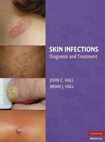
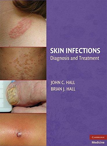
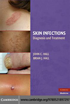
SKIN INFECTIONS
Diagnosis and Treatment
EDITED BY
John C. Hall
University of Missouri–Kansas City School of Medicine
Brian J. Hall
St. Louis University School of Medicine
CAMBRIDGE UNIVERSITY PRESS
Cambridge, New York, Melbourne, Madrid, Cape Town, Singapore, São Paulo
Cambridge University Press
The Edinburgh Building, Cambridge CB2 8RU, UK
Published in the United States of America by Cambridge University Press, New York
www.cambridge.org
Information on this title: www.cambridge.org/9780521897297
This publication is in copyright. Subject to statutory exception and to the provision of relevant collective licensing agreements, no reproduction of any part may take place without the written permission of Cambridge University Press.
Cambridge University Press has no responsibility for the persistence or accuracy of urls for external or third-party internet websites referred to in this publication, and does not guarantee that any content on such websites is, or will remain, accurate or appropriate.
Francisco G. Bravo and Salim Mohanna
John C. Hall
1
2
3
Tammie Ferringer
Alejandra Varela, Anne Marie Tremaine, Aron Gewirtzman, Anita Satyaprakash, Natalia Mendoza, Parisa Ravanfar, and Stephen K. Tyring
Aditya K. Gupta and Elizabeth A. Cooper
4
5
6
7
8
9
Bhushan Kumar and Sunil Dogra
Arturo P. Saavedra and Samuel L. Moschella
Francesca Prignano, Caterina Fabroni, and Torello Lotti
Dirk M. Elston
Evandro Ararigbóia Rivitti and Paulo Ricardo Criado
Francisco G. Bravo and Salim Mohanna PART
Joseph C. Pierson and David J. DiCaudo 11
Marcia Ramos-e-Silva, Paula Pereira Araújo, and Sueli Coelho Carneiro
Domenico Bonamonte and Gianni Angelini
PART IV INFECTIONS IN SELECTED PATIENT
Joseph S. Susa and Clay J. Cockerell 14 INFECTIONS IN ORGAN
Daniela Kroshinsky, Jennifer Y. Lin, and Richard Allen Johnson
15
16
Michelle R. Wanna and Jonathan A. Dyer
17 SKIN INFECTIONS IN THE ELDERLY
Noah S. Scheinfeld
18 SKIN INFECTIONS IN ATHLETES
Brian B. Adams
19 SKIN INFECTIONS IN DIABETES MELLITUS
Nawaf Al-Mutairi
PART V INFECTIONS OF SPECIFIC SKINASSOCIATED BODY SITES
20 INFECTIONS OF THE SCALP 255
Shannon Harrison, Haydee Knott, and Wilma F. Bergfeld
21 INFECTIONS OF THE NAIL UNIT 268 Gérald E. Piérard, Claudine Piérard-Franchimont, and Pascale Quatresooz
22 INFECTIONS OF THE MUCOUS MEMBRANES 275
Julia S. Lehman, Alison J. Bruce, and Roy S. Rogers, III
PART VI SPECIAL DISEASE CATEGORIES
23 INFECTIONS IN SKIN SURGERY
Jean-Michel Amici, Anne-Marie Rogues, and Alain Taïeb
24 VENEREAL DISEASES 309
Travis Vandergriff, Mandy Harting, and Ted Rosen
25 LIFETHREATENING SKIN INFECTIONS: SKIN SIGN S OF IMPORTANT BACTERIAL INFECTIOUS DISEASES 322
Lisa A. Drage
Contributors
Brian B. Adams , MD, MPH
Associate Professor of Dermatology
Director of Sports Dermatology Clinic
Chief of Dermatology, VAMC Cincinnati University of Cincinnati Cincinnati, OH
Nawaf Al-Mutairi , MD, FRCPC
Associate Professor Faculty of Medicine
Kuwait University Safat, Kuwait
Jean-Michel Amici , MD
Service de Dermatologique
Groupe Hospitalier Saint-Andre Bordeaux Cedex, France
Gianni Angelini , MD
Professor of Clinical Dermatology
Head of the Dermatology Clinic
Department of Internal Medicine, Immunology and Infectious Diseases
University of Bari, Italy
Paula Pereira Araújo , MD
Former Post-Graduation Student, Sector of Dermatology
University Hospital, Federal University of Rio de Janeiro Rio de Janeiro, Brazil
Wilma F. Bergfeld , MD
Head of Dermatology and Pathology
Department of Dermatology
Cleveland Clinic Foundation Cleveland, OH
Kumar Bhushan , MD
Department of Dermatology, Venereology and Leprology
Postgraduate Institute of Medical Education and Research Chandigarh, India
Domenico Bonamonte , MD
Research Fellow
Department of Internal Medicine, Immunology and Infectious Diseases
Dermatologic Clinic
University of Bari Bari, Italy
Francisco G. Bravo , MD
Associate Professor of Dermatology and Pathology
Facultad de Medicina Alberto Huardo
Instituto de Medicina Tropical Alexander von Humbold Universidad Peruano Cayetano Heredia Lima, Peru
Alison J. Bruce , MBChB
Consultant, Department of Dermatology
Mayo Clinic
Associate Professor of Dermatology
College of Medicine, Mayo Clinic Rochester, MN
Sueli Coelho Carneiro , MD, PhD
Dermatologist, Sector of Dermatology
University Hospital, Federal University of Rio de Janeiro
Associate Professor, Sector of Dermatology
University Hospital, State University of Rio de Janeiro
Rio de Janeiro, Brazil
Clay J. Cockerell , MD
Professor of Dermatology and Pathology
University of Texas Southwestern Medical Center Dallas, TX
Elizabeth A. Cooper , B.E.Sc., H.B.Sc. Mediprobe Research, Inc. London, Ontario, Canada
Paulo Ricardo Criado , MD
Dermatologist, Division of Dermatology
Hospital das Clinicas School of Medicine
San Paulo University, Brazil
San Paulo, Brazil
David J. DiCaudo , MD
Associate Professor
Department of Dermatology and Pathology
Mayo Clinic Scottsdale, AZ
Sunil Dogra , MD, DNB
Assistant Professor
Department of Dermatology, Venereology and Leprology
Postgraduate Institute of Medical Education and Research Chandigarh, India
Lisa A. Drage , MD
Assistant Professor of Dermatology Mayo Clinic Rochester, MN
Jonathan A. Dyer , MD
Assistant Professor of Dermatology and Child Health University of Missouri School of Medicine Columbia, MO
Dirk M. Elston , MD
Director, Department of Dermatology
Geisinger Medical Center Danville, PA
Caterina Fabroni , MD
Specialist in Dermatology and Venereology Department of Dermatological Sciences
Florence University Florence, Italy
Tammie Ferringer , MD
Associate Departments of Dermatology and Pathology Geisinger Medical Center Danville, PA
Aron Gewirtzman , MD Center for Clinical Studies Houston, TX
Aditya K. Gupta , MD, PhD
Mediprobe Research, Inc. London, Ontario, Canada Professor of Dermatology Sunnybrook Health Sciences Center University of Toronto Toronto, Ontario, Canada
Brian J. Hall , MD Resident Department of Pathology
St. Louis University School of Medicine St. Louis, MO
John C. Hall , MD
Associate Professor of Dermatology
University of Missouri–Kansas City St. Luke’s Hospital
Kansas City Free Health Clinic Kansas City, MO
Shannon C. Harrison , MBBS, MMed, FACD
Clinical Research Fellow Department of Dermatology
Cleveland Clinic Foundation Cleveland, OH
Mandy Harting , MD Assistant Professor Department of Dermatology
Baylor College of Medicine Houston, TX
Richard Allen Johnson , MDCM Department of Dermatology
Massachusetts General Hospital
Harvard Medical School Boston, MA
Haydee Knott Department of Dermatology
Cleveland Clinic Health Sciences Center Cleveland, OH
Daniela Kroshinsky, MD Department of Dermatology
Massachusetts General Hospital Harvard Medical School Boston, MA
Julia S. Lehman , MD
Resident of Dermatology Mayo School of Graduate Medical Education Rochester, MN
Jennifer Y. Lin , MD Dermatologist
Department of Dermatology
Brigham and Women’s Hospital
Harvard Medical School Boston, MA
Torello Lotti , MD Chairman Department of Dermatological Sciences
Florence University Florence, Italy
Natalia Mendoza , MD, MSc Center for Clinical Studies Houston, TX
Salim Mohanna , MD Clinical Research Associate Instituto de Medicina Tropical Alexander von Humbold Universidad Peruano Cayetano Heredia Lima, Peru
Samuel L. Moschella , MD
Clinical Professor (Emeritus) of Dermatology
Harvard Medical School
Staff Dermatologist Lahey Clinic Burlington, MA
Gérald E. Piérard , MD, PhD
Professor and Head of the Dermatopathology Department University Hospital of Liège Liège, Belgium
Claudine Piérard-Franchimont, MD, PhD
Associate Professor, Chief of Laboratory Dermatopathology Department University Hospital of Liège Liège, Belgium
Joseph C. Pierson , MD
Dermatopathologist, Anatomic and Clinic Pathologist Uniformed Services University of Health Sciences Bethesda, MD
United States Military Academy West Point New York, NY
Francesca Prignano , MD, PhD
Assistant Professor Department of Dermatological Sciences Florence University Florence, Italy
Pascale Quatresooz , MD, PhD Master of Conference, Chief of Laboratory Dermatopathology Department
University Hospital of Liège Liège, Belgium
Marcia Ramos-e-Silva , MD, PhD
Associate Professor and Head, Sector of Dermatology University Hospital, Federal University of Rio de Janeiro Rio de Janeiro, Brazil
Parisa Ravanfar, MD, MBA, MS
Dermatology Clinic Research Fellow Center for Clinical Studies Houston, TX
Evandro ArarigbÓia Rivitti , MD
Dermatologist and Chairman of Department of Dermatology School of Medicine San Paulo University, Brazil San Paulo, Brazil
Roy S. Rogers III, MD Professor of Dermatology Mayo Medical School Consultant in Dermatology Mayo Clinic Rochester, MN
Anne-Marie Rogues , MD, PhD Service d’Hygiene Hospitaliere Groupe Hospitaliere Pellegrin Bordeaux, France
Ted Rosen , MD Professor of Dermatology
Baylor College of Medicine Chief of Dermatology
Michael E. DeBakey VA Medical Center Houston, TX
Arturo P. Saavedra , MD, PhD Instructor in Dermatology
Harvard Medical School Boston, MA
Anita Satyaprakash Dermatology Clinic Research Fellow Center for Clinical Studies Houston, TX
Noah S. Scheinfeld , MD, JD Assistant Clinical Professor Department of Dermatology
Columbia University New York, NY
Joseph S. Susa , DO Assistant Clinical Professor Department of Dermatology University of Texas Southwestern Medical Center Dallas, TX
Alain Taïeb, MD, PhD Head
Department of Dermatology and Pediatric Dermatology University Hospitals of Bordeaux National Reference Center for Rare Skin Disorders Bordeaux Cedax, France
Anne Marie Tremaine , MD Clinical Research Fellow Center for Clinical Studies Houston, TX
Stephen K. Tyring , MD, PhD, MBA Clinical Professor of Dermatology University of Texas Health Science Center Houston, TX
Travis Vandergriff , MD Department of Dermatology
Baylor College of Medicine Houston, TX
Alejandra Varela , MD University of Texas Health Science Center Houston, TX
Michelle R. Wanna , MD Resident Physician, Dermatology University of Missouri School of Medicine Columbia, MO
Acknowledgments
My wife, Charlotte, gave me all the encouragement anyone could ask.
The late Richard Q. Crotty, MD, gave me the chance to be a dermatologist. The late Clarence S. Livingood, MD, taught me to be true to my science and my patients. The late David Gibson, MD, and Ken Watson, DO, led my way in dermatopathology. My office manager, Christa Czysz, has kept me afloat. My nurses,
Brandi DelDebbio and Kelly Hudgens, and my office staff, Kelly Howell and Jennifer Phillips, have helped in enumerable ways.
A special thanks is due to my son, Brian J. Hall, MD, for endless hours helping edit and organize this text. The incredible group of authors in this book will speak for themselves through these pages. It would be an honor to be half the physician these dermatologists have proven to be.
INTRODUCTION
John C. Hall
A comprehensive melding of the fields of dermatology and infectious diseases is long overdue. It is the skin that is often the first sign of infection and the easiest organ to quickly access with an educated eye, culture, scraping, and histopathologic evaluation. It is the observation of the skin that can hold the chance for the earliest diagnosis and thus the most timely attempts at therapy. This same observation can guide the clinician through the maze of enumerable, often confusing, and sometimes costly follow-up confi rmatory tests. Let us now, in these pages, take this opportunity the integument has given us to lead us through the ever-increasingly important field of infectious diseases.
TECHNIQUES IN DIAGNOSING DERMATOLOGIC MANIFESTATIONS OF INFECTIOUS DISEASES
Francisco G. Bravo and Salim Mohanna
GETTING THE SAMPLE
A vital step toward making the right diagnosis when dealing with infectious diseases is ordering the appropriate test. That implies having a certain idea of the range of possible organisms involved and directing your workup toward ruling in or out a specific agent. Of course, there will be cases where a more blind approach is in order and a large range of diagnostic possibilities should be considered. In those situations, smears and cultures for bacterial, mycobacterial, and fungal microorganisms are indicated. Also viral diseases should be considered in specific situations, such as febrile patients with disseminated maculopapular or vesicular rashes. However, just for practical purposes, it is better to take a syndromic approach, considering a range of possibilities regarding the etiology of the lesions and then, selecting the appropriate test. Let us take an example such as a patient with a sporotrichoid pattern of lesions. If the diagnosis to confirm is sporotrichosis, a fungal culture will be very sensitive and very specifi c. Pyogenic bacteria such as Staphylococcal aureus can also produce such a pattern. In these cases, a Gram stain and routine culture will be helpful. But, if the patient likes fishing, swimming, or diving besides gardening an atypical mycobacterial infection (M. marinum) also has to be listed in the differential. In such cases, a biopsy, acid-fast stain, and mycobacterial culture should also be considered, although recognizing this is a difficult diagnosis to make because of the low sensitivity of each individual test. In the same line of thought, the same patient just came back from a trip to the Amazon: leishmaniasis is then another possibility. In leishmaniasis, there is no test with high sensitivity, so a panel approach is indicated (direct exam, culture, intradermal reaction, histopathology, and PCR, when available). Nocardia, another disease capable of giving such a pattern, can only be detected if the laboratory takes special precautions while culturing. Then, it is better to direct our workup toward a specific diagnosis. Of course, that also implies having some knowledge regarding the sensitivity and specifi city of each test for a specific etiological agent.
Nobody other than the clinician will know best where to take the sample from. Unfortunately for regulatory or administrative reasons, the task is commonly left to a technician.
As a rule, purulent or oozing secretions are considered excellent samples and should be regularly submitted for direct examination with Gram, fungal, and acid-fast staining. Abscesses should be punctured and the pus submitted under sterile conditions. Taking a biopsy of an abscess is usually not rewarding, but clinically if there is a thick wall surrounding the cavity, it may reveal a granulomatous infiltrate when biopsied.
Solid lesions, such as those suspicious for granulomatous diseases, are better studied submitting the tissue for culture; even then, the appropriate area should be sampled. In mycetoma, for example, unless the biopsy is taken from areas containing granules, the yield of histology and culture will be very low.
When dealing with dry, scaly lesions, such as in tinea cases, the scraping is very sensitive. However, in hairy areas (scalp, beard), getting some hairs may reveal the presence of spores in the absence of superficial hyphae. This is usually the case when a tinea barbae has been previously treated blindly with a topical antifungal. In cases where there is a possibility of tinea incognita, it is advisable to microscopically examine the proximal portion of the hairs. When dealing with white onychomycosis, scrape the surface. If the subungeal area is affected, the detritus under the nail are most likely to reveal the hypha or spore. Nail clippings are considered good samples, even suitable for histological study. The diagnosis of microscopic ectoparasites, such as scabies, requires taking the sample from the most commonly affected areas. Blindly scrapping off different areas is not very rewarding. In contrast, scrapping a whole scabies burrow will frequently reveal the presence of the mites, eggs, or feces.
Moist ulcers can be swabbed and the secretions submitted for direct examination and culture; dry ulcers can be sampled by touch preparation. Aspirating the fluid under the border of the ulceration with a micropipette is useful for leishmania; in leprosy, examining the fluid obtained by slitting the ear lobe under pressure is an excellent method to visualize the mycobacteria.
TECHNIQUES
Smears
Direct examination of material obtained from lesions is a vital first step to orient the clinician toward a specific etiology. Gram and acid-fast stains are now routinely performed by laboratory technicians, and the techniques themselves are beyond the scope of this book. However, the results provided are of vital importance. Gram stain is regularly done in urethral secretions to look for Neisseria gonorrhoeae; its absence in the presence of neutrophiles is indicative of nongonococcal chlamydial urethritis. Acid-fast staining of smears from leprosy patients may help establish the bacterial load.
Some tests are more easily done, on a daily basis, at dermatology offices around the world. Examples of such tests are potassium hydroxide (KOH) preparations and the Tzanck test. The KOH preparation is usually done using a 5% to 40% concentration. The idea is that the reaction will dissolve most of the normal host cells, sparing the infectious agent. The condenser of
the microscope is lowered, to facilitate observation by light contrast. The test is done on skin scrapings to detect the presence of hyphae in dermatophytes or candida. Yeast of invasive fungi such as Blastomyces and Paracoccidioides can also be detected by this method in purulent secretions. Hairs and nails can also be examined under the microscope in a similar manner. The same preparation can be used to examine scrapings while looking for scabies mites and Demodex. A variation on the theme is adding colored stains to facilitate viewing of fungal structures. Using mineral oil instead of KOH may allow the assessment of viability and motility of ectoparasitic mites. Instead of regular scraping, one can use adhesive tape to take the sample, a technique especially useful when dealing with rapidly moving targets, such as the face of a small child. Surprisingly, a similar technique will allow detection of large fungal structures, such as the agents of chromoblastomycosis and lobomycosis when the tape is applied on top of the clinical lesion. This is possible because of the phenomenon of transepidermal elimination of the microorganisms. Tzanck test implies the examination of cells at the base of an unroofed blister in suspected cases of Herpes simplex or Herpes Zoster infection. Once air dried, the slide is stained with Wright, Giemsa, or methylene blue. The goal is to detect multinucleated giant keratinocytes that are indicative of herpetic infections. A more sophisticated technique utilizes an immunofluorescent antibody against the virus, allowing species-specific identification.
Touch preparation of a genital ulcer can be examined under dark field for the presence of treponemal spirochetes. The same preparations, if stained as a PAP smear may allow detection of the presence of multiple intracellular bacteria in cases of granuloma inguinale. Touch preparation of the bottom of a large ulcer with undermined borders and acid-fast staining will be extremely useful in detecting large amount of mycobacterium in a Buruli ulcer (Table T-1).
Culture
The purpose of cultures is isolation of the infectious agent to comply with one of the Koch postulates. If the diagnosis is uncertain, samples should be sent for bacterial, fungal, viral and acid-fast bacterial cultures. Culturing requires special media, depending on the microorganism suspected (see Table T-2). One has to keep in mind that certain areas of the body are heavily contaminated, such as the mouth and perianal region. The skin, by no means an aseptic organ, can be colonized by different bacteria and fungi. The result of cultures should be interpreted appropriately, with correlation to the clinical lesion. Some culture media are designed to facilitate the growth of the microorganism (such as the Thayer-Martin media for gonococcal infection or oxygen depleted systems for anerobes). Others, such as the Mycosel, will restrict the growth of saprophytic fungi while allowing the growth of dermatophytes. Most bacteria will grow in a matter of days; some, such as Brucella, may require weeks in specially designed media (such as Ruiz Castañeda). Candida will also grow fast, even on bacterial culture media, in days. Regular fungi, cultured in Sabouraud’s agar may grow as fast as in 1 week (Sporothrix), 2 weeks (dermatophytes), or 4 weeks (Histoplasma and Actinomyces). Mycobacteria may be fast growers (M. fortuitum, M. chelonae, or M. abscessus) or take several weeks (M. tuberculosis and M. ulcerans), with additional specific temperature requirements.
Table T-1: Selected Microorganism with Special Culture Requirements
H. influenza
N. gonorrhea
B. pertussis
C. diphtheriae
M. tuberculosis
Chocolate agar with factors V and X
Thayer-Martin media
Bordet-Gengou agar
Tellurite plate, Loffler’s medium, blood agar
Lowenstein-Jensen agar
Lactose-fermenting entericsMacConkey’s agar (pink colonies)
Surprisingly enough, even in the 21st century, some famous pathogens are still unable to be isolated in culture media. Treponema pallidum, Mycobacterium leprae, and Loboa Loboi are three examples. The isolation of virus by culture is not routinely done (except for herpes simplex and varicella-zoster viruses). Modern diagnosis relies more on molecular techniques or serologic assays.
Intradermal reactions
Intradermal reactions are widely used to support the diagnosis of some dermatological and nondermatological diseases. They are mainly indicated for the detection of type I (immediate hypersensitivity) and type IV (delayed hypersensitivity) reactions toward exogenous or endogenous antigens. Intradermal reactions for the diagnosis of infectious diseases are indicated to detect previous contact with the agent as revealed by delayed hypersensitivity to the whole organisms or their antigens.
The intradermal reaction is a localized inflammatory reaction with marked proliferation of lymphocytes, monocytes, and small numbers of neutrophiles, with a tendency toward cellular accumulations arround small vessels. The induration results from fibrin formation.
The principle of an intradermal reaction is the inoculation of an antigen into the superficial layer of the dermis through a
— Francisco G. Bravo and Salim Mohanna
fine-bore needle with its bevel pointing upward. The quantity injected may vary from 0.01 to 0.1 mL, but 0.1 mL is universally used. Although the test could be done at any site, the proximal part of the flexor aspect of the forearm is conventionally used. Corticosteroids or immunosuppressive agents should be discontinued before testing for intradermal reactions because they may inhibit the delayed hypersensitivity reaction. Intradermal reactions for the detection of delayed hypersensitivity are read at 48 hours, although they can be read as early as 12 hours and as late as 4 days. The size of the induration is more important than erythema when interpreting delayed hypersensitivity reactions.
The tuberculin test (also called PPD [purified protein derivative], Pirquet test, or Mantoux test) is a diagnostic tool used to detect latent infection or recent infection (as shown by conversion from negative to positive) and as part of the diagnosis of tuberculous disease. A standard dose of tuberculin is injected intradermally on the flexor aspect of the forearm and a reading is taken after 48 hours. The reaction is read by measuring the diameter of induration in millimeters. The interpretation of the test result will depend on all relevant clinical circumstances. An induration measuring more than 10 mm in diameter is considered to be a positive response while that measuring less than 5 mm is considered negative. A positive test indicates past or present infection with M. tuberculosis or vaccination with bacillus Calmette-Guérin (BCG). An induration of more than 15 mm is unlikely to be due to BCG vaccination and is strong evidence in favor of active tuberculosis. In the absence of specific risk factors for tuberculosis, an induration between 6 and 10 mm is more likely to be due to previous BCG vaccination or infection with environmental mycobacteria than to tuberculosis infection. When there is a higher probability of tuberculosis, such as recent contact with an infectious case, a high occupational risk or residence in a high prevalence country, an induration of 6 mm or more is more likely to be due to tuberculosis. Anergy is present in AIDS patients. Other factors that can weaken the reaction include severe tuberculous disease, renal failure, diabetes, immunosuppressive drugs, and old age. Initial skin tests may have a booster effect on reactions to subsequent doses. More sophisticated tests based on interferon production by stimulated cells are also available. Intradermal reactions for atypical mycobacteria have also been prepared. They include PPD-Y for Mycobacterium kansasii, Scrofulin for Mycobacterium scrofulaceum, and Burulin for Mycobacterium ulcerans.
The leishmanin test was first done by Montenegro in 1926, in Brazil. This test (also called the Montenegro test) is indicative of the delayed hypersensitivity reaction to leishmania, which plays a major role in disease resolution and wound healing. It usually becomes positive early in the course of cutaneous or mucocutaneous leishmaniasis (except in diffuse cutaneous leishmaniasis) and only after recovery from visceral leishmaniasis. It is highly sensitive for cutaneous leishmaniasis. The test is considered positive when induration is more than 5 mm in diameter after 48 to 72 hours. The test is not species specific. A negative test may be attributed to an anergic state, decreased cell mediated immunity, early treatment, or presence of an unusual serotype of leishmania, whereas a positive test favors active disease if the patient is not a resident of the area. The same positive reaction does not have the same relevance for natives and current residents.
The lepromin test classifies the stage of leprosy based on the reaction and differentiates tuberculoid leprosy, (in which there is
a positive delayed reaction at the injection site) from lepromatous leprosy (in which there is no reaction despite the active infection). The test is not diagnostic since normal uninfected persons may react. Two types of antigens are available: Mitsuda lepromin, an autoclaved suspension of tissue (whole bacilli) obtained from experimentally infected armadillos; and Dharmendra lepromin, a purified chloroform-ether extracted suspension of M. leprae (fractionated bacilli with a soluble protein component). The response after intradermal injection is typically biphasic, with an early Fernandez reaction (in the form of a tuberculin reaction with Dharmendra antigen) and a late Mitsuda reaction (in the form of erythematous, papular nodules with Mitsuda antigen).
Other important tests used for diagnostic aid, or to evaluate the cellular immune response in patients suspected of having reduced cell-mediated immunity, or in epidemiological studies include the anthraxin test, the onchocerca skin test, the candidin test, the coccidioidin or spherulin test, the histoplasmin test, and the trichophytin test. Finally, there are some tests of historical importance only, as they are no longer used for diagnostic purposes: Lymphogranuloma venereum (Frei’s test), Chancroid (Ito Reenstierna test), Bartonellosis (Foshay test) and Scarlet fever (Dick’s test).
Serology
Some tests are based on the detection of the infectious agent antigens in serum or by the detection of the circulating antibodies generated by the host. Agglutination tests (latex agglutination test) are based on the capturing the antibody from a suspected patient with whole bacteria or antigen absorbed to latex particles. The presence of circulating antibodies will then be detected by the agglutination phenomenon. N. meningitidis and Cryptococcus can be detected by latex agglutination. The complement fixation (CF) test measures complement-consuming (complement-fixing) antibody in the serum or CSF of the patient. The serum to be tested is mixed with known quantities of complement plus the antigen targeted by the antibodies to be measured. The degree of complement fixation indicates the amount of antibody in the specimen. CF is used for diagnosis of some viral and fungal infections, particularly coccidioidomycosis.
Enzyme immunoassays are based on detection of antibody binding to a substrate linked to an enzyme. They are very sensitive and for that reason, commonly used for screening. They include the enzyme immunoassay (EIA) and the enzyme-linked immunosorbent assay (ELISA). ELISA is available for Chlamydia infections, herpes virus, and human immunodeficiency virus (HIV). On the other hand, Western blot test detects the specific antibodies by measuring its union to antigens fixed to a membrane by blotting. It is quite specific and commonly used as a confirmatory test in tandem with ELISA as the screening test (i.e. in HIV, HTLV-1, and Borrelia burgdorferi).
Other examples of humoral responses that can be tested include the treponemal serology test (including VDRL, RPR, and the most specific FTA-Abs), as well as antibodies against Borrelia, Legionella, Bartonella, and Leptospira. Also many viral infections, such as hepatitis A, B and C, Epstein-Barr virus (EBV), dengue, CMV, coxsackie, and parvovirus can be tested. Rising titers of four times the normal baseline over a 2-week period are especially useful in viral diseases with occasional skin involvement. Even parasitic diseases such as enteric amebiasis, cysticercosis,
and fasciolasis, as well as toxocariasis and toxoplasmosis can be studied via serology.
Molecular biology
The current concept in the use of molecular biology techniques is based on the detection of DNA and RNA material from specific organisms, providing an extremely reliable method of diagnosis with high specificity and sensitivity. The current method can rely either on the amplification of the material and posterior identification (polymerase chain reaction) or on the direct detection of the material in tissues (in situ hybridization).
Polymerase chain reaction (PCR) consists of denaturalization of nuclear material (DNA), with posterior addition of complementary primers and synthesis of new chains by adding an enzyme such as a polymerase. By using repeated cycles of high and low temperatures one can obtain an amplification of nuclear material (amplicon) until reaching amounts detectable by gel eletrophoresis or enzyme assay base using color detection. Real time PCR is a more sophisticated method, whereas the newly synthesized amplicons can be detected as they are produced by using immunofluorescent methods. Ligase-based method (LCR) amplifies the probe rather than the microorganism’s nuclear material. Some methods, like transcription-mediated amplification (TMA) and nucleid acid sequence-based amplification (NASBA), rely on the amplification of RNA material, which is usually more abundant than DNA. The current application for those techniques includes several pathogens listed in Table T-3. For example, both PCR and TMA are FDA approved for the diagnosis of pulmonary and extrapulmonary tuberculosis. By this technique, some researchers have been able to demostrate the presence of bacillus in cutaneous tuberculosis and tuberculids. These findings are, however, not consistent with other similar studies. The variable sensitivity and specificity of the method may be related to tissue inhibitors or fixation methods. PCR is also available for the second and third most common mycobacteriosis around the world, which are, Mycobacterium leprae and Mycobacterium ulcerans. The PCR technique has been very helpful in the study and discovery of new bartonella species as well.
Several viruses are suitable for PCR testing, with some variations in methodology such as the multiplex reverse transcriptase PCR used in varicella zoster infections. The cause–effect relation between HHV-8 and Kaposi’s sarcoma has been demostrated by PCR. The role of different HPV in cutaneous neoplasia, such as HPV-16 in verrucous carcinoma of the foot and HPV-6 and -11 in anogenital verrucous carcinoma have been possible by this amplification method. PCR allows not only identification but also quantification of viral loads in in HIV infections. In some parasitic diseases such as leishmaniasis, the PCR techniques are considered quite sensitive and quite specific. The gene targets include 18S-rRNA, small subunit rRNA, mini-exon gene repeat, β-tubulin gene, transcriber spacer regions, and microsatellite DNA of the kinetoplast. The sensitivity is so high that it may allow detection of as little as half a parasite. Many fungal infections can now be precisely identified, as is the case for mycetomas by Madurella mycetomatis
Another useful technique now available is in situ hybridization (ISH). It is based on the detection of nucleic acid material from different microorganisms directly in tissues, even if they are fixed in formalin. Fluorescent ISH (FISH) is a more sophisticated
Table T-3: Skin infectious diseases where PCR can be a useful diagnostic technique
Bacterial pathogens
Mycobacterium tuberculosis
M. leprae
M. ulcerans
Rickettsia rickettsii
R. prowazekii
Baronella henselae
Spirochetes
Treponema pallidum
Borrelia burgdorferi
Virus
Herpes simplex 1 and Herpes simplex 2
Varicela zoster virus
Human herpesvirus 8
Human papillomavirus
HIV
Hepatitis C virus
Parvovirus B19
Epstein-Barr virus
Parasites
Leishmania infantum
Leishmania braziliensis
Balamuthia mandrillaris
Fungi
Aspergillus fumigatus
Aspergillus versicolor
Aspergillus flavus
Blastomyces dermatitides
Cryptococcus neoformans
Candida albicans
Candida dubliniensis
Coccidioides immitis
Histoplasma capsulatum
Sporothrix schenckii
Madurella mycetomatis
Various dermatophytes
Modified from Sra KK, Torres G, Rady P, Hughes TK, Payne DA, Tyring SK. Molecular diagnosis of infectious diseases in dermatology. J Am Acad Dermatol. 2005 Nov;53(5):749–65.
Table T-4: Infectious Agents that can be Identified by ISH
Varicella Zoster
Molluscum contagiosum
HIV
HPV
EBV
Hepatitis C virus
HHV-8
Mycobacterium tuberculosis
Mycobacterium leprae
Leishmania spp.
Candida albicans
Cryptococcus neoformans
Modified from Sra KK, Torres G, Rady P, Hughes TK, Payne DA, Tyring SK. Molecular diagnosis of infectious diseases in dermatology.
J Am Acad Dermatol 2005;53(5):749–765.
method that is used not only to identify infectious agents but also in gene mapping and chromosome analysis. ISH has been used to confirm the presence of HPV in epidermodysplasia verruciformis and EBV in hydroa-like lymphomas. Table T-4 details infectious agents that can be identified by ISH.
The development of DNA microarray technology, also known as DNA chip technology, will allow testing for multiple microorganisms or genetic defects, all at once. In this method, DNA from a clinical sample is first amplified by PCR, converted to RNA or more DNA, mixed with fluorescent dyes, and then applied over a plate containing many different oligonucleotides or cDNA libraries. After the hybridization between sample material and fixed probes on the plates takes place, stimulation by laser will light up the hybridized labeled probes. The total image then obtained will reflect the DNA or RNA contents in the original sample and compare them to control arrays. This technique has allowed identification of M. tuberculosis variants with gene mutations conferring resistance to isoniazid and rifampin. As more of these sophisticated methods become available, our understanding of disease mechanisms and the interactions between humans and pathogens will be complemented by knowledge about microorganism genetic variations, drug resistance, and disease epidemiology.
Skin biopsy as a diagnostic technique
While dealing with cutaneous infectious diseases the accessibility of skin as a source of sampling makes the biopsy an important diagnostic tool. Like any other method, one can determine the sensitivity and specificity of the biopsy for diagnostic purposes. Specificity can be very high, as in a case of molluscum contagiosum or rhinoscleroma, or very low, such as in a grossly superinfected ulcer, where one can see many bacterial colonies on the surface. The sensitivity can be very high, in a case of South
American blastomycosis (paracoccidioidomycosis), or very low in a case of sporotrichosis.
Biopsies can be considered suggestive of, compatible with, or diagnostic of a specific infectious disease. How we use these terms, although subjective, may be based on the frequency of specific histological patterns seen associated with a specific agent, or the findings of the microorganism itself in the histological cuts. We think the term “suggestive” should be used if the histological pattern is commonly seen in a condition unsuspected by the clinician. The term “compatible with” should be used if the pattern seen coincides with one of the entities considered in the differential listed by the clinician, although the microorganism itself is not seen. The term “diagnostic of” should imply visualizing the agent itself, and carries the most certainty. Biopsies also allow establishing the inflammatory or neoplastic nature of a lesion.
The prevalence of a specific cell in a pattern will also direct our diagnosis:. For example a plasma cell–rich liquenoid infiltrate with histiocytes will be in favor of a spirochete-induced disease (either syphilis or borreliosis). Vacuolated histiocytes accompanied by a lymphoplasmacytic infiltrate are commonly seen in leishmaniasis, rhinoscleroma, and granuloma inguinale. Suppurative granulomas are indicative of mycobacteria and deep fungal infection. A dense perivascular and interstitial, superficial and deep inflammatory infiltrate rich in eosinophils, with extension into the subcutaneous tissue, is suggestive of a deep larva migrans or gnathostomiasis.
An important fact obtained from the skin biopsy is the granulomatous nature of an infiltrate. Many chronic infectious diseases are characteristically granulomatous. Granulomas can be divided into five types: tuberculous, suppurative, foreign body, palisaded, or sarcoidal. It should be mentioned that tuberculous granulomas are not exclusively seen in tuberculosis but in several other entities. Caseation necrosis is a more reliable sign of tuberculosis, but in its absence, other diagnostic possibilities for tuberculoid granulomas may include leishmaniasis, tuberculoid leprosy, and sporotrichosis. Even caseation necrosis may not be that specific as it can be seen in nontuberculous processes such as rosacea.
Suppurative granulomas, containing neutrophils in the center, are commonly seen in deep fungal infections such as North American blastomycosis, paracoccidioidomycosis, chromoblastomycosis, and sporotrichosis, as well as in some atypical mycobacterioses. A pseudocarcinomatous hyperplasia may be seen on top of the granuloma. Stellate necrosis in a granuloma is suggestive of cat scratch disease. Sarcoidal granulomas can be seen in sarcoidosis but also as a reaction to foreign materials such as silica and beryllium. Rarely, leprosy and paracoccidioidomycosis can be quite sarcoidal in appearance.
The diagnosis by histological patterns of the inflammatory diseases of the skin, as was outlined by Dr. Ackerman, can also be applied to these infections. There are some patterns that are quite specific for certain diseases like rhinoscleroma, bartonellosis, lepromatous leprosy, and Buruli ulcer.
In rhinoscleroma there is a diffuse infiltrate that occupies all of the dermis. The infiltrate is made up of foamy histiocytes, arranged in mantles. Plasma cells are intermixed in the infiltrates. Some of these cells accumulate such an amount of immunoglobulin within their cytoplasm that they develop into eosinophilic globules, so-called Russell bodies. Occasionally, rod-shaped
Random
documents with unrelated content Scribd suggests to you:
MCCORD (VERA) PRODUCTIONS, INC.
*Good-Bad Wife. 1920.
MCCORMICK, JOHN.
Ella Cinders. 1926.
Footlights and Fools. 1929. Happiness Ahead. 1928. Her Wild Oat. 1927. It Must Be Love. 1926. Lilac Time. 1928. Lost at the Front. 1927. Naughty but Nice. 1927. Oh Kay! 1928.
Orchids and Ermine. 1927.
Smiling Irish Eyes. 1929. Synthetic Sin. 1928. Tender Hour. 1927. Twinkletoes. 1926. We Moderns. 1925.
MACDONALD, WILLIAM COLT. Call the Mesquiteers. 1938. Come on Cowboys. 1937. Ghost-Town Gold. 1936. Gunsmoke Ranch. 1937. Heart of the Rockies. 1937. Heroes of the Hills. 1938. Hit the Saddle. 1937. Kansas Terrors. 1939.
New Frontier. 1939. Night Riders. 1939. Outlaws of Sonora. 1938.
Overland Stage Raiders. 1938. Pals of the Saddle. 1938. Powdersmoke Range. 1935. Range Defenders. 1937. Red River Range. 1938. Riders of the Whistling Skull. 1937. Roarin' Lead. 1936.
Santa Fe Stampede. 1938. Three Mesquiteers. 1936. Three Texas Steers. 1939. Trigger Trio. 1937. Wild Horse Rodeo. 1937. Wyoming Outlaw. 1939.
MCDONELL, GORDON. When Thief Meets Thief. 1937.
MACDONOUGH, GLEN. Babes in Toyland. 1934.
MCDOUGALL, WILLIAM B. Mitsi-Adazi, Nature's Workshop. 1937.