Cover Story


THE WORLD’S FIRST FUNKY OPHTHALMOLOGY MAGAZINE
One of Ophthalmology’s First Post-Lockdown Conferences p14 THE BUSINESS ISSUE Dec 2020/Jan 2021 piemagazine.org 16
nAMD DME RVO
EYLEA® is indicated for adults for the treatment of neovascular (wet) age-related macular degeneration (AMD), visual impairment due to macular edema secondary to retinal vein occlusion (branch RVO or central RVO), visual impairment due to diabetic macular edema (DME), and visual impairment due to myopic choroidal neovascularization (myopic CNV).


REFERENCES: 1 EYLEA® approved package insert Singapore March 2019, Bayer (South East Asia) Pte Ltd. 2. Wells JA, Glassman AR, Ayala AR, et al. Aflibercept, bevacizumab or ranibizumab for diabetic macular edema: two-year results from a comparative effectiveness randomized clinical trial. Ophthalmology. 2016;123:1351-1359 3 Korobelnik JF, Do DV, Schmidt-Erfurth U et al. Intravitreal aflibercept for diabetic macular edema. Ophthalmology 2014;121:2247-2254. 4 Eleftheriadou M, Gemenetzi M, Lukic M, et al. Three-year outcomes of aflibercept treatment for neovascular age-related macular degeneration: evidence from a clinical setting. Ophthalmol Ther. 2018;7:361-368. 5 Pielen A, Clark WL, Boyer DS, et al. Integrated results from the COPERNICUS and GALILEO studies. Clin Ophthalmol. 2017;11:1533-1540.
ABBREVIATED PRESCRIBING INFORMATION
EYLEA SOLUTION FOR INJECTION IN VIAL 2MG. Approved name(s) of the active ingredient(s) One ml solution for injection contains 40 mg aflibercept. Each vial provides a usable amount to deliver a single dose of 50 µl containing 2 mg aflibercept. Indication EYLEA is indicated for the treatment of neovascular (wet) age-related macular degeneration (AMD), macular edema secondary to retinal vein occlusion (branch RVO or central RVO), diabetic macular edema (DME) and myopic choroidal neovascularization (myopic CNV). Dosage Regimen wAMD: The recommended dose for EYLEA is 2 mg aflibercept, equivalent to 50 µl. EYLEA treatment is initiated with one injection per month for three consecutive doses, followed by one injection every two months. Based on the physician’s judgement of visual and/or anatomic outcomes, the treatment interval may be maintained at two months or further extended, such as with a treat-and-extend dosing regimen, where treatment intervals are increased in 2- or 4- weekly increments to maintain stable visual and/or anatomic outcomes. If visual and/or anatomic outcomes deteriorate, the treatment interval should be shortened accordingly to a minimum of two months during the first 12 months of treatment. There is no requirement for monitoring between injections. Based on the physician’s judgement the schedule of monitoring visits may be more frequent than the injection visits. Treatment interval greater than 4 months between injections have not been studied. Branch RVO or central RVO: The recommended dose for EYLEA is 2 mg aflibercept, equivalent to 50 microliters. After the initial injection, treatment is given monthly until visual and/or anatomic outcomes are stable. Three or more consecutive, monthly injections may be needed. The interval between two doses should not be shorter than one month. If there is no improvement in visual and anatomic outcomes over the course of the first three injections, continued treatment is not recommended. If necessary, treatment may be continued and the interval may be extended based on visual and/or anatomic outcomes (treat and extend regimen). Usually, monitoring should be done at the injection visits. During treatment interval extension through to completion of therapy, the monitoring schedule should be determined by the treating physician based on the individual patient’s response and may be more frequent than the schedule of injections. DME: The recommended dose for EYLEA is 2 mg aflibercept, equivalent to 50 microliters. EYLEA treatment is initiated with one injection per month for five consecutive doses followed by one injection every two months. There is no requirement for monitoring between injections. After the first 12 months of treatment with EYLEA, and based on visual and/or anatomic outcomes, the treatment interval may be extended, such as with a treat-and-extend dosing regimen, where the treatment intervals are gradually increased to maintain stable visual and/or anatomic outcomes; however there are insufficient data to conclude on the length of these intervals. If visual and/or anatomic outcomes deteriorate, the treatment interval should be shortened accordingly. The schedule for monitoring should therefore be determined by the treating physician and may be more frequent than the schedule of injections. If visual and anatomic outcomes indicate that the patient is not benefiting from continued treatment, EYLEA should be discontinued. Myopic CNV: The recommended dose for EYLEA is a single intravitreal injection of 2 mg aflibercept, equivalent to 50 microliters. Additional doses should be administered only if visual and anatomic outcomes indicate that the disease persists. Recurrences are treated like a new manifestation of the disease. The monitoring schedule should be determined by the treating physician based on the individual patient’s response. The interval between two doses should not be shorter than one month. Method of administration Intravitreal injections must be carried out according to medical standards and applicable guidelines by a qualified physician experienced in administering intravitreal injections. Following intravitreal injection patients should be instructed to report any symptoms suggestive of endophthalmitis (e.g., eye pain, redness of the eye, photophobia, blurring of vision) without delay. Each vial should only be used for the treatment of a single eye. Contraindications Hypersensitivity to the active substance aflibercept or to any of the excipients, active or suspected ocular or periocular infection, active severe intraocular inflammation. Special warnings and special precautions for use Endophthalmitis, increase in intraocular pressure, immunogenicity, systemic adverse events including non-ocular haemorrhages and arterial thromboembolic events. As with other intravitreal anti-VEGF treatments for AMD, the safety and efficacy of Eylea therapy administered to both eyes concurrently have not been systematically studied. When initiating Eylea therapy, caution should be used in patients with risk factors for retinal pigment epithelial tears. The dose should be withheld and treatment should not be resumed earlier than the next scheduled treatment in the event of: a decrease in best-corrected visual acuity (BCVA) of ≥30 letters compared with the last assessment of visual acuity; a subretinal haemorrhage involving the centre of the fovea, or, if the size of the haemorrhage is ≥50%, of the total lesion area. The dose should be withheld within the previous or next 28 days in the event of a performed or planned intraocular surgery. EYLEA should not be used in pregnancy unless the potential benefit outweighs the potential risk to the foetus. Women of childbearing potential have to use effective contraception during treatment and for at least 3 months after the last injection of aflibercept. Undesirable effects Very Common: Conjunctival hemorrhage, eye pain. Common: Retinal pigment epithelial tear, detachment of the retinal pigment epithelium, retinal degeneration, vitreous haemorrhage, cataract (cortical, nuclear, subcapsular), corneal erosion, corneal abrasion, intraocular pressure increased, vision blurred, vitreous floaters or detachment, injection site pain, foreign body sensation in eyes, lacrimation increased, eyelid edema, injection site hemorrhage, punctate keratitis, conjunctival hyperemia, ocular hyperemia. For a full listing of precautions and undesirable effects, please refer to the full product insert. For further prescribing information, please contact: Bayer (South East Asia)Pte Ltd. 2 Tanjong Katong Road #07-01 Paya Lebar Quarter 3 Singapore 437161. Date of revision of text March 2019.
Bayer (South East Asia) Pte Ltd
2, Tanjong Katong Road #07-01, Paya Lebar Quarter 3, Singapore 437161. Tel: +65 496 1888 Fax: +65 6496 1491 Website: www.bayer.com
PP-EYL-SG-0054-1(09/19)
ded dosing1–5
TRE A T W I T H Achieve uns


21 26 Matt Young CEO & Publisher Robert Anderson Media Director Hannah Nguyen Production & Circulation Manager Gloria D. Gamat Chief Editor Brooke Herron Editor International Business Development Ruchi Mahajan Ranga Brandon Winkeler Writers Andrew Sweeney April Ingram Chow Ee-Tan Joanna Lee Hazlin Hassan Olawale Salami Sam McCommon Tan Sher Lynn Maricel Salvador Graphic Designer Media MICE Pte. Ltd. 6001 Beach Road, #19-06 Golden Mile Tower, Singapore 199589 Tel: +65 8186 7677 / +1 302 261 5379 Email: enquiry@mediamice.com www.mediaMICE.com Published by
Friends Asia-Pacific Vitreo-retina Society Vitreo-Retinal Society - India IN THIS ISSUE... We are looking for eye docs who can contribute articles to PIE magazine. Interested? Let's talk! Send us an email at editor@mediamice.com. To place an advertisement, advertorial, symposium highlight, video, email blast, or other promotion in PIE magazine contact CEO Matt Young at matt@mediamice.com. Dr. Nancy Holekamp: Advancing Women in Ophthalmology Posterior Segment Enlightenment Case Reviews: When to Consider Ozurdex for DME in Pseudophakic Patients Retinal Experts Share Tools and Pearls in Managing Ophthalmic Trauma Identifying PCV without ICGA and Self-diagnosing AMD at Home All Eyes on the Retina Innovation Showcase Trauma Management on Spotlight at APOTS Virtual Conference Data from AAO’s IRIS Registry: The Key to Better Treatments in the Future? Innovation Conference Highlights 08 24 12 10 08 26 28 14 Cover Story m a g a z i n e poster or segmen nnova on en gh enmen Ophthalmology Innovation Summit All India Ophthalmological Society One of Ophthalmology’s First Post-Lockdown Conferences
Society
Dear Readers,

was the year that Media MICE, as a company, entered the beginning of its second decade. The team, as we had our reunion in Da Nang, Vietnam, late in January 2020, was at an all time high. We made great plans of moving the company forward and leveling up.


And we thought the coronavirus wasn’t going to make it out of China ... until it did. By mid-March the rest of the world went on pandemic lockdown … and the rest was COVID-19 history.
Having two magazines (PIE and CAKE) that revolve around the global ophthalmic conference calendar, we were terrified (to say the least), when one conference after another were first postponed, and then eventually cancelled. Even our clients who have pre-booked advertisements and other editorial projects were at a loss on what to do. Everything went on hold.
Then our CEO Matt Young, upon his return from a business trip in Europe got quarantined and tested COVID-19 positive. This was when things went a bit dark. But not for long … because we realized that we are designed and prepared for this situation
We have been operating as a virtual office since 2015. The magazines, although printed on-site at all conferences we bring them to, always had an e-book counterpart. Our magazines’ websites were already in progress before 2019 ended. So we just had to scramble a little bit to put them up. We took advantage of the pandemic lockdown and the additional time on our hands. Before we knew it, piemagazine.org and cakemagazine.org were live. You see, we were always ready for the virtual world! With the coming of the pandemic, the rest of the world joined the Media MICE-normal.
Then the ophthalmic conferences went virtual. From ASCRS in May all the way through AAO in November, we’ve never published this many show dailies done in one year! CAKE POST, PIE POST, and CAKE & PIE POST were born in various time zones around the world. And we’re operating from Southeast Asia. So imagine the “jetless lag” the team had to endure. (Yes, jetless lag is real. But we prevailed.)
And webinars galore, 24/7! Virtual Media MICE turned Media MICE without Borders. Need we say more?


And as second wave and flattening the curve became household names, we at Media MICE are riding on a tidal wave of new platforms for our content: our ophthalmic database got into full use for electronic distribution; our websites (and social media accounts) got fully utilized for digital content; and by the end of October, our optometry magazine – COOKIE − was launched. We can’t
thank our industry partners enough, for trusting us despite these uncertain and challenging times!
2020: It may have been the year of the COVID-19 pandemic and the worst string of super typhoons in Southeast Asia, but it was definitely our busiest year ever. So, just as we agreed to do at our team reunion in the beginning of the year, we indeed moved the company forward, leveled up… and more.

While we can’t wait for a COVID-free world, we remain excited for whatever the future brings.
See you in 2021 (or 22?),
Chief Editor, Media MICE
| Dec 2020/Jan 2021 4 LETTER TO READERS
January
2020: Media MICE Team at Bana Hills, Da Nang, Vietnam.

| Dec 2020/Jan 2021 5
Dr. Cheung currently serves as deputy head and senior consultant of the medial retina service for Singapore National Eye Centre (SNEC), as well as senior clinician investigator for the Singapore Eye Research Institute (SERI). Her research interests include the study of risk factors and clinical features of macular diseases that may be unique in Asian populations.

Dr. Cheung has published more than 150 articles, mostly regarding age-related macular degeneration, including polypoidal choroidal vasculopathy, and conducted several clinical trials in anti-vascular endothelial growth factor therapies. Dr. Cheung has also been actively involved in training and education, and has served as an instructor on Asia-Pacific Academy
of Ophthalmology (APAO) and American Academy of Ophthalmology (AAO) courses and many other educational programmes. In addition, she is also a volunteer faculty member for the ORBIS Flying Eye Hospital Programme.
Dr. Cheung has received a number of prestigious awards, including the Macula Society Young Investigator Award (2017), APAO achievement award (2017), APAO Nakajima Award (2014), APAO Outstanding Service in Prevention of Blindness Award (2013), the Bayer Global Ophthalmology Research Award (2012), the Roper-Hall Medal (2005) and the Elizabeth Hunt Medal (Royal College of Ophthalmologists, UK).
gemmy.cheung.c.m@singhealth.com.sg
Prof. Gillies presently holds a number of positions including: director of research and director of the Macula Research Group for the Save Sight Institute; foundation fellow for the Sydney Medical School; professor in the Department of Clinical Ophthalmology at the University of Sydney; head of the Medical Retina Unit at the Sydney Eye Hospital; deputy chair for the Ophthalmic Research Institute of Australia; and director of Eye Associates in Sydney.
Prof. Gillies has served as a principal investigator or associate investigator in more than 70 clinical trials, and his research regarding macular degeneration and drug safety and efficacy has been published in 188 journals. He has also received a number of grants to study treatments for age-related macular degeneration, retinal disease and Muller cell dysfunction
– among other treatments and studies. Prof. Gillies has also appeared in national media on numerous occasions, including the evening news of all major networks, on ABC radio as a local expert, as well as in print media.
His dedication and research has resulted in multiple awards. Most recently, he received Gerard Crock trophies for the best papers at the Royal Australian and New Zealand College of Ophthalmologists (RANZCO) Annual Scientific Meeting (2013 and 2015), an achievement award from the Asia-Pacific Academy of Ophthalmology (APAO) in 2014, and an achievement award from the American Academy of Ophthalmology (AAO) in 2015.
mark.gillies@sydney.edu.au
Dr. Gupta currently serves as a professor of ophthalmology at Postgraduate Institute of Medical Education and Research (PGIMER), Chandigarh in India. Throughout her career, she has completed original work in the fields of intraocular tuberculosis, optical coherence tomography, diabetic retinopathy, and fungal endophthalmitis. In addition, she is actively studying vitreoretina and uveitis diseases.

She has been published in 65 per-reviewed journals, and has authored 17 book chapters and four complete books. Dr. Gupta also holds a US patent for the development of
multiplex PCR for uveitis. In addition, she is a sought after speaker, and has made more than 350 presentations in various national and international meetings.
Dr. Gupta has received several awards for her work, including the first JN Pahwa award from the Vitreo Retinal Society of India, the first NA Rao Award from the Uveitis Society of India, and the first NA Rao award from All India Ophthalmological Society (AIOS).
| Dec 2020/Jan 2021 6
Dr. Gemmy Cheung
Prof. Mark Gillies
Dr. Vishali Gupta
BOARD
vishalisara@yahoo.co.in ADVISORY
MEMBERS


APVRS 2021 Taipei December 10 -12, 2021 nd The 62 Annual Meeting of TOS http://2021.apvrs.org/
Identifying PCV without ICGA and Self-diagnosing AMD at Home
by Hazlin Hassan
Polypoidal choroidal vasculopathy (PCV), a subtype of neovascular age-related macular degeneration (nAMD), is a disease primarily affecting the vascular layer of blood vessels in the choroid, resulting in damage to the overlying retina. The condition tends to occur in individuals over the age of 60, and affects those of Asian and African descent more than Caucasians.
Up to 50% of nAMD cases in Asia are estimated to be of the PCV subtype, compared to 10-20% in Caucasian populations.
Anti-vascular endothelial growth factor (anti-VEGF) therapy has been shown to be effective in controlling disease activity associated with PCV. However, its effect on closing polypoidal lesions has been variable, leaving clinicians uncertain as to the long-term outcomes of PCV treatment with anti-VEGF monotherapy. Furthermore, in clinical series describing the outcomes of treatment of nAMD, eyes that were observed to be poor responders to antiVEGF monotherapy subsequently were found to have the PCV variant.
Therefore, it is important to distinguish PCV from typical nAMD. Unfortunately, the current gold standard for diagnosing


PCV requires indocyanine green angiography (ICGA), an invasive imaging procedure that may not be widely available.
On the other hand, spectral-domain optical coherence tomography (SDOCT) provides high-resolution images, is quick and noninvasive, and is the mainstay imaging method for nAMD diagnosis and monitoring.

One of the questions posed during a webinar by the Asia-Pacific Ocular Imaging Society (APOIS) PCV Workgroup in September was whether or not PCV can be diagnosed without ICGA. APOIS aims to advance the research and education of scientific knowledge relevant to ocular imaging, with a focus on common Asian ocular diseases.
The APOIS PCV Workgroup is a newlyformed associate member society of the Asia-Pacific Academy of Ophthalmology (APAO) to promote the application of ocular imaging in the understanding and management of PCV worldwide.

Non-ICGA diagnostic criteria
During the session chaired by Professor
Gemmy Cheung and Professor Won Ki Lee, Dr. Kelvin Teo from the Singapore National Eye Centre (SNEC) presented on a practical real-world set of non-ICGA diagnostic criteria for differentiating PCV from typical nAMD — and this can reduce the need for ICGA in many settings.
Nine shortlisted features of PCV include extensive subretinal hemorrhage; orange nodules; sharp peaked pigment epithelial detachment (PED); en face OCT-complex RPE elevation; sub-RPE ring-like lesion; complex or multilobular PED; double layer sign; thick choroid with dilated Haller’s layer vessels; and fluid compartment.
The three major criteria are a sub-RPE ring-like structure on cross-sectional OCT, complex RPE elevation on en face OCT, and sharp-peaked PED on cross-sectional OCT. Validation of this new scheme in 80 independent eyes from Singapore and Italy achieved 82% accuracy.
To sum up, the APOIS PCV Workgroup proposed a set of practical and easily adopted non-ICGA diagnostic criteria based on OCT for diagnosing PCV. “ICGA remains the gold standard for diagnosing PCV but these findings are
| Dec 2020/Jan 2021 8 RETINAL IMAGING POSTERIOR SEGMENT
If it looks like
nAMD,and
so u nds like nAMD . . . it could still be PCV.
relevant in practices that lack this capability,” said Dr. Teo. This allows for early identification of patients that may be resistant to monotherapy and warrant further attention.

Personalizing wet AMD treatment in the future with home OCT
Patients with AMD could successfully self-operate an OCT device at home, according to study findings presented at the same webinar.
The benefits of at-home OCT diagnosis testing allows recurrence or progression to be detected as soon as it happens, enabling patients to get treatment as soon as fluid is seen, resulting in improved visual acuity outcomes, said Judy E. Kim, MD, professor of ophthalmology and visual sciences at Medical College of Wisconsin.
Office visits can be customized to the individual patient’s needs, which leads to increased patient satisfaction. This development is also particularly relevant now with the coronavirus outbreak. “People may not want to come into the doctor’s office during the COVID-19 pandemic,” Dr. Kim noted.
Talking about results from the HARBOR study, she said: “What we found was that some patients needed only 3 treatments in the 24 months while others required 24 injections or monthly injections. The average, or median, was 14 injections over two years.” Therefore, patients’ treatment needs to be personalized.
Dr. Kim and colleagues conducted a prospective clinical trial comparing a patient-operated Notal Vision Home OCT device to a commercial in-office OCT device in consecutive eyes with dry and wet AMD and visual acuity of at least 20/400, to determine the usability and performance of a home OCT system.
Non-dilated subjects were imaged by a commercial OCT (ZEISS Cirrus or Heidelberg Spectralis).
Following a two-minute video tutorial, subjects self-operated the Home OCT by Notal Vision (Tel Aviv, Israel) to capture
OCT images of their own eyes.
Images from the Home OCT were compared to commercial OCT images to determine the presence of intra- and/or subretinal fluid in the center 10 degrees of the macula. Out of 196 patients, 90% successfully self-completed the Home OCT test and captured gradable images in at least one eye. Patients also reported that their experience was consistently positive regarding their ability to use the machine and scan their eyes.
Notal Vision’s Home OCT device had comparable imaging to the commercial OCT device.
Compared with the commercial office OCT, researchers found that fluid detection with the Home OCT had a sensitivity of 91.5% and specificity of 97%.
“Home OCT may be the answer going forward. Diagnostic tests at home using the Home OCT will give us the opportunity to catch recurrence of the fluid as soon as it happens and it would also give us a better idea of what is happening with the treatments,” said Dr. Kim.
Fluid data is tracked by Notal Vision Diagnostic Clinic to enable individualized remote patient monitoring and alerts. The physician will review monthly interactive surveillance reports to manage the
patient. The physician can set patientspecific fluid alert criteria that generate emergency alert reports which are then sent to the physician to decide on whether to bring the patient to the office and to treat.
She also noted that the deep learningbased AI algorithm for automatic fluid quantification of OCT images performed comparably to human readers. The OCT fluid evaluation system analyzes fluid status and volume dynamics, and visualizes the locations of fluid down to sub-nano-liter volumes of fluid.
“The system has the potential to accurately monitor AMD disease activity in a patient self-operated home-use environment,” Dr. Kim concluded.
Editor’s Note:
The PCV webinar organized by the Asia-Pacific Ocular Imaging Society (APOIS) was held on September 19, 2020. Reporting for this story took place during the event.
| Dec 2020/Jan 2021 9
For best outcomes, distinguish between
PCV and nAMD.
Retinal Experts Share Tools and Pearls in Managing Ophthalmic Trauma
 by Tan Sher Lynn
The retina and uvea play a central role in the function of the eye. It is therefore essential to treat any injury to these parts with diligence in order to preserve vision. Also, ocular trauma is the most common cause of reversible and preventable visual morbidity, with 55 million incidences globally each year, leading to blindness in 1.6 million people. During the 5th Asia Pacific Ophthalmic Trauma Society (APOTS) virtual conference, retinal experts shared their experience in managing retinal and uveal trauma, and discussed their views on tackling the global issues contributing to blindness.
Managing trauma of the retina and uvea
Dr. Felipe Morera, from the San Juan
de Dios Hospital (Chile), shared a case where a 36-year-old female patient suffered a bilateral projectile injury, resulting in vision loss, eyelid wounds, orbital open fractures, ruptured globe (OD) and probable open globe injury. She underwent a 6-hour surgery involving eyelid repair without grafts, upper lacrimal duct repair, scleral sutures and implant placement. He noted that severe ocular trauma like this always involves multidisciplinary and aesthetic management, which is important not only for the patient, but also the family. “Management includes psychological intervention and rehabilitation, in which the treating physician must accompany the patient throughout the present and future processes. In severe ocular trauma with frontal involvement, always consider taste, smell and behavior disruptions,” he emphasized.
Traumatic macular hole (TMH) is a complication of ocular injury which may be vision-threatening. Dr. Gitalisa Andayani, from the faculty of Medicine Universitas Indonesia, said that high-resolution OCT is an important diagnostic tool as it can show complete retinal thickness loss at the fovea, cystic retinal changes and other pathologies. “The issue regarding TMH is whether to observe or do surgery right away. Since spontaneous closure rate is high, observation is prudent in young patients with smaller holes and no other pathologies. Nevertheless, surgical management with vitrectomy and ILM peeling achieves excellent outcomes,” she said, adding that photoreceptor and RPE damage may limit functional outcomes, despite successful anatomical closure.
| Dec 2020/Jan 2021 10 TRAUMA MANAGEMENT POSTERIOR SEGMENT
In trauma, always assess the bigger picture.
Meanwhile, Dr. Wai Ching Lam, from the University of Hong Kong, said that physicians need to be aggressive in the management of eye trauma in children. “It is important to check the other eye, which often has similar trauma. Silicone oil is preferred over gas as it is more stable and less dependent on positioning. We have to be wary of using retinectomy in children because due to their thick retinas, there’s a tendency for the retina to curl in,” he said. He also noted that clues of occult open globe injury include an IOP of less than 10mmHG, abnormal AC depth, peaked pupil, nonspecific redness and irritation. “The use of imaging is important to rule out IOFB if history is suggestive. If the eye is difficult to examine, examination under anesthesia may be required. Surgery for cataract, penetrating keratoplasty (PKP), clear vitreous hemorrhage, etc., can be performed in stages. You do not need to do all things at the same time.”
Sympathetic ophthalmia (SO) is an autoimmune disease causing inflammation of the uveal tract after trauma or surgery to the other eye. According to Dr. Peter McCluskey, from the University of Sydney (Australia), the incidence of SO has progressively decreased over the years, and today it is considered a rare disease due to expert microsurgical repair and control of inflammation with steroids and other therapies. Multi-modal imaging, such as B-scan, EDI-OCT, FFA and ICGA, has also made it easier to detect and diagnose the disease. “Removal of the injured eye does not prevent SO. Whether or not to keep or remove the injured eye should be decided on a patient-to-patient basis, based on clinical and surgical factors. Immunosuppressive therapy for SO provides a good outcome in many patients,” he said.
Ophthalmic trauma: Global perspectives
Electrophysiology comprises a series of tests which record electrical signals produced by the visual system. The primary means of electrophysiologic testing are electroretinogram (ERG), pattern ERG, electro-oculogram (EOG), and visually evoked potential (VEP) testing. Dr. Graham Holder,
from the National University Hospital (Singapore), presented various cases where the use of electrophysiology facilitates decision-making in terms of management of head/ophthalmic trauma. “Electrophysiology provides objective assessment of function, and localization and quantification of dysfunction. It assists in diagnosis and decision-making in regard to ocular trauma management. Remember to always take a full history,” he said.
Damage control ophthalmology (DCO) focuses on minimizing immediate damage to the eye during an emergency with the goal of restoring ocular integrity and maintaining critical structures and functions. Dr. Robert Mazzoli, from the Uniformed Services University of the Health Sciences, Bethesda (USA), said: “DCO principles apply in situations when we face limitations of time and capability, when the ophthalmologist is not in charge (such as during polytrauma where you need to operate simultaneously with others), when resources are overwhelmed, and other in crises. DCO is primarily reliant on teamwork and involves minimal surgery with the goal of stabilizing the globe and setting a good foundation for later repairs or revisions. It is rarely a definitive surgery, though sometimes it can be. It is equally important to know what not to do when under DCO conditions.”
Sharing about ophthalmology trauma in Africa, Dr. Jan Talma from South Africa, said that the blindness-causing eye diseases to population ratio is high in the continent. These diseases are cataract, glaucoma and diabetes. On the other hand, the comparable doctor to population ratio is low.
“The dilemma of South Africa is that we have a dual system which differentiates between the rich (20% of the population who receive first world medicine) and the poor (the remaining 80% who receive second/third world care). On the positive side, 100% of our doctors have excellent skills, and the facilities and quality of healthcare at private and university hospitals are excellent. Even so, the differentiation between training facilities and peripheral areas is a problem; access to ophthalmologists is very limited; many areas have long referral times;
and trauma care is delivered mostly by residents with limited direct supervision. These challenges, including logistical obstructions in the referral system, must be addressed by the respective governments,” he said.
Ferenc Kuhn, of the Helen Keller Foundation (USA), told of his experience as a young resident in 1977, when a patient came in with IOFB but had full vision and clear media. The IOFB was extracted using an external electromagnet while the patient was awake. Immediately after the extraction, the patient screamed and said he could not see. By measuring success by IOFB removal instead of vision, every colleague of his considered the procedure a success — but Dr. Kuhn did not, as the patient eventually lost his eye. This experience led him to realize that doctors must always question everything.
“The most important thing is to question everything, no matter who said it. Don’t believe everything they tell you but ask ‘is this really good?’ and ‘does it really work?’ Progress comes from questioning the status quo and finding new answers to improve on it. As the father of vitrectomy, Dr. Robert Machemer said: ‘Progress comes from doing the unconventional.’ Nevertheless, we must also accept the fact that the unconventional doesn’t always work. We will have failures, but don’t be discouraged, and keep on asking questions and doing the right thing,” he advised.
Editor’s Note:
The 5 th Asia Pacific Ophthalmic Trauma Society (APOTS) virtual conference was held on September 5 and 6, 2020. Reporting for this story took place during the event.
| Dec 2020/Jan 2021 11
Case Reviews
When to Consider Ozurdex for DME in Pseudophakic Patients
It’s time to consider the time aspect of DME. “There is time sensitivity to DME, in general, and we sit on it too long and do not realize there are long term effects with that,” said Dr. Christina Weng, associate professor at Baylor College of Medicine (USA). Part of this is pseudophakic diabetic macular edema (DME) patients tend to delay treatment as they present later. Further, previous cataract surgeries provide better vision before DME rears its head.
Sponsored by Allergan, the satellite symposium held at the virtual American Society of Retinal Specialists Annual Meeting (ASRS 2020) included moderator, Prof. Tarek Hassan from the Oakland University William Beaumont School of Medicine and fellowship director at Associated Retinal Consultants in Royal Oak, Michigan. He is also the current president of the ASRS. During the symposium, three doctors discussed their experiences and their case reviews on using Ozurdex while incorporating lessons from the MEAD trial1 into their practice.
Assistant Clinical Professor from the Shiley Eye Institute at UC-San Diego Dr. Eric Nudleman explained, “The longer the swelling is there, the more inflammatory cytokines are released. So, the less responsive it is to antiVEGFs and the more responsive it is to steroids.”
Learning from the MEAD trials
This pivotal trial allowed Ozurdex to be approved for market. It was compared at a ratio of 1:1 with the sham with the primary endpoint of 3 years. Patients were treated with Ozurdex every 6 months, a time point generated from previous data thought to be a reasonable period for this drug.
by Joanna Lee
The primary endpoint of the study was BCVA gain of more than 15 letters (or 3 lines).
In the real world, Dr. Weng said many patients would not make it to the 6-month time point before fluid begins to recur during the use of steroids. In her clinical experience, 12 weeks has been the average.
“We learned from this trial that 6 months was too long,” Dr. Nudleman said.
The MEAD trial involved mostly male patients with type 2 diabetes; most of them have had DME for 2 years on average. Only 25% receiving Ozurdex were pseudophakic.
“The trial’s results were biased by a largely phakic population which could develop cataracts. And almost 70% of patients in this trial have had prior DME treatment with macular laser,” he said.
The trial results showed Ozurdex patients had a 6-letter gain compared to 2.6-letters in sham patients after one injection. By the end of 3 years, 20% of patients on the steroid gained >15 letters (3 lines).
Cataract concerns
About 80% of patients came into the study with lens opacity and phakic at baseline, while an average period of 14 to 15 months went by before cataracts were seen in patients in this study, Dr. Hassan explained, adding 68% of phakic Ozurdex patients experienced cataracts in the MEAD trials.
For Dr. Weng, there’s no age cut-off for prescribing Ozurdex to patients with DME, even for young patients. “When I’m considering a patient for Ozurdex, I’ll bring up cataracts very early on.”
She tells them their cataract may likely grow faster because of the steroid.
Dr. Weng added that MEAD postoperative results showed those who developed cataract and had it removed do get back to their visual gains.
Dr. Nudleman agreed. “Cataracts, to me, are not a reason to refrain from treatment. I think it’s more important that we treat the disease and achieve the best possible outcomes we can, than to withhold treatment.”
Effects of intraocular pressure increase
From the study, we know that 28% of Ozurdex patients had intraocular pressure (IOP) greater than 10mmHg from baseline versus 4% with sham. Their IOP would return to baseline in between the treatment cycles.
Dr. Weng said one thing she has learned from MEAD and still adopts in her everyday clinical practice is this: “When the IOP peaks it tends to be around 6 weeks. I like bringing them back within 6 weeks for two reasons. The predictability of Ozurdex is a huge advantage compared to other intravitreal steroids and you can really count on that timepoint to catch a great majority of people who are going to have the response.”
She said doctors would then have that security of knowing when to prescribe topical drugs or decide if that might not be a suitable treatment.
Dr. Nudleman said, “Somebody with a history of glaucoma has a higher risk of steroid response.” However, he also pointed to the MEAD study that showed two-thirds of patients did not have a steroid response. “Of the third that did, the percentage of patients that needed to have an incisional surgery was very low, like one patient in the MEAD.
“Even when they have a response, it’s manageable with topical therapy and I think that is tolerable for most patients,” he said.
If the patients don’t develop any steroid responses within 3 to 4 injections, he said he would try to spread out the
| Dec 2020/Jan 2021 12 DME TREATMENT POSTERIOR SEGMENT
subsequent visits. This, he said, might be relevant in a time of the current pandemic situation.
Case Reviews: Ozurdex considerations
Case 1 is about Ozurdex for a phakic patient with DME who had previously received other treatments, an average representation of patients from the MEAD data. He’s a 62-year-old diabetic patient with a little lens opacity (+0.5 NS [nuclear sclerosis] in the right eye). He has 20/80 vision in the right eye and 20/25 in the left.
Both Drs. Weng and Nudleman said for patients like this, they would usually start with an anti-VEGF treatment of 3-monthly injections if he had laser previously, before switching to steroids.
Dr. Nudleman said Protocol I2-3(from DRCR4) showed that after 3 anti-VEGF injections people usually maintain the visual gains that they started with. “The longer they’ve had edema, the longer the limited vision there will be in the long run. If you can get rid of the edema earlier, you can hopefully achieve a better outcome,” he said.
Dr. Weng said Protocol T5 shows that at this level, in terms of visual acuity, aflibercept may be a preferable antiVEGF.
Case 2, was about treating a pseudophakic patient for DME. Dr. Nudleman’s 49-year-old patient had a history of poorly controlled type 2 diabetes, metabolic syndrome, hypertension and tractional retinal detachment in the right eye (OD). She had received anti-VEGF, laser and membrane peel; she was also pseudophakic with center-involving edema at 20/50 (OD). At that point, he switched her to Ozurdex. After the first injection, the swelling subsided and her vision improved.
He was able to show the predictability of the drug. However, after 2 years of treatment, the edema’s swelling and exudation would increase after 4.5 months from the last injection. This patient preferred to inject only when her vision was really down.
Dr. Weng agreed that the predictability of Ozurdex on her patients — averaging every 12 weeks — indicated its efficacy. She also found the same predictability with IOP increases and said doctors and patients seem to like that they know when they would need to come in again for injections.
Case 3 presented by Dr. Weng involved a 41-year-old type 2 diabetic man diagnosed with bilateral DME (severe, nonproliferative diabetic retinopathy) and was treatment naive. The patient’s chief complaint was blurred vision. He seldom checked his blood glucose levels and is on anti-diabetic medication. He had subretinal (SRF) and intraretinal fluid (IRF) exudations, with +0.5 nuclear sclerosis in one eye.
He refused bilateral injections. Also, his response to anti-VEGF was suboptimal compared to other patients (he was still exudating SRF and IRF), causing her to think if he might benefit with a switch to Ozurdex.
This patient disappeared for three months but came back looking worse than ever, she said. He apologized and was on board when she suggested Ozurdex to him along with counseling for possible worsening of his cataract. After the first injection and after 6 weeks, she said, “Such a ‘wow’ effect, with dramatic drying. He still had pockets of SRF and IRF but he could feel better even with his vision.”
After 12 weeks, his eyes were still dry and his vision had improved to 20/25. Surprisingly, six months after his first injection, his vision and eyes were still holding up well. It wasn’t only until the 30th month that his eyes started exudating again.
Dr. Hassan said he would go back to anti-VEGF at some point to see if it works. “It’s become increasingly clear that DME has multiple mechanisms at play and there are so many connecting aspects of these mechanisms to gain control of one, it may impact the other. It’s worth trying an anti-VEGF to see if it works,” he said.
For Dr. Nudleman, if there’s evidence that this patient will develop neovascularization, then he’d need
to brave an anti-VEGF. “I think combination therapy is a reasonable option, like keeping some anti-VEGF, even if quarterly as part of the regimen would be a benefit.”
He continued: “It’s interesting when you have this severe of an edema, it’s kind of the opposite of AMD — with choroidal neovascularization you get SRF first and later the IRF. Since this is the retinal circulation, you have the edema and the IRF first and when it’s really bad, you get SRF. When I see that the SRF has been there for a while and when they have a lot of ischemia, I think a steroid is appropriate.”
Dr. Hassan summed up the session’s perspectives about the use of steroids together with the use of anti-VEGFs in treating DME: “I think the ability to work it all into our paradigm is really key.”
References:
1 Augustin AJ, Kuppermann BD, Lanzetta P, et al; Ozudex MEAD Study Group. Dexamethasone intravitreal implant in previously treated patients with diabetic macular edema: subgroup analysis of the MEAD study. BMC Ophthalmol. 2015;15:150.
2 Elman MJ, Aiello LP, Beck RW, et al; The Diabetic Retinopathy Clinical Research Network. Randomized Trial Evaluating Ranibizumab Plus Prompt or Deferred Laser or Triamcinolone Plus Prompt Laser for Diabetic Macular Edema. Ophthalmology. 2010; 117(6): 1064–1077.e35.
3 Sun Jk, Jampol LM. The Diabetic Retinopathy Clinical Research Network (DRCR.net) and Its Contributions to the Treatment of Diabetic Retinopathy. Ophthalmic Res. 2019;62(4):225230.
4 The DRCR.net Public Website. Available at: https://public.jaeb.org/drcrnet Accessed on 14 December 2020.
5 Cai S. Bressler NM. Aflibercept, bevacizumab or ranibizumab for diabetic macular oedema: recent clinically relevant findings from DRCR.net Protocol T. Curr Opin Ophthalmol. 2017;28(6):636-643.
Editor’s Note:
The Allergan-sponsored satellite symposium at the virtual ASRS 2020 was held on July 30, 2020. Reporting for this story took place during the event.
| Dec 2020/Jan 2021 13
In early March 2020, I was so sick I wondered if I might die.
I was cloistered in an Ethiopian hotel room wracked with a volcanic headache, fever and aching muscles — the combination confining me to a bed that was fast turning into a swimming pool, thanks to my sweat. Endless stomach cramps rolled across my abdomen in waves and forced a heaving dry cough from my asthmatic lungs.
I did not eat for days, I barely slept, and when I wasn’t stuck to the bed, I was chained to the toilet.
The doctor I managed to visit told me it was nothing to worry about, and that I could not possibly have contracted the novel coronavirus. I was told that even if I was tested, I wouldn’t learn anything, as the antibodies don’t linger forever. I will never know if what I had in that dank Addis Ababa hotel room was COVID-19. But at the time, I was certain that COVID-19 was the only thing it could be.

If it was, I could have been the first recorded case in Ethiopia, possibly even in Africa. So apologies to Ethiopia, if
One of Ophthalmology’s First Post-Lockdown Conferences
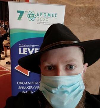
by Andrew Sweeney (a.k.a. Andyana Jones)
| Dec 2020/Jan 2021 14 COVER STORY
you are reading this ... my bad, please let me in again someday as I am rather fond of your food. Also, to Media MICE CEO and Publisher of PIE Matt Young, who I met for the first time after my possible COVID-19 sojourn, apologies: I really do hope it was not me who gave you the virus.
Fortune and glory kid, fortune and glory...
So when I had the opportunity to travel to the only major ophthalmology conference proceeding in person this year, I wondered if I was tempting fate. Was I taking an unnecessary risk by traveling from the Czech Republic? My adopted home was one of the worst affected countries in Europe at that point. And alternately, if my Ethiopian illness was something else — was I running a risk of catching COVID-19?
Was this an elaborate prank — revenge for possibly infecting the boss, designed to leave me stranded in the desert with half a liter of whiskey and a compass? Somehow he does not seem like the vengeful type but who knows ... it is always the quiet ones you have to watch, after all. Still, I decided to take the risk.

Cue “Andyana Jones and the Quest for EPOMEC”. And I’m beginning to think that Matt’s idea of revenge was making me wear an Indiana Jones hat across three continents. If that was the case — kudos — I looked ridiculous.
The 7th Evolving Practice of Ophthalmology Middle East Conference (EPOMEC) 2020 was held in Dubai, the largest city in the United Arab Emirates (UAE) from December 10 to 11. Hundreds of ophthalmologists, clinicians, pharmaceutical representatives and more, attended from the Middle East and surrounding regions. The conference contained all the usual trappings with concession
stands, in-depth seminars and hobnobbing.
EPOMEC’s defining feature was that it offered a glimpse into the future of ophthalmology, where hybrid conferences will become the norm. Live streams of surgery at the Magrabi Eye Hospital Dubai took place on the first day and interactivity was the watchword, which allowed featured guests to ask the surgeons questions intraoperatively.
Corneal issues were featured prominently at EPOMEC, with some highlights including Cornea: My Best Tips in Managing Corneal Disease in 2020, and Cornea Hot Chair: What Would You Do if You Were Me? So too did retina; one of my favorite sessions was Retina: Oscar Video Competition for Best Surgical Video. Multimedia and multi-focused, EPOMEC’s content did not disappoint.
The conference also held an online event, which we have all become familiar with. These online symposiums and webinars will be the new norm as they allow more people to attend conferences who otherwise would be unwilling or unable due to health concerns, financial constraints, border closures or other difficulties. We should appreciate the opportunity to have our collective cakes and eat them, too.
For all of that, it was good to be back among the people and the ophthalmic community, to meet new contacts — and of course, receive free food. You cannot meet wonderful maverick doctors from Yemen, based in Abu Dhabi, who received their instruction in the Russian language in Moscow, and have a chat in the aforementioned language, when peering through an online portal. Or have an engaging conversation about the future of ophthalmology and Dubai’s role in its progress with a pharmaceutical representative, while standing next to an astronaut ... you simply cannot.
But the story was not just the conference, it was getting there too.
The Egyptian layover
One of the earliest mentions of
ophthalmic practice was a reference to the treatment of chronic trachoma in a papyrus, dating back to approximately 1550 BC. It was therefore fitting to visit the Giza Pyramids, though I barely had a moment to film. Such is the paucity of tourists caused by coronavirus that the usual touts and scammers are subsisting on scraps, making them rather forthright in their activities.
“Ah yes my friend, habibi, welcome to Cairo. Please let me give you this gift, it is a traditional headdress, made from traditional polyester … yes, yes, the finest material imported from China, it is absolutely free for you, my friend with nice tattoos. Let me put it on your head, yes, no, no it is a gift, I swear. I know that you do not want it but please, it is Egyptian hospitality, please take it. It’s for free, yes, yes. See, does that not look good on your head? Thank you, thank you for taking my gift. It is free, free, okay, thank you! Give me money, I want 50 Egytpian pounds. Money yes, you took my headdress. I want money, now give it, money, money…”
I was not really ready for this somewhat aggressive sales pitch, and frankly I was a little confused, but the chap did take on a progressively menacing air. Luckily, the driver I had hired and the tour guide (turns out they were in cahoots and scamming me anyway) managed to shake my “friend” off. Better the devil you know I suppose.
Welcome to Cairo, habibi. History does not repeat itself but it does follow a rhythm: Here I am, back in Africa, on a trip associated with COVID-19. At least I could go for a walk this time.
Start the engine!
At this point, you may be asking yourself what I was doing in Cairo: Was the conference not in Dubai, on the other side of the Middle East? Well, booking flights was a challenge, too. I was already slated to be in Turkey prior to the conference and we were unable to book a direct flight.
That left us with the option of finding a layover, which was no easy task in itself. For various reasons including time, cost, quarantines and bans, Lebanon, Jordan, Kuwait and Oman were all out.
| Dec 2020/Jan 2021 15
The only option we had was to go to Dubai from Istanbul via Egypt.

Istanbul was at the other end of the spectrum to Egypt’s capital. A total curfew was declared a few days before I arrived, meaning Turkish citizens were not allowed out at all, except in limited circumstances, from 9 p.m. Friday until 5 a.m. Monday. This however, did not apply to tourists, meaning I had one of the world’s great metropolises almost to myself.
One of my favorite places in the world, Istanbul is a hot mess of a city, usually pulsing with frenetic energy. To see it so subdued, under curfew and without the usual sights and sounds of the largest city in Europe, was surreal. I had plenty of time to take in the sights, hop on one ferry to another between Asia and Europe, and plenty more to dwell on the year that was.
The adventure continues...
For many of us, 2020 was a year of superlative horror, whereas for myself, the COVID-19 cloud certainly had silver linings. Joining Media MICE and taking
part in this adventure was certainly one of those, which to my family is utterly hilarious. Ever since a childhood eye infection, I have endured a crippling anxiety about eyes — to the extent I could never look at someone taking a contact lens out. Imagine my reaction watching eye surgery for the first time.
I was living in Palestine before COVID-19 hit, indulging an ambition to become a respected war zone correspondent, while somehow skipping the intermediate stages and the experience they would imbibe. As my year began in the Middle East so it ended too, sentimentally at least, in Dubai. My first ever ophthalmology conference and hopefully the first of many (Serena, Media MICE’s absolutely wonderful accountant, please take note).

If you want to be a good ophthalmologist you gotta get out of the clinic
If anything really sticks out from my adventure, or from the whole of benighted 2020 really, it’s actually something that sticks up. Never before
have my nostrils been so assaulted — and for those of us who have taken at least one COVID-19 test — the sensation of a Q-tip launched with the speed of a cruise missile into one’s sinuses is a memorable one. If you are planning on attending any ophthalmology events in 2021, be prepared for it.
For you will be tested, repeatedly and with aplomb. I was only able to enter Egypt with the negative COVID-19 test I took in Istanbul, and that should have been enough for Dubai. However, I had to take another test on arrival in Dubai and remain in quarantine until I received my results, which nearly scuppered everything.
Make sure you plan for every eventuality and leave wiggle room in your schedule. As the emergence of a new, far more contagious form of COVID-19 in England’s Southeast highlights, borders can close and plans change with remarkable speed. At least thanks to the hybrid conference model, you will not miss out on everything if you cannot attend.
Soon enough, conferences will resume with something approaching regularity, and you will be able to gaze in awe at the sight of our own Matt Young cutting about in a new outlandish costume. You might even see me make another appearance in the bloody Indiana Jones hat, who knows.
The point is that while many things will be different, ophthalmology will persist… And Media MICE will be there to report on it, wearing outlandish costumes.
Editor’s Note:
EPOMEC 2020 is the first ophthalmic conference during the pandemic which allowed onsite participation of delegates.


| Dec 2020/Jan 2021 16 COVER STORY
Company stalls still made an appearance.
I finally made it!
The infamous (totally a gift) headdress.
Socially distanced seating in the main conference room.
A rare moment of calm in Egypt
COVID-19 Pandemic and the Vitreo-retina Practice
by Tan Sher Lynn
Patient volume — it’s a real issue since the pandemic hit. According to Dr. Alay Banker, director of Banker’s Retina Clinic in Ahmedabad, Gujarat, India, the number of patients has been reduced by about 60% since the pandemic began. Due to the need for social distancing and disinfecting chairs and equipment after each patient, he has had to reduce the number of appointments and keep surgeries to a maximum of two or three per day.
Income has also gone down by about 50-60% compared to the previous year. This was due to the decrease of patients and increase in expenses, such as purchasing personal protective equipment (PPE) and cleaning and sanitizing products, which served as a double blow to the business.
“In order to reduce the time that patients spent in the hospital, we came out with a very good system. Fifteen minutes are set for each appointment. Everything is done prior to the patient’s arrival — history is taken via phone and reports and previous investigations are sent via email. Patients are instructed to stay in the car and wait for their turn to get their retina checked, which is essentially done within five to 10 minutes,” he said.
“Only one person is allowed to accompany the patient and personal belongings have to be deposited outside the clinic. There are only two chairs, placed six feet apart, in the sitting area. The entire staff wears full PPE. Sometimes we also do a video call with relatives to explain what’s happening.”
People tend to have a habit of pulling their mask down when they want to talk. To prevent this, they apply tape over the patient’s nose and mask at the hospital entrance. This simple act
also prevents the examination lens from fogging.
In order to avoid physical contact, the windows and doors of the hospital are open to provide complete ventilation. This also keeps people from touching the doors. All of these measures have made his patients feel safe.
To Dr. Banker, the positive side of the pandemic is the increasing opportunities for discussion among colleagues and getting in touch with international faculty through webinars, where a lot of useful ideas are exchanged in regard to treating and caring for patients.
During the pandemic, people tend to refrain from going to places like hospitals. Therefore, he thinks that more new long-lasting anti-VEGF drugs, such as brolucizumab, will emerge as they reduce the number of injections needed and thus, hospital visits. “Another innovation which will probably be increasingly used is widefield photography, which takes an image of the entire eye and reduces the time of contact with people, as opposed to physically examining the eye,” he said.
were mainly emergencies and urgent cases. The reduction of patients has taken a toll on his practice, causing him to seek help from the private banking system. And as the hospital was directed to cut down on the number of employees, the staff are required to maintain the quality of work with less manpower. There are also other challenges, such as the need to be extra-careful not to break new policies in clinics and at the hospital, and respecting the practice of wearing masks, washing hands and physical distancing.
Yet, in the midst of all this, excellent opportunities for knowledge sharing and deepening of relationships have emerged, taking on the form of online lectures and webinars. “All of these brought us closer together, even though we are physically distant,” he said.
Moving into 2021, Dr. Nakamura thinks that there are now better structures to deal with the challenges that COVID-19 brings, and to avoid its spread. “In terms of innovation, we are now more ready and capable of carrying out new ideas and finding new purposes and objectives in our career, with the ultimate aim of achieving the best results for everyone,” he said.
To Dr. Hudson de Carvalho Nakamura, an ophthalmologist at the Eye Bank Foundation in Goias, Brazil, the pandemic has really affected medical practice. “We needed to reduce our team, change our schedules, and organize only emergency surgeries and space them in a broader way. We also needed to screen patients on a daily basis, observe suspicious patients, and have patients examined alone, unless they were unable to see due to poor visual acuity,” he said.
Due to COVID-19, his hospital received, on average, less than 40% of its usual patients, and procedures

| Dec 2020/Jan 2021 17
2020 YearEnd Wrap-Up
Let’s Hear it from the Industry KOLs & CEOs
We can all surely say that 2020 has been a year like no other, thanks to the COVID-19 pandemic. In ophthalmology, both business and practice has shifted (for better or worse). So, throughout 2020, PIE magazine CEO Matt Young interviewed many of the industry’s KOLs for their insight into this tumultuous year. Below, we share some of these thoughts.
On pandemic induced changes:
“There is so much innovation in ophthalmology, but will there be enough resources for those companies to continue operations? Before, business was moving along nicely for everyone and now it’s kind of falling off a cliff in terms of funding. Over a period of time, that will come back. But this will delay innovations for ophthalmology in the R&D pipeline . . . ultimately, some won’t make it. There’s just not enough resources.”
“There will be a change in how patients are managed. It won’t matter based on your specialty [if you’re anterior or posterior specialist], you will have to change your way of handling patients. And this is applicable for ophthalmologists, optometrists and opticians. Our OCT, especially OCT-A, can be used from one room to another. So you can definitely respect social distancing rules. In fact you can use the OCT on your tablet to keep patients distanced.”
“Telemedicine is really the way to carry on now . . .telemedicine means right now, we are organizing our department with one triage that can be done now with a phone call — it begins as a general triage and then an ophthalmology triage. And then we can choose if the patient goes for telemedicine or a face-to-face visit. And telemedicine also means home delivery care, also specific imaging that (we) can get remotely.”
| Dec 2020/Jan 2021 18
— Thomas Dunlap, ophthalmic medical device consultant, Orange County, California, USA
— Adel Bencheikh, director of eye care, Canon Medical Systems Europe B. V., Zoetermeer, The Netherlands
— Dr. Mario Romano, Bergamo, Italy
On annual meetings:
“If you look at all the academy, ASCRS, and ESCRS, the greatest penetration is in the speciality days. It is in the mind of the doctors that they want speciality. So, I think you are still going to see the speciality days. The small meetings, which have been proliferating over the past years, they are just not going to be able to continue. Financially, doctors can go, some companies can survive and change dramatically.”
— Jim Mazzo, CEO and president of Avellino, California, USA
“[In the future] I think a few major international meetings will have to continue like APAO, AAO and the main European meetings . . . but a lot of the smaller satellite meetings or single specialty meetings will probably be scaled down.”
“I truly believe that with virtual engagement you can reach a much broader audience than ever before. If you ask me about the future of congresses, digital is not going to go away and it’s here to stay. Personally, I’m looking forward to supporting societies and to really strive for excellence in delivering this, because it’s going to be part of the new normal.”
— Jan Voss, VP and Head of Therapeutic Area Ophthalmology, Bayer Consumer Care AG
| Dec 2020/Jan 2021 19
— Dr. Kenneth Fong, managing director of OasisEye Specialists in Kuala Lumpur, Malaysia


2 18 November 2022 | Issue #1 SHOW DAILY by DIGITAL MARKETING + ADVERTISING + VIDEO PRODUCTION + MEDICAL WRITING + EVENTS Request our 2023 Media Kit Now! Write enquiry@mediamice.com for a copy HQ Office: 6001 Beach Road, #09-09 Golden Mile Tower, Singapore 199589 Phone: +65 8186 7677 Satellite Office: 2 Nuoc Man 2 Street, Da Nang City, Vietnam 50506 Phone: +84 868 063 773 E-mail: enquiry@mediamice.com Web: www.mediamice.com
All Eyes on the Retina Innovation Showcase
by Joanna Lee
From novel functional vision tests, to treating genetic diseases and discoveries on inhibiting protein cells, innovations are forging ahead despite the challenging environment — and giving a glimpse of hope in the near future.
Despite 2020 being a disruptive year in terms of ophthalmologists’ personal and professional lives, the retina space is continuing to thrive in terms of the development of drugs and devices,
said Dr. Firas Rahhal, a partner at Retina-Vitreous Associates Medical Group (Los Angeles, CA, USA) and ExSight Ventures (New York, NY, USA) during the opening of the OIS Retina Innovation Showcase.
Novel vision test
First in the showcase was Ora (Andover, MA, USA) introducing its Ora-VNC™ Mobility Courses. It’s a
novel functional vision test designed to address various levels of visual deficit (acuity and visual field) to address areas of unmet need, particularly in the areas of inherited retinal diseases (e.g., LCA, Stargardt’s, Achromatopsia).
Ora’s Executive Director and Therapeutic Area Head of Posterior Segment, Keith Lane, said the company behind 47 ophthalmic product approvals so far, shared how they saw the need to create consistency in the form of modular courses that can be installed in any setting.
They developed four different mobility courses designed to address various

| Dec 2020/Jan 2021 21 INNOVATION INDUSTRY UPDATE
levels of visual deficits, as well as the capability to facilitate multi-site clinical trials. Features include wellcontrolled lighting systems, lighting rigs and independently dimmable bulbs controlled via smartphone or computer. There are also multiple iterations which facilitate repeat testing and prevent memorization with light levels validated for consistency across each course tile for all four courses. To keep the consistency, Ora also offers services to install the course, for light level validation, site training and certification, and reading center grading services.
So far the company has done 24 installations in North America and European locations.
“There are classrooms, auditoriums or hospital room settings, so the flexibility in our lighting control system we use enables us to standardize conditions in all these settings,” Mr. Lane said.
“We’re quite comfortable that we’ve now created an endpoint that supports the development of products for inherited retinal diseases. We’ll hopefully allow these products to make it to market where they can actually help patients with blinding genetic diseases,” he added.
Next on its innovation table is Ora’s AMD endpoint program. It is still in its early stages of development. “The goal of this program is to be able to identify a functional deficit in AMD patients prior to reaching the point where geographic atrophy develops,” explained Mr. Lane.
Ora then developed the Ora-VCF™ Contrast Test (a variable contrast flicker test).
“What we found is that this test is able to differentiate between normal and non-advanced AMD populations, while traditional endpoints such as visual acuity and Pelli-Robson contrast sensitivity tests are not able to do so,” said Mr. Lane.
These developments add hope to identify a functional deficit in these earlier stage patients that may be augmented by potential therapies. Currently, the company is discussing and negotiating with the FDA on the contrast flicker test.
Regenerative medicine and gene therapy
Regenerative medicine has also been propelling forward in its momentum. Chief Medical Officer Dr. Aaron Osborne of Adverum (Redwood City, CA, USA), spoke on its lead clinical program called ADVM-022, which is an intravitreal (IVT) novel gene therapy candidate for VEGF-driven retinal diseases. It is designed to deliver longlasting efficacy via continuous delivery of aflibercept in a single injection with a goal to maintain vision, while reducing the treatment burden. So far, trials on three cohorts over the past year (the longest being 84 months for cohort 1) have shown impressive results with no adverse events associated with ADVM-022. There have been zero supplementary anti-VEGF injections when administered at a high dose (of ADVM-022) and significantly reduced supplementary anti-VEGF injections at a low dose with a dose response being apparent. Any intraocular inflammation has been responsive to and manageable with steroid eye drops. There’s no sign of posterior inflammation, vasculitis, retinitis, choroiditis, vascular occlusions, nor endophthalmitis reported.
This IVT gene therapy can also be used for diabetic macular edema (DME) to prolong the effects of aflibercept and potentially prevent sight loss from this condition, as seen in their INFINITY trials.
Next, Dr. Magali Taiel, chief medical officer of Gensight Biologics (Paris, France) spoke about Lumevoq, a gene therapy for Leber Hereditary Optic Neuropathy (LHON) patients. LHON is the most common mitochondrial disease and can cause bilateral blindness. Patients with this disease
experience a rapid and sequential drop in vision once it occurs, and within one year, they can become blind.
Lumevoq gene therapy specifically targets the mutations that cause this genetic disease.
Gensight’s RESCUE & REVERSE studies, in collaboration with the EMA (European Medicines Agency), included patients with vision loss of less than 6 months (REVERSE) and patients with vision loss of more than 6 months (RESCUE). They found close improvement patterns in both Lumevoq and sham treated eyes. To compare with the natural history of the disease, they also completed another study called REALITY.
The results were remarkable, with patients reaching close to 6 lines of improvement (28 letters) for REVERSE and about 5 lines (26 letters) for RESCUE.
“These improvements are far above the three line threshold commonly accepted at the clinically meaningful level of visual improvement,” Dr. Taiel said. She added that in subsequent non-human primate studies they discovered there was DNA transfer of Lumevoq from the injected eyes into the non-injected eyes.
They will be filing for EMEA registration in September this year based on the REVERSE and RESCUE study results, while their U.S. registration is expected next year (in Q1 2020) based on their REFLECT study results, which are ongoing to assess the efficacy and safety of bilateral injections (phase 3 trial).
Application of immunology technology
In another interesting development, Aviceda Ophthalmics (Cambridge, MA, USA) Chairman, CEO and Founder Dr. Mohamed Genead introduced their glyco-immunology technology, which can be used to develop drugs to modulate the innate immune system.
| Dec 2020/Jan 2021 22 TREATMENT REGIMEN INNOVATION INDUSTRY UPDATE
The innate immune system is related to problems in the eye and may be relevant to macular degeneration with regard to the innate immune system.
“Our immune technology is based on a nanoparticle platform to deliver and optimize on our biological response,”
Dr. Genead said. He added, “We have a lead asset called AVD-104 (glycan-coated nanoparticle) which is a lead optimized nanoparticle in INDreadiness phase for dry AMD.”
Dr. Genead explained how macrophage plays a role in the innate immune activation of AMD, where no therapies thus far have directly targeted the macrophage activation.
“We try to deactivate the cells and re-polarize the activated cells to the resolution healing state of the cell,” he said. At the same time, they try to deactivate the alternative complement pathway.
He said they try to target the macrophages via Siglecs (sialic acid binding immunoglobulin-type lectins) modulation while explaining the biology of their mechanism.
Dr. Genead further said Aviceda is the only therapeutic platform with the flexibility to fully address the innate immune system.
In another development, clinical stage company, Iveric Bio (New York, NY, USA) has developed Zimura, which blocks the C5 cleavage complementary pathway in an attempt to treat dry AMD and geographic atrophy. The company’s Executive Vice President and Chief Business and Strategy Officer Dr. Pravin Dugel, said there is positive data for the first two phase 3 trials with a statistically significant reduction (27%) in geographic atrophy growth over 12 months, with potential expansion into intermediate and wet AMD areas.
“Preserving C3 while inhibiting C5, we believe is key to the success of Zimura,” he said. C3, Iveric deems, plays roles in ischemia-reperfusion.
Stem Cell Technology and Protein Inhibitors
Meanwhile, jCyte (Newport, CA, USA), which was founded out of UC-Irvine, introduced their stem cell technology, which delivers stem cell therapy through a simple intravitreal injection for retinitis pigmentosa, and is undergoing clinical trials. CEO of jCyte Paul Bresge said their phase 2b shows promising results in retinitis pigmentosa (RP). They are currently preparing for a global phase 3 trial for RP in collaboration with Ex-U.S. licensing partner Santen (for Q4-2021). How it works is that their proprietary jCell Intravitreal Cell Therapy can remain in the vitreous for extended periods, up to 12 months, after injection. The jCell does not target a particular genotype, but releases neurotrophic factors to reduce photoreceptor cell death and promote the function of existing photoreceptors.
Also advancing on is ONL Therapeutics (Ann Arbor, MI, USA). Its President and CEO, David Esposito shared the exciting development of its Fas inhibitors “It’s the first-inclass neuroprotective platform to inhibit the Fas receptor and protect retinal cells,” he said. Fas inhibition prevents cell death signaling at the initiation point (surface receptor) to protect vision. ONL1204, a peptide delivered through intravitreal injection, blocks apoptosis to improve macular detachment.
This protein inhibitor has relevance and possible indications for rhegmatogenous retinal detachment (RRD), acute primary angle closure glaucoma (APAC), dry AMD/geographic atrophy, and inherited retinal degeneration (IRD), among other conditions involving the retinal cells. Mr. Esposito revealed they have been issued a U.S. patent for ONL1204 with earliest possible expiry in 2036.
In summary, all eyes are looking toward these innovations coming into fruition in the near future. Dr. Rahhal also mentioned there are other innovations in the pipeline like drug delivery technology — and large and small companies are aware of this need. As a heads up, Genentec is developing a surgical implant while other companies like Re-vana, EyePoint and others, are developing solid and gelatinous carrier-type molecules to make the drugs last longer.
These discoveries and innovations will subsequently be featured on OIS so stay tuned for the next showcase.
Editor’s Note:
OIS Retina Showcase, organized by Ophthalmology Innovation Summit (OIS), was held on September 10. Reporting for this story took place during the event. A version of this article was first published online at piemagazine.org on September 11.
| Dec 2020/Jan 2021 23
Dr. Nancy Holekamp Advancing Women in Ophthalmology
by April Ingram
Here at PIE, we love to shine a spotlight on the exceptional work being done in ophthalmology and profile those retina specialists who are leading the way. We especially enjoy highlighting those who are successful leaders and mentors, and also happen to be women. In this issue of PIE, we consider it a privilege to introduce our readers to Dr. Nancy Holekamp – although I’m certain that no introduction is necessary.
Nancy M. Holekamp, MD, is the director of Retina Services at the Pepose Vision Institute in St. Louis, Missouri, USA. Prior to that, she was professor of clinical ophthalmology and visual sciences at the Washington University School of Medicine in St. Louis (Missouri, USA).
“No” to a desk job
Dr. Holekamp received her undergraduate Bachelor of Arts degree from Wellesley College, Summa cum Laude and studied medicine at Johns Hopkins School of Medicine Alpha Omega Alpha. She knew she wanted to be a physician while in college and recalls, “I knew I didn’t want a desk job. I was seeking a job where I would be continually moving, interacting and helping people.”
During the summer after her second year at Johns Hopkins School of Medicine, Dr. Holekamp worked at the Retinal Vascular Center at the Wilmer Eye Institute under the mentorship of Dr. Stuart Fine,
“So I was always destined to be a retina specialist.” When asked Dr. Holekamp if for some reason she was not a retina specialist, what career path she may have taken, she said: “I would have been a college professor. It would be entirely consistent with my need

to passionately teach and continually learn.”
Dr. Holekamp completed an internship in internal medicine and a residency in ophthalmology at the Washington University School of Medicine. At that time, there were not a lot of women in these programs, she shares. “As an ophthalmology resident, I was grateful to have another woman in my residency class, so I always had an empathetic ear or a shoulder to lean on.” Her fellowship training in vitreoretinal surgery was with the Retina Consultants in St. Louis, where all of the attending physicians and all her peers were men, however Dr. Holekamp did not see this as a negative. “They were 100% supportive of my career. In fact, being a woman allowed me to stand out.”
Championing women in retina
Dr. Holekamp has always been a mentor and champion for women in ophthalmology, and more specifically in retina, and she is pleased to see more women taking on the specialty. She and four other women retina specialists cofounded Women in Retina in 2007. She explains: “At that time, only 11% of all retina specialists were women. That number has risen to 17% so things are slowly changing.” In 2018 she was awarded the Suzanne Verronneau-Troutman award for her contributions to advancing women in ophthalmology.
| Dec 2020/Jan 2021 24 WOMEN IN OPHTHALMOLOGY ENLIGHTENMENT
Dr. Holekamp is a member of the major subspecialty societies, including the Retina Society and the Macula Society. She acts as a reviewer for all the major ophthalmology journals and as a consultant to several ophthalmic pharmaceutical companies. After six years on the American Academy of Ophthalmology (AAO) Ethics Committee, she has developed an interest in medical ethics.
Many of us have had the pleasure of hearing Dr. Holekamp at retina conferences around the world as a highly sought-after expert and speaker, receiving more than 120 speaking invitations both nationally and internationally. This is in addition to her 80+ peer-reviewed publications and 22 book chapters she has authored.

Even though this year we have not had the opportunity to meet with our colleagues face-to-face at all the big ophthalmology conferences, like AAO or ASRS, even in their virtual state, Dr. Holekamp shares her favorite part of these meetings. “I look forward to seeing data presented for the first time, whether it is from large clinical trials or small, innovative investigator-initiated studies. We have so many smart people in our field. I like to see what they are doing and how they are thinking. I can always learn something new.”
We all recognize Dr. Holekamp as being highly active in clinical research, having been principal investigator or sub-investigator in over 38 national clinical trials dealing with age-related macular degeneration, retinal vascular occlusion, and diabetic retinopathy. When asked to share what it was about being involved in research that she enjoyed so much, she replied, “There is nothing in my practice of retina that is the same as when I trained 25 years ago. Thus, I am keenly interested in learning new things and being on the cutting-edge of our field. I am an early adopter of technology that is validated by large randomized clinical trials. They move our field forward.”
After treating thousands of patients over her 25 years as a retina specialist, Dr. Holekamp explains that the best thing a patient can say is “thank you,” and adds: “That sentiment is still the most meaningful part of my job.” When we
asked what the worst thing might be, she shared: “The worst thing a patient and I can go through together is the irrecoverable loss of vision despite our best efforts. Sadly, it still happens from time to time. Retinal disease can be unrelenting.”
Keeping the inner nerd alive
In addition to her clinical practice, busy surgical schedule and research activities, Dr. Holekamp founded a vision mission to La Gonave, Haiti, inspired by one of her wet AMD patients, “We have supported annual trips (until the pandemic) in which 1,000 pairs of donated glasses were given to some of the poorest, underserved people of that impoverished nation.” This important work is also a family affair, “During these charitable trips I was able to bring my two teenage children, allowing them to feel the benefits of helping people quite different from themselves. It has been an incredibly rewarding experience.”
Dr. Holekamp’s work has been honored with numerous honors and awards, including being named in the “Best Doctors in America” annually since 2005 and is included in the US News & World Report Best Doctors list and Castle Connolly Top Doctors list. She has received the Achievement Award, the Senior Achievement Award, and the Secretariat Award from the American Academy of Ophthalmology (AAO), and the Honor Award and Senior Honor Award from the American Society of Retina Specialists (ASRS).
Aside from all her busy practice, research, accolades and accomplishments, did you know that Dr. Holekamp is also a huge ice hockey fan? Check out the photo of Dr. Holekamp and her son Jack and daughter Katie during the Stanley Cup playoffs, cheering on their St. Louis Blues. She also loves a good laugh, and in keeping with the science and research theme, when we asked if Dr. Holekamp had a favorite TV show, she shared, “My favorite TV show of all time is Big Bang Theory. I still laugh out loud while watching reruns. I guess the show speaks to my

inner nerd.” We love this about her! She likes to read comedy as well. “One of my favorite books is Born a Crime by Trevor Noah.” Also, as a PIE breaking news exclusive, did you know that Dr. Holekamp has an identical twin? She does! “I call her the ‘smart twin’. She retired from investment banking at a very early age. But, she never loved her job like I do,” she shared.
Finally, we asked Dr. Holekamp if she had any words of wisdom to offer women that may be beginning or considering a career in retina and her message is clear: “Go for it! There are few other careers in medicine that are as rewarding as being an ophthalmologist or a retina specialist. And, if you don’t think you can do it, you can. There are a lot of women ahead of you showing you the way. It is possible!”
Contributing Doctor
Nancy M. Holekamp , MD, is the director of Retina Services at the Pepose Vision Institute in St. Louis, Missouri, USA. She is a highly sought-after expert and speaker in retina conferences worldwide. Dr. Holekamp, a holder of numerous honors and awards in her field is a member of the major subspecialty societies, including the Retina Society and the Macula Society.
nholekamp@peposevision.com
| Dec 2020/Jan 2021 25
Dr. Holekamp (R), with her children.
Trauma Management on Spotlight at APOTS Virtual Conference
 by Tan Sher Lynn
From treating orbital trauma to various neuro and pediatric trauma cases, experts shared their experiences during the 5th Asia-Pacific Ophthalmic Trauma Society (APOTS) virtual conference.
Pearls of orbital trauma management
Orbital fractures account for a significant portion of traumatic facial injuries — and quick diagnosis and management prove fundamental for treatment success. Dr. Priti Udhay from the ORR Eye Care and Oculoplasty Hospital (India), shared a case where a patient had hyperglobus due to a roof fracture. “Diplopia due to hyperglobus is a problem usually seen in fracture patients. The globe position is the first important thing you must check in a trauma patient,” said Dr. Udhay. In this case, titanium mesh is used to reconstruct the roof and correct his hyperglobus and dissolve the diplopia. “Another reason for consistent diplopia is because the surgeon has not done forced duction properly. As such, forced duction tests should be done in various stages during fracture repair surgery — not only when you start the surgery, but also after removing the interorbital content and after placing the implant,” she stressed.
Speaking on optic canal decompression (OCD) in traumatic optic neuropathy (TON), Dr. Farzad Pakdel, from the Tehran University of Medical Sciences (Iran), said that currently, OCD cannot be recommended as a first-line treatment option for all TON patients. “No well-designed study has shown whether surgical decompression or steroids have any better outcome than observation alone. OCD is done right over the optic nerve and can potentially
induce more injury. Nevertheless, OCD is still viable and can be offered to patients with TON and severe or bilateral visual loss with neuroprotective strategies,” he said.
Sharing about his experience in treating eyes with orbital foreign bodies (OFBs), Dr. Mehbub Ul Kadir, from the Bangladesh Eye Hospital, noted that identifying OFBs requires a high index of suspicion. “CT scan of the orbit is the first choice, but MRI is better at detecting wooden foreign bodies than a CT scan. If you find one OFB, search for another. Remove all organic OFB. But an inert, small OFB may be left as it is,” he said.
Meanwhile, Dr. Mariel Parulan, from the Quirino Memorial Medical Centre
(Philippines), discussed an unusual pediatric trauma case where a 5-yearold girl, after being hit on the head by a broom, had orbital subperiosteal hematoma — a rare pathology where blood pools within the space between the periosteum and orbital bones. On the first consultation, VA (visual acuity) was 20/400 and IOP (intraocular pressure) was 20mmHg for both eyes. There was also chemosis with mucopurulent discharge, and the cornea was hazy with superficial defects. Rapid swelling and proptosis occurred within a few days and the patient was admitted on day five. Surgical evacuation via needle aspiration under general anesthesia was performed, resulting in 3cc of blood from the right eye and 2.5cc from the left.
| Dec 2020/Jan 2021 26 APOTS 2020 COVERAGE CONFERENCE HIGHLIGHTS
“Don’t let mild trauma deceive you, especially in pediatric patients — it can lead to significant subgaleal hematoma with orbital extension and result in rapid ophthalmic deterioration. Typical symptoms of this condition are a swollen forehead and periorbital area. It’s important to make the diagnosis early with clinical findings, confirmed by imaging tests. And when vision starts deteriorating, immediately go for evacuation to avoid permanent complications,” she said.
Last but not least, Dr. Suzanne Freitag, from the Harvard Medical School (USA), shared her 25 years of experience in dealing with orbitofacial trauma. “Lesson one, always take a good history. Second, speak to the victim alone, away from family and accomplices, to give them the opportunity to tell the truth or ask for help. Thirdly, there is almost never any tissue missing in trauma cases (except gunshot wound and dog bites),” she said, explaining that lacerations typically come together perfectly. She stressed that surgeons should clean the wound well to prevent infections and avoid tattooing of the skin, adding that titanium doesn’t have a role in the orbit. “I prefer porous polyethylene (PPE) implants for orbital fractures. For very small fractures that need minimal, short-term support, I use the 0.075mm gelatin film, which is absorbed in weeks to months,” she said.
Diagnosing and treating traumatic neuro- and pediatric ophthalmology cases
According to Dr. Alia Arianti, from JEC Eye Hospitals and Clinics (Indonesia), ophthalmic trauma accounted for 1.6 million cases of blindness globally, usually caused by falls, assault or vehicle accidents. “Imaging modalities can help in early detection and management of ophthalmic trauma, which are sometimes initially neglected due to other life-threatening injuries sustained,” noted Dr. Arianti.
CT scans, which utilize X-rays to produce cross-sectional images of the body, are superior in the evaluation of bony structures, according to Dr.
Arianti. MRIs utilize magnetic fields and radio waves, using non-ionizing radiation to produce three-dimensional images in any plane and are superior in the evaluation of soft tissues, including the optic nerve and periorbital soft tissues, and organic foreign bodies. “In summary, CT scans remain the gold standard for imaging in the acute setting of trauma; while MRIs are invaluable for secondary assessment of visual pathway post-trauma or subacute/chronic traumatic brain injury,” she said.
Dr. Haoyu Chen, from the Joint Shantou International Eye Center (China), presented two cases of preexisting posterior capsular rupture (PCR) in pediatric traumatic cataract. Both patients, a male and a female, were 4-years-old and had unilateral white cataract. Both could not tell the history of ocular trauma. “However, the ultrasound B-scan identified PCR, suggesting that the cause may be trauma, which was subsequently confirmed during operation. The first case involved anterior capsular laceration and posterior iris synechia; while the second patient had an intact anterior capsule, suggesting blunt trauma. Identification of PCR preoperatively helped us in diagnosis and operation planning, reducing bottle height and aspiration, thereby increasing patient safety,” he said.
Meanwhile, speaking on the management of pediatric eyelid lacerations, Dr. Emily Wong, from the Hong Kong Eye Hospital, said that common causes of eye trauma in children are dog bites, handlebar injuries and collisions with sharp objects. Physicians should also watch out for child abuse. “If an animal or human bite is involved, there is a higher chance of involving the lacrimal apparatus, including the inferior canaliculus. Vigorous cleansing and irrigation of the wound with saline, checking of rabies status of the animal, and antibiotics coverage are definitely needed. During the ocular exam, we need to rule out life-threatening and emergency ocular conditions. Good choices for repairing wounds in children include fast-absorbing sutures and tissue adhesive as it is fast-acting and no needles are required,” she said.
Optic nerve avulsion (ONA) is the separation of the optic nerve fibers from the globe at the level of the lamina cribrosa, without rupture of the optic nerve sheath or the adjacent sclera. Dr. Bikram Thapa, from the Lumbini Eye Institute (Nepal), presented a case where a 17-year-old man was diagnosed with right eye traumatic ONA (OD) due to slingshot injury. Pulse steroid was given to him for three days but there was no improvement. “ONA is a rare form of traumatic optic neuropathy (TON) which is currently untreatable and has poor visual outcome. Using a B-scan is very helpful for investigation where ophthalmoscopy is not possible, while MRI of the brain and orbit confirm the diagnosis. Pulse steroid therapy and optic nerve sheath decompression are not helpful, so differentiating ONA from other TON helps us to prevent unnecessary treatments and provide the necessary counseling to the patient,” he said.
According to Dr. Prem Subramanian, from the University of Colorado School of Medicine (USA), TON is related to force transmission. “Currently, models are being recreated in the lab so that we can come out with better treatment strategies and validate them. Secondly, TON and TBI can co-occur and different visual concerns in TBI are still poorly understood. Unfortunately, TON treatment remains unproven. So, prevention is the key, especially because patients who had one episode of TON are at risk of getting a second one. Preventive measures can include wearing a helmet when riding a motorcycle or face shields when playing certain sports,” he said.
Editor’s Note:
The 5 th Asia Pacific Ophthalmic Trauma Society (APOTS) virtual conference was held on September 5 and 6, 2020. Reporting for this story took place during the event.
| Dec 2020/Jan 2021 27
Data from AAO’s IRIS Registry
The Key to Better Treatments in the Future?
by Hazlin Hassan
Research from the American Academy of Ophthalmology’s IRIS Registry was presented at the AAO 2020 Virtual annual meeting in November, with the world’s largest specialty clinical data registry revealing fresh insights into eye disease prevention and treatment.
The registry powered a number of big data studies this year, which can only pave the way for better diagnostics and treatment.
“With a growing pool of aggregated data, the IRIS Registry empowers ophthalmologists to recognize disease patterns, better define risk factors, and improve prevention and treatment options for their patients,” said Flora Lum, MD, vice president of Quality and Data Science at the American Academy of Ophthalmology (AAO).
“All IRIS Registry studies presented at AAO 2020 this year reveal interesting and promising results for a better future in eye care,” she added.

Launched in 2014, the IRIS Registry is the first comprehensive eye disease registry in the United States. It currently includes data from nearly 60 million unique patients and 350 million patient visits.
Here are some of the IRIS Registrypowered researches presented at the AAO 2020 Virtual.
COVID-19 and the anti-VEGF injection rate
In a study on the impact of the COVID-19 pandemic on anti-VEGF injection rates, Dr. Stephen D.
Schwartz, from the University of California (Los Angeles, USA) and colleagues, sought to find out if the coronavirus affected anti-VEGF injection rates, and if patients suffered vision loss as a result.
During the 2020 pandemic, visits were interrupted for a period of time. The slow-down was both doctor- and patient-driven.
On March 18, 2020, the AAO issued a statement that all ophthalmologists should cease providing any treatment other than urgent or emergent care due to the outbreak.
Prior to the COVID-19 outbreak, the daily average number of patient visits was 214,000 per day, but this dropped to a trough in April at 34,000 visits.
The number of anti-VEGF injections in the IRIS Registry data between October and March was 3,070,382 injections, or 511,730 a month. The total number of anti-VEGF injections in the 12 weeks after March 18 came to 1,196,341 or an average of 99,695 a week.
Analysis showed that ophthalmologists’ response to the pandemic was measured and appropriate, and that patients’ vision was not adversely affected.
| Dec 2020/Jan 2021 28 AAO 2020 COVERAGE CONFERENCE HIGHLIGHTS
Along with socioeconomic status, race and gender can impact surgical results.
“Overall, vision was stable across all injection groups. The 10% of patients who did not receive injections lost approximately one line of vision,” said Dr. Schwartz.
Vision quality after corneal transplant
Endothelial keratoplasty (EK) is the procedure of choice for endothelial disorders due to faster visual recovery, more predictable refractive error and favorable complication profiles compared to penetrating keratoplasty. But a recent study has found that race and lower socioeconomic status were risk factors associated with worse visual outcomes following corneal transplants.
Divya Srikumaran, MD, who is a consultant for Alcon, and co-authors Stephen Hsu and Sudeep Pramanik, MD, conducted a retrospective analysis of IRIS Registry data involving 26,267 eyes that underwent EK surgery between 2013 and 2018.
It revealed that 70% of patients had improvement in visual acuity (VA) postoperatively with a median VA of approximately 20/40.
The risk of worsening VA was associated with female gender versus male; Black
race versus White; Medicaid insurance versus private; and the presence of ocular comorbidities, including glaucoma, macular degeneration and diabetic retinopathy.

In this large national cohort, female gender, Black race and lower socioeconomic status as indicated by insurance type were independent factors associated with worse VA outcomes after EK in analyses adjusting for multiple other factors.
Patients with the above risk factors need to be advised that they are at risk for worse vision after surgery. However, disparities identified in this study warrant further investigation so that strategies may be developed for mitigation.
Race, health insurance and diabetic eye disease
A study to investigate the influence of social determinants on baseline characteristics and health-care outcomes in diabetic macular edema (DME) treated with anti-VEGF injections, found significant disparities among patients with DME.
Black patients were more likely to present with proliferative diabetic
retinopathy (PDR) compared to white patients, while Hispanic patients appeared to receive the fewest antiVEGF injections and suffered poorer visual acuity, according to Nisha Malhotra, MPH, and colleagues from the Cleveland Clinic (USA).
The study involved 203,707 DME patients in the IRIS Registry who had received at least one anti-VEGF injection.
Regression analyses were carried out on how race, ethnicity and insurance status influenced presentation features and clinical outcomes such as severity of diabetic retinopathy, anti-VEGF injection utilization and VA.
The results showed that Black patients were more likely to present with PDR compared to White patients (46.7% versus 41.3%).
Being of Hispanic ethnicity had the strongest association with receiving fewer anti-VEGF injections and having poorer VA after 12 months of treatment.
Relationship between IOL Type and PCO diagnosis
Bret L. Fisher, MD, from the Eye Center, North Florida (USA) and co-authors,
| Dec 2020/Jan 2021 29
conducted a study on the relationship between intraocular lens (IOL) type and posterior capsule opacification (PCO) diagnosis to find out which IOL types are associated with a shorter time to PCO development.

The study, which was sponsored by Alcon Vision, compared the mean time to PCO diagnosis in different types of IOLs with two hydrophobic acrylic materials: AcrySof (A) and Tecnis (T). A retrospective analysis was carried out on 43,023 eyes between 2016 and 2018 which had at least two years of follow-up after cataract surgery.
Results showed that 35.7% of eyes had PCO diagnosis, with mean diagnosis at 268 days.
Earlier PCO diagnosis was observed in eyes with multifocal or diffractive EDOF IOLs compared with monofocal IOLs. Hence, IOL biomaterial impacts time to PCO diagnosis, and AcrySof IOLs may be more resistant to PCO formation compared to Tecnis.
Effectiveness of brolucizumab in treating AMD
Phase III trials have shown that brolucizumab is an effective treatment option in wet AMD, with the potential to extend injection intervals. Brolucizumab 6mg provides robust visual gains and greater anatomical outcomes with over 50% of patients on a q12w dosing interval after the initial loading doses until week 48.
A study on the effectiveness of brolucizumab in treating AMD found that the majority of wet AMD patients initiating brolucizumab switched from a prior anti-VEGF agent.
Therefore, unmet needs continue to exist for many wet AMD patients, as demonstrated by those switching to brolucizumab, according to Marco
A. Zarbin, MD, from the Institute of Ophthalmology and Visual Science, Rutgers-New Jersey Medical School, Rutgers University (USA), and coauthors.
A total of 10,594 brolucizumab patient eyes (9,547 patients) were included,
with 93.7% of eyes previously having received a different antiVEGF agent: aflibercept (70.8%), ranibizumab (14.6%) or bevacizumab (14.6%).
Patients with intraocular inflammation (IOI) and/or occlusion in the 12 months prior to the first brolucizumab injection had the highest observed risk rate for an event of IOI and/or RO among patient eyes in the six months post first brolucizumab treatment. Further analyses are ongoing.
Retinal detachment in patients with severe myopia
The risk of retinal detachment (RD) is higher among people with severe myopia, but male, White, and ophthalmic-comorbid patients with myopia are at a notably greater risk.
Danielle Fujino, MPH, from the California Department of Public Health (USA) and colleagues, sought to assess baseline characteristics associated with the development of RD in patients with severe myopia.
A retrospective cohort analysis of severe myopia patients from 2013-2018 in the IRIS Registry who were never diagnosed with RD previously, involving 461,281 eligible eyes from 249,381 patients, saw a total of 12,402 (2.69%) experiencing RD.
In a Cox proportional hazards model adjusted for sociodemographics and comorbid conditions, eyes with severe myopia had a decreased risk of developing RD if they were female versus male, or Asian compared to white.
An increased risk of developing RD was seen among patients in the U.S. Midwest versus the South, or with glaucoma and vitreous degeneration.
Patients from the U.S. Midwest region, and those with a history of glaucoma and vitreous degeneration, had a greater risk of developing RD than
eyes without these ocular conditions, while female or Asian patients were at decreased risk of RD.
Corneal transplant failure
An analysis by Divya Srikumaran, MD, and co-authors, showed that Black patients are at higher risk for graft failure following endothelial keratoplasty (EK).
Other factors contributing to graft failure include history of glaucoma, prior glaucoma surgery or prior graft failure or bullous keratopathy.
The retrospective analysis of IRIS Registry data involved 26,267 eyes that underwent EK surgery from 2013 to 2018.
Kaplan-Meir survival analysis was used to determine probability of repeat keratoplasty.
A multivariable Cox proportional hazards regression model adjusting for demographics, surgical indication, comorbidities and complications were used to assess risk factors for repeat keratoplasty.
In conclusion, this large national study identified the Black race as an independent factor associated with repeat keratoplasty and other clinical factors at baseline, such as history of glaucoma, prior glaucoma surgery or prior graft failure or bullous keratopathy.
Further studies are needed to explore the underlying causes of the racial disparities identified in this analysis so that strategies can be developed to improve outcomes.
Editor’s Note:
The AA0 2020 Virtual was held on November 14 to 17, 2020. Reporting for this story took place during the event.
| Dec 2020/Jan 2021 30
AAO 2020 COVERAGE CONFERENCE HIGHLIGHTS
Heidelberg Hire Scientific Director to Strengthen Product Development
Charles Reisman has joined Heidelberg Engineering as scientific director. This newly created role is dedicated to planning clinical research to directly feed the product development pipeline.
“Charles brings excellent credentials and a deep understanding of the ophthalmic research and development landscape. We are confident that his expertise will help us bring more and even better products to the market. We are specifically focused on early detection of diseases by supporting the confident identification of clinical biomarkers,” said Arianna Schoess Vargas, managing director of Heidelberg Engineering.
With decades of experience of ophthalmic industry and high-tech expertise, Reisman is well-positioned for this task. He is an accomplished clinical validation expert, has published in numerous peer-reviewed journals, and is well-versed in deep learning and artificial intelligence. He holds an electrical engineering degree from the Massachusetts Institute of Technology (MIT) and has several patents on signal and image processing methodologies within the field of ophthalmic imaging.
As scientific director, Reisman will focus on Heidelberg’s clinical trial and research, with the ultimate goal of furthering the company’s development pipeline. He will not
only associate closely within the departments of R&D and Clinical and Regulatory Affairs, but also with external researchers from around the world.
“I look forward to contributing to Heidelberg Engineering’s product development goals within our heavily regulated environment. I am particularly inspired by the collaborations with leading clinicians and researchers across the world as well as the impressive team of R&D experts at Heidelberg Engineering, that are among the brightest in the industry,” said Reisman.
Researchers Develop First Biohybrid Artificial Retina
The first biohybrid artificial retina built with silk fibroin and retinal cells has been developed by researchers from the Universidad Complutense de Madrid (UCM).
According to Fivos Panetsos, director of the Neuro-computation and Neuro-robotics Group of the UCM and member of the Institute of Health Research of the Hospital Clínico San Carlos de Madrid (IdISSC): “The biohybrid retina is a cell therapy for the reconstruction of the damaged retina by implanting healthy cells in the patient’s eye.”
In order to build this artificial retina, very thin silk fibroin biofilms — with similar characteristics to Bruch’s membrane — were developed. These
biofilms are made from a biomaterial that is 100 percent biocompatible with human tissue. In addition, they are covered by a gel that offers protection during eye surgery — thus, allowing the cells of the artificial retina to survive until they are integrated into the surrounding tissue following transplantation.
These cells are biofunctionalized, so that retinal cells can adhere — and on them they can grow epithelial and neural cells. Finally, the researchers conducted an in vitro study* of the structural and functional characteristics of the biohybrid. Further, the transplanted retina also contains mesenchymal cells that function as producers of neuroprotective and neuroreparative molecules and facilitate functional
integration between implanted and patient cells, added an UCM researcher and director of the study.
This technology is important for patients who suffer from incurable retinal pathologies like age-related macular degeneration (AMD).
Panetsos concluded that “this research is an important step towards solving the problem of blindness faced by AMD patients.”
* Jemni-Damer N, Geudan-Duran A, Cichy J, et al. First steps for the development of silk fibroin-based 3D biohybrid retina for age-related macular degeneration (AMD). J Neural Eng. 2020;17(5):055003.
| Dec 2020/Jan 2021 31 INDUSTRY UPDATE
INDUSTRY UPDATE


6001 Beach Road, #19-06 Golden Mile Tower, Singapore 199589 Phone: +65 8186 7677 | US: +1 302 261 5379 Email: enquiry@mediamice.com | Web: www.mediamice.com DIGITAL MARKETING + ADVERTISING + MEDICAL WRITING
